Chapter 1.
Discussion
Mitosis
To study cell division, you need cells that are actively dividing. Where are these located? In flowering plants, the formation of new cells (i.e., growth) occurs only in special regions called meristems, which are found at the tips of branches (shoots) and roots. In animal cells, division can occur throughout the body’s cells (i.e., somatic cells) to replace either old or damaged cells. Cell division occurs more frequently in juvenile animals, whose bodies are increasing in mass and size and therefore in cell number. Growing tips in plants and developing embryos in animals represent ideal places to observe cell division, since many cells are rapidly dividing.
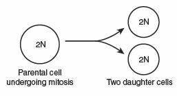
Mitosis involves the division of one cell (parent cell) into two new cells called daughter cells. A cell involved in mitosis contains the normal or diploid (2N) complement of chromosomes for that organism. At the end of mitosis, each daughter cell contains the same number of chromosomes as the parental cell (2N). As you will see shortly, this contrasts with meiosis, which is another type of cell division for the production of gametes.
A large portion of a cell’s life is in a period called interphase. This phase occupies the major portion of the cell cycle when the cell is manufacturing proteins, breaking down harmful products of cell metabolism, and any other special functions it must do. A point is reached when the cell has grown and it must divide. During interphase, the chromosomes (which carry the genetic material, DNA) and organelles are duplicated in preparation for mitosis. One or more nucleoli are visible inside the membrane-bound nucleus. Animal cells also contain structures called centrioles, which also duplicate prior to mitosis. In lab, use the following text along with the available models and posters to better visualize this process. Pay particular attention to the differences between plant and animal cells undergoing mitosis.
Cell division (mitosis) has four main stages and each of these stages will be discussed below. In order of sequence, they are prophase, metaphase, anaphase, and telophase. Collectively they are often abbreviated as “PMAT.” The last event in cell division is cytokinesis, division of the cytoplasm. This process somewhat overlaps with the latter part of telophase and results in the production of two smaller, but functionally complete “daughter” cells.
The Stages of Mitosis:
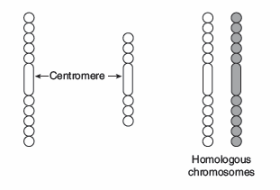
Prophase: The chromosomes become distinct, gradually thickening and shortening. Each chromosome is comprised of two chromatids (referred to as “sister chromatids”), which are connected at the centromere. Under high power you may be able to see these features of the chromosome. During prophase, the nuclear membrane breaks down. In animal cells, the two centrioles separate and organize formation of the mitotic spindle, a scaffold made up of fine fibers called microtubules. Most higher plant cells lack centrioles but a mitotic spindle does form in much the same way as in animal cells.
Metaphase: The chromosomes migrate on the spindle and align at the equatorial plane of the cell. The centromeres are replicated.
Anaphase: The sister chromatids separate as the centromeres are pulled toward opposite poles of the cell. The chromatids move along the spindle toward the opposing poles.
Telophase: In this last stage of mitosis, the chromosomes return to their uncoiled state and the nuclear membrane reappears. The latter part of telophase is marked by the separation of the cell into two daughter cells; this is accomplished by the process of cytokinesis.
Cytokinesis is not a stage of mitosis, but it is the final step in cell division (i.e., division of the cytoplasm). In most animal cells, separation of the two daughter cells is accomplished by the formation of a cleavage furrow. Imagine if a lasso were pulled tight around the midline of the cell to pinch it into two parts. In most plant cells, this separation is achieved by the formation of a new cell wall, called the cell plate, at the midline of the cell. At the end of cytokinesis, the two daughter cells are separated and begin entering interphase thus marking the completion of the cell cycle.
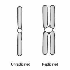
Below is a diagram of a single chromosome and a replicated chromosome. Where appropriate, LABEL the centromere and the sister chromatids.
Lab Overview
Modeling Mitosis—Simulate the events of the four main stages and cytokinesis. You will use plastic pop-it beads and magnetic centromeres to represent chromosomes. To simplify matters, you will assume that the diploid number is four (2N=4). You will need to assemble four replicated chromosomes (or eight sister chromatids). In other words, you will have 2 long red chromatids, 2 short red chromatids, 2 long blue chromatids, and 2 short blue chromatids. A long chromosome/chromatid has 10 beads total (5 beads at each end), a short chromosome/ chromatid has 6 beads total (3 beads at each end). For each homologous pair, red represents the maternal (egg) contribution, blue the paternal (sperm contribution).
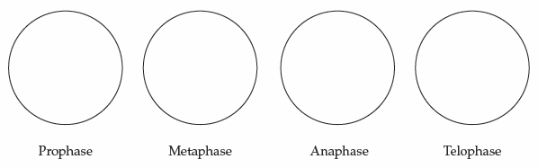
Work through the events of mitosis (PMAT) with your lab partners. Align and move the chromosomes through each of the four stages and be sure you understand what is happening at each stage. When ready, your lab instructor will ask you to demonstrate this process. If it is helpful, make a quick sketch of the basics in the cell diagrams below.
Mitosis in Plant Cells—Onion Root Tips
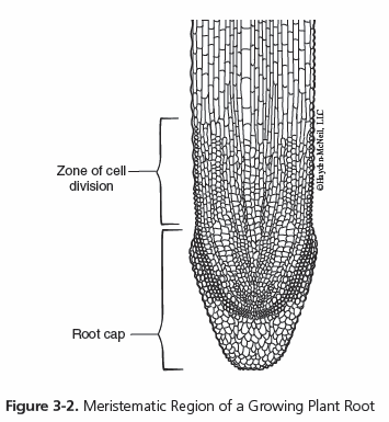
In this exercise you will view prepared slides of onion root tips. As mentioned earlier, the growth regions in plants (meristems) are excellent sources of actively dividing cells. These regions are close to the growing tip (see Figure 3-2).
Self-Check
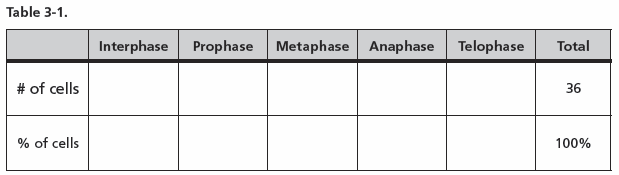
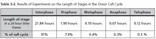
After completing the information on the chart above, answer the following questions about the length of each stage of the cell cycle.
1.
Which stage do yout hink is the longest in mitosis?
2.
Which stage do you think is the shortest in mitosis?
3.
Do you think any of the stages take about the same amount of time? If so, list them below?
4.
See Table 3-2 below for the results of some experiments on the length of the onion cell cycle. Do those results support your conclusions above?