Chapter 10. Modeling Protein Synthesis; Mitosis Review and Meiosis
Objectives
By the end of the period, students will be able to:
- model the process of protein synthesis.
- review the stages of the cell cycle and the process of mitosis.
- understand the importance of and the process of meiosis in animals and plants.
- review the principles of Mendelian genetics using human traits.
Protein Synthesis
We previously modeled the structure of DNA and used that to understand how DNA is replicated. The first part of the lab this week will be to model protein synthesis, or the means that the information in DNA gets transcribed to mRNA, and from there, how it is translated to amino acids, thus forming proteins.
Transcription is the process of the DNA serving as a template for mRNA. There are several steps to transcription, each an important part of the process. Transcription starts when the enzyme RNA polymerase binds to the promotor, a specific site on the DNA. This binding is a key piece to the DNA unwinding. The enzyme then moves along the DNA in a 3' to 5' direction, and as it moves, it builds the complementary strand of mRNA. Remember all new nucleotide strands are built 5’ to 3’.
In lab, we will use the DNA Puzzle Kit to model the creation of the mRNA strand.
Once we have the mRNA strand, it can be read as a series of “codons,” each of which codes for a specific amino acid.
In lab, we will practice reading the chart that determines which amino acid is added to a growing amino acid chain.
Translation is the process of building the amino acid sequence based on the codons on the mRNA.
In lab, we will model this process as well.
Meiosis
Last week we studied how cells divide through the process of mitosis. In mitosis, the DNA in a cell replicates and the cell divides, resulting in two daughter cells that are identical to the original cell. In mitosis, the number of chromosomes in the daughter cells is exactly the same as the number of chromosomes in the parent cell. In meiosis, however, the number of chromosomes is reduced by half, allowing for the important process of reproduction.
MEIOSIS IN ANIMALS
In most animals, the process of meiosis is associated with gamete formation. In most animals, the cells of the body, or the somatic cells, are diploid. A diploid cell is a cell that has two copies of every chromosome. In humans, the somatic cells contain 46 chromosomes, or two copies of each of 23 pairs of chromosomes. Your text shows a karyotype of human chromosomes, showing how the chromosomes occur in pairs.
The sex cells or gametes of animals are haploid, or have only one copy of each chromosome. In humans, the gametes contain 23 chromosomes, or one of each pair. The process of forming gametes includes meiosis and the special processes involved in forming the sperm and the eggs. Spermatogenesis is the process of forming sperm; oogenesis is the process of forming eggs or ova. Fertilization occurs when gametes join. The process of fertilization in animals restores the diploid number of chromosomes. Thus the individual animal, from zygote to adult, is diploid. In most animals, only the gametes are haploid.
Spermatogenesis
Spermatogenesis is the process that involves meiosis and results in sperm cells. Four sperm cells are produced by each spermatogonia. This process occurs in the seminiferous tubules, and in mammals happens continuously once an individual is sexually mature. In this lab we will look at a cross section of the testis to see the process of spermatogenesis. You will make a sketch of this cross section in your Hand-In for this lab.
Oogenesis
Oogenesis is the process that involves meiosis that results in ova or eggs. Unlike spermatogenesis, the cell divisions involved in oogenesis are unequal, and while four cells are produced, only one cell gets much cytoplasm and that cell becomes the ovum. This process occurs in the ovaries, and begins in the fetus. The developing ova are held in the ovaries and continue maturation one at a time starting at puberty. Curiously, in mammals, the process of oogenesis is only completed if fertilization takes places. We will look at a cross section of the ovary to see oogenesis. You will make a sketch of this as part of your Hand-In.
MEIOSIS IN PLANTS
Unlike animals, plants show an alternation of generations: A portion of the life cycle of the plant is haploid and a portion of the life cycle of the plant is diploid. The haploid stage of the plant is called the gametophyte and the gametophyte produces gametes by mitosis. When gametes fuse during the process of fertilization, a diploid individual is formed called the sporophyte. This diploid individual produces a special structure called the sporangium. In the sporangium, meiosis occurs and haploid spores are produced. The haploid spores develop into the haploid gametophyte. You should be able to compare the generalized life cycle of plants and animals and know what process produces gametes for each. We will cover more on life cycles next semester.
THE STAGES OF MEIOSIS
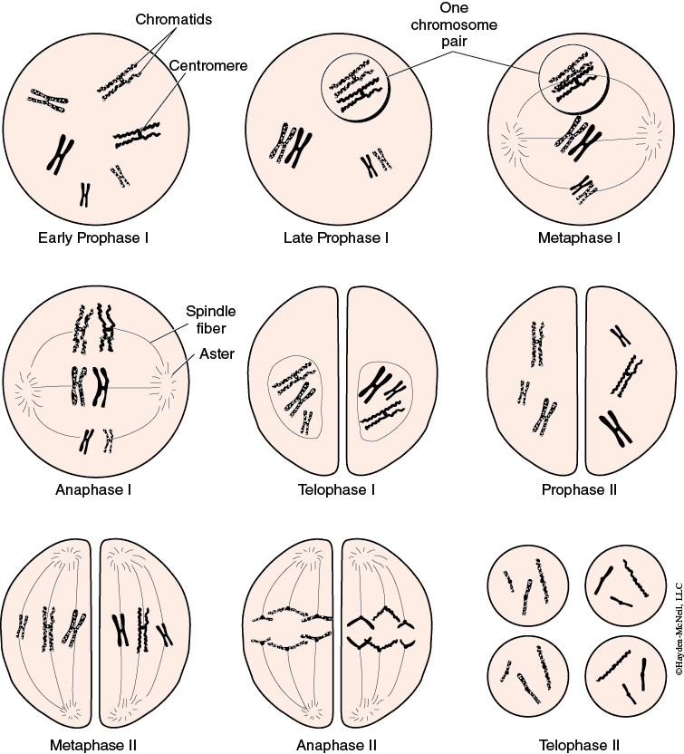
Meiosis involves two separate divisions, resulting in four haploid cells. DNA is replicated only before the first division. The first division is called meiosis I and is sometimes called the reduction division, as it is in this division that the number of chromosomes is reduced to half. In this division, the homologous pairs of chromosomes line up on the equatorial plate. During anaphase I, the homologous pairs split, with one of each pair going to the first daughter cells. The second division is call meiosis II or the equational division. This division is much more like mitosis. Figure 10.1 summarizes the process. We will use “pop beads” to demonstrate this process.
Laboratory Work
Using the compound microscopes, you will look at cross sections of mammalian testes and ovaries as well as lily ovaries and anthers. After that, you will work on understanding what is happening with the chromosomes in meiosis (using an animal model). Each group of 3–4 students should make a nuclear envelope with 4 chromosomes: 1 red and 1 yellow chromosome, each of which is 8 beads long, and 1 red and 1 yellow chromosome, each of which is 6 beads long.
First, simulate the S phase of the cell cycle. Students should replicate each chromosome to make sister chromatids.
Students should demonstrate metaphase of mitosis, telophase of mitosis, anaphase, and cytokinesis.
Next, students should again show a cell that completes the S phase of the cell cycle. This cell will now go through meiosis.
Now show:
Metaphase I:
Telophase I:
Anaphase I:
Metaphase II:
Telophase II:
Anaphase II:
How is meiosis important in fertilization?
Are gametes haploid or diploid?
What happens (genetically) when fertilization takes place?