2.3 Prenatal Development
Now that we understand the starting point, let’s chart prenatal development, tracing how the microscopic, fertilized ovum divides millions of times and differentiates into a living child. This miraculous transformation takes place in three stages.
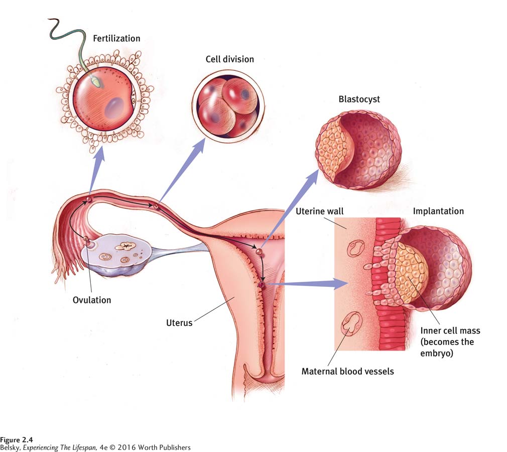
First Two Weeks: The Germinal Stage
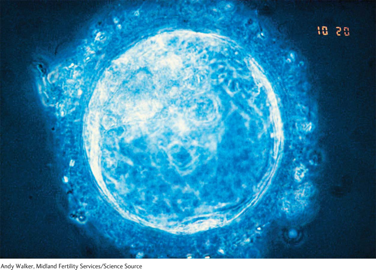
The first approximately two weeks after fertilization—
40
The blastocyst seeks a landing site on the upper uterus. Its outer layer develops projections and burrows in. From this landing zone, blood vessels proliferate to form the placenta, the lifeline that passes nutrients from the mother to the developing baby. Then, the next stage of prenatal development begins: the all-
Week 3 to Week 8: The Embryonic Stage
Although the embryonic stage lasts roughly only six weeks, it is the most fast-
One early task is to construct the conduit responsible for all development. After the baby hooks up to the maternal bloodstream—
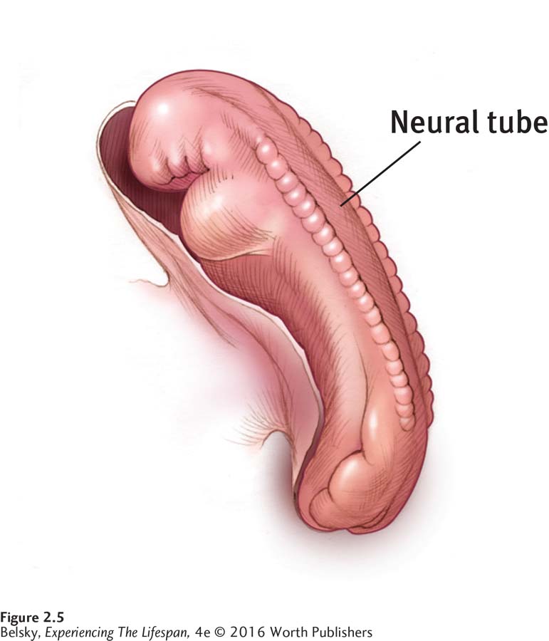
At the same time, the rudiments of the nervous system appear. Between 20 and 24 days after fertilization, an indentation forms along the back of the embryo and closes up to form the neural tube (see Figure 2.5). The upper part of this cylinder becomes the brain. Its lower part forms the spinal cord. Although it is possible to “grow” new brain cells throughout life, almost all of those remarkable branching structures, called neurons, which cause us to think, respond, and process information, originated in neural tube cells formed during our first months in the womb.
Meanwhile, the body is developing at an astounding rate. At day 26, arm buds form; by day 28, leg swellings erupt. At day 37, rudimentary feet start to develop. By day 41, elbows, wrist curves, and the precursors of fingers can be seen. Several days later, raylike structures that will become toes emerge. By about week 8, the internal organs are in place. What started out looking like a curved stalk, then an outer-
Principles of Prenatal Development
In scanning the photographs of the developing embryo below, can you spell out three guiding principles related to the sequence of development I just described?
Notice that from a cylindrical shape, the arms and legs grow outward and then (not unexpectedly) the fingers and toes protrude. So growth follows the proximodistal sequence, from the most interior (proximal) part of the body to the outer (distal) sides.
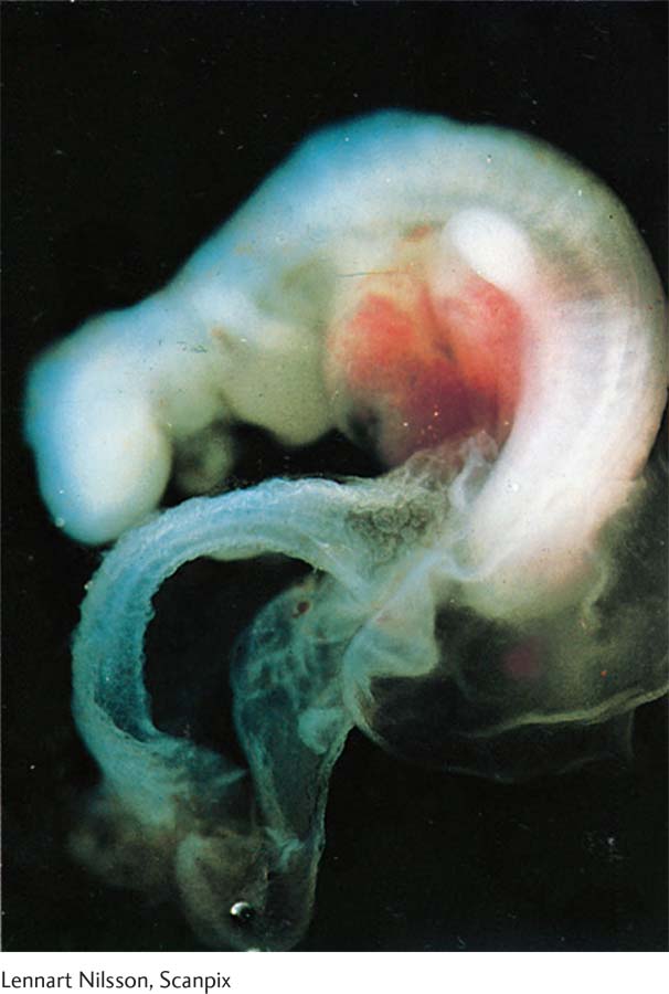 At about week 3, the embryo (the upside-
down U across the top) looks like a curved stalk. Lennart Nilsson, Scanpix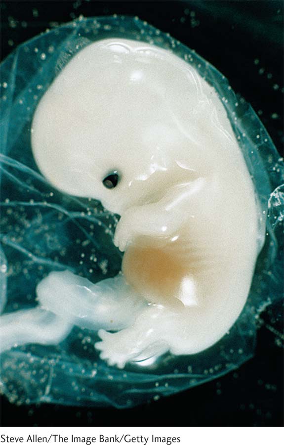 At week 4, you can see the indentations for eyes and the arms and legs beginning to sprout.Steve Allen/The Image Bank/Getty Images
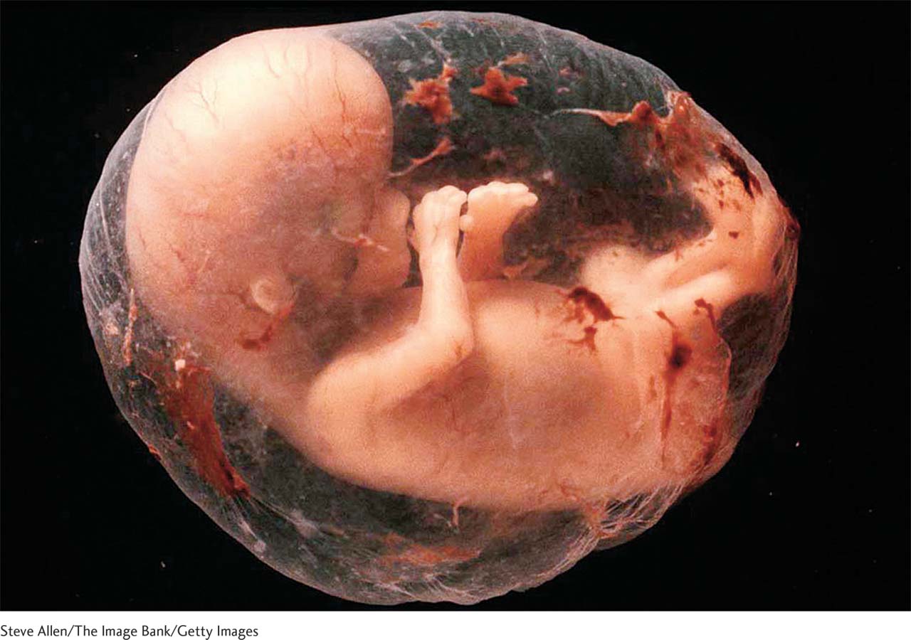 At week 9, the baby-
to- be has fingers, toes, and ears. All the major organs have developed, and the fetal stage has begun! Steve Allen/The Image Bank/Getty Images41
Notice that from a huge swelling that makes the embryo look like a mammoth head, the arms emerge and the legs sprout. So, development takes place according to the cephalocaudal sequence, meaning from top (cephalo = head) to bottom (caudal = tail).
Finally, just as in constructing a sculpture, nature starts with the basic building blocks and then fills in details. A head forms before eyes and ears; legs are constructed before feet and toes. So the mass-
to- specific sequence, or gross (large, simple) structures before smaller (complex) refinements, is the third principle of body growth.
Keep these principles in mind. As you will see in the next chapters, the same patterns apply to growth and motor skills after the baby leaves the womb.
Week 9 to Birth: The Fetal Stage
During the embryonic stage, body structures literally sprout. In the fetal stage, development occurs at a more leisurely pace. From the eyebrows, fingernails, and hair follicles that develop from weeks 9 to 12, to the cushion of fat that accumulates during the final weeks, it takes seven months to transform the embryo into a resilient baby ready to embrace life.
Why does our species need this long refining period? One reason is to allow ample time for that masterpiece organ—
During the late embryonic stage, a mass of cells accumulates within the neural tube that will eventually produce the more than 100 billion neurons composing our brain (Stiles & Jernigan, 2010). From this zone, the neurons migrate to a region just under the top of the differentiating tube (see Figure 2.6). When the cells assemble in their “staging area,” by the middle of the fetal period, they lengthen, develop branches, and interlink. This interconnecting process—
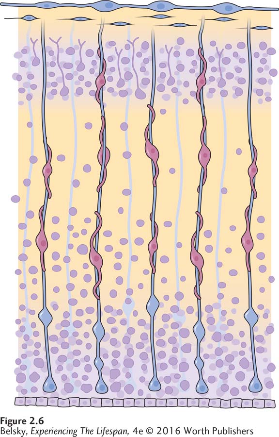
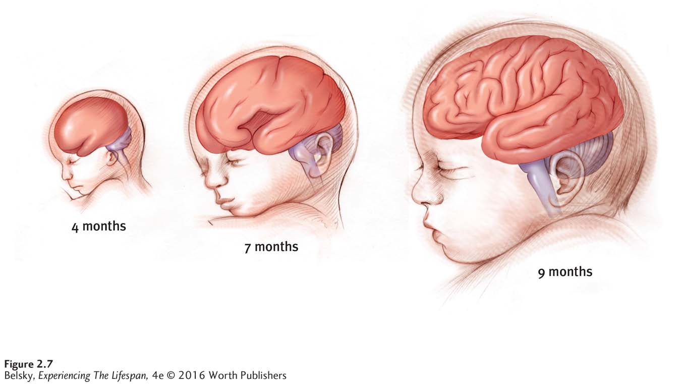
Figure 2.7 shows the mushrooming brain. Notice that the brain almost doubles in size from month 4 to month 7. By now, the brain has the wrinkled structure of an adult.
This massive growth has a profound effect. At around month 6, the fetus can hear (Crade & Lovett, 1988). By month 7, the fetus is probably able to see (Del Giudice, 2011). And by this time, with high-
42
However, it is vitally important that the fetus stay in the uterus as long as possible. As I will describe later, being born too early (and too small) can make a lifelong impact in health.
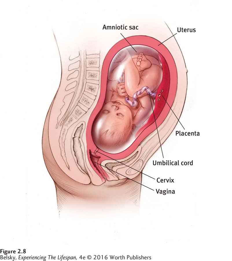
Figure 2.8 shows the fetus during the final month of pregnancy, when its prenatal nest is cramped and birth looms on the horizon. Notice the baby’s support structures: the placenta, projecting from the uterine wall, which supplies nutrients from the mother to the fetus; the umbilical cord, protruding from what will be the baby’s bellybutton, the conduit through which nutrients flow; the amniotic sac, the fluid-
At this stage, parents may be running around, buying the crib or shopping for baby clothes. Middle-
Tying It All Together
Question 2.5
In order, name the three stages of prenatal development. Then, identify the stage in which the organs are formed.
germinal; embryonic; fetal. Organs are formed during the embryonic stage.
Question 2.6
A pregnant friend asks you, “How does my baby’s brain develop?” Describe the process of neural migration—
From the neural tube, a mass of cells differentiates during the late embryonic phase. During the next few months, the cells ascend to the top of the neural tube, completing their migration by week 25. In the final months of pregnancy, the neurons elongate and begin to assume their mature structure.
Question 2.7
Match the following in utero descriptions to the correct names. (Choose from cephalocaudal/proximodistal/mass-
(a) The fingers form before the fingernails; (b) The head forms first and the feet last; (c) the neural tube develops and then the arms.
(a) mass to specific (b) cephalocaudal (c) proximodistal
Question 2.8
You are horrified to learn that your friend went into premature labor yesterday. Pick the minimum pregnancy age that she might be able to have a live birth: around 12 weeks; around 22–
Around 22-