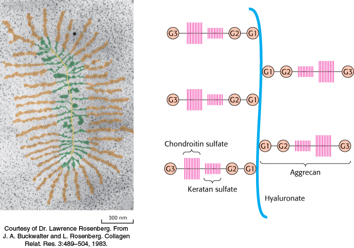
FIGURE 11.21 Structure of proteoglycan from cartilage.3(A) Electron micrograph of a proteoglycan from cartilage (with false color added). Proteoglycan monomers emerge laterally at regular intervals from opposite sides of a central filament of hyaluronate. (B) Schematic representation. G globular domain. [(A)
Courtesy of Dr. Lawrence Rosenberg. From J. A. Buckwalter and L. Rosenberg. Collagen Relat. Res. 3:489– 504, 1983.]
[Leave] [Close]