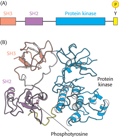
FIGURE 14.33 Src structure. (A) Cellular Src includes an SH3 domain, an SH2 domain, a protein kinase domain, and a carboxyl- terminal tail that includes a key tyrosine residue. (B) Structure of c- Src in an inactivated form with the key tyrosine residue phosphorylated. Notice how the three domains work together to keep the enzyme in an inactive conformation: the phosphotyrosine residue is bound in the SH2 domain and the linker between the SH2 domain and the protein kinase domain is bound by the SH3 domain.
[Drawn from 2PTK.pdb.]
[Leave] [Close]