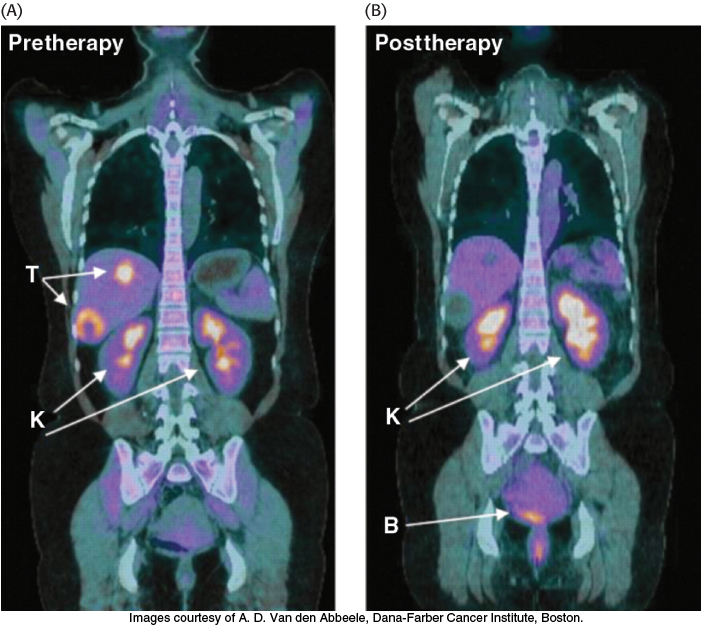
FIGURE 16.22 Tumors can be visualized with 2- 18F- 2- D- deoxyglucose (FDG) and positron emission tomography. (A) A nonmetabolizable glucose analog infused into a patient and detected by a combination of positron emission and computer- aided tomography reveals the presence of a malignant tumor (T). (B) After 4 weeks of treatment with a tyrosine kinase inhibitor (Section 14.5), the tumor shows no uptake of FDG, indicating decreased metabolism. Excess FDG, which is excreted in the urine, also visualizes the kidneys (K) and bladder (B).
[Images courtesy of A. D. Van den Abbeele, Dana- Farber Cancer Institute, Boston.]
[Leave] [Close]