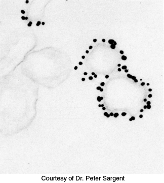
FIGURE 3.26 Immunoelectron microscopy. The opaque particles (150- Å, or 15- nm, diameter) in this electron micrograph are clusters of gold atoms bound to antibody molecules. A gold- labeled antibody against a channel protein (Section 13.4) identifies membrane vesicles at the termini of neurons that contain this protein.
[Courtesy of Dr. Peter Sargent.]
[Leave] [Close]