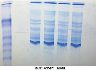
FIGURE 3.9 Staining of proteins after electrophoresis. Mixtures of proteins from cellular extracts subjected to electrophoresis on an SDS– polyacrylamide gel can be visualized by staining with Coomassie blue. The first lane contains a mixture of proteins of known molecular weights, which can be used to estimate the sizes of the bands in the samples.
[©Dr. Robert Farrell.]
[Leave] [Close]