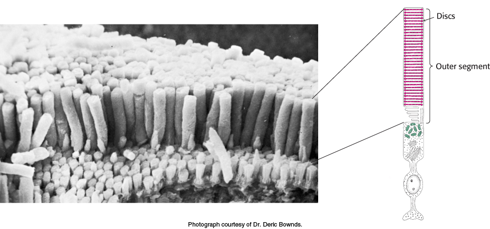
FIGURE 33.19
The rod cell.
(Left) Scanning electron micrograph of retinal rod cells. (Right) Schematic representation of a rod cell.
[
Leave
] [
Close
]
Next