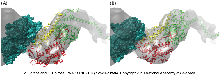
FIGURE 35.13 The structure of myosin bound to actin. (A) The gray surface represents the structure observed by cryoelectron microscopy, with the green space- filling model representing one actin subunit. The ribbon diagram shows the structure of the S1 fragment of myosin docked into the cryoelectron microscopic structure. Notice that some of the myosin structure lies outside the gray surface. (B) The structure after the myosin S1 fragment has been allowed to adjust to more closely match the structure observed by cryoelectron microscopy. Notice that the myosin structure now more closely matches the gray surface.
[Leave] [Close]