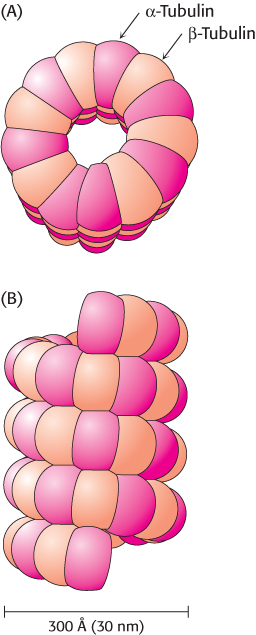
FIGURE 35.20
Microtubule structure.
Schematic views of the helical structure of a microtubule. α-Tubulin is shown in dark red and β-tubulin in light red. (A) Top view. (B) Side view.
[
Leave
] [
Close
]
Next