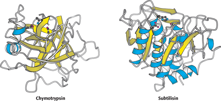
 FIGURE 6.19 Structures of mammalian chymotrypsin and bacterial subtilisin. The overall structures are quite dissimilar, in stark contrast with the active sites, shown at the top of each structure. The β strands are shown in yellow and the α helices in blue.
[Drawn from 1GCT.pdb. and 1SUP.pdb.]