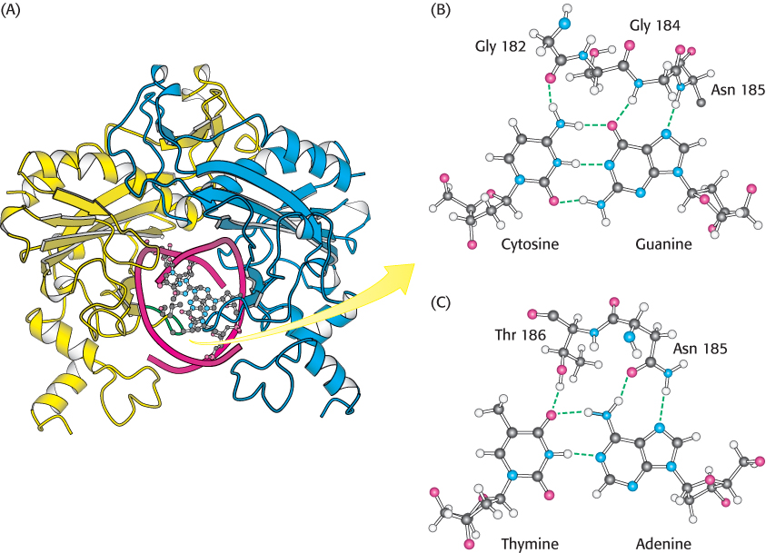
EcoRV embracing a cognate DNA molecule. (A) This view of the structure of EcoRV endonuclease bound to a cognate DNA fragment is down the helical axis of the DNA. The two protein subunits are in yellow and blue, and the DNA backbone is in red. Notice that the twofold axes of the enzyme dimer and the DNA are aligned. One of the DNA- A-
[Drawn from 1RVB.pdb.]