18.1 Eukaryotic Oxidative Phosphorylation Takes Place in Mitochondria

Recall that a biochemical role of the citric acid cycle, which takes place in the mitochondrial matrix, is the generation of high-energy electrons. It is fitting, therefore, that oxidative phosphorylation, which will convert the energy of these electrons into ATP, also takes place in mitochondria. Mitochondria are oval-shaped organelles, typically about 2 μm in length and 0.5 μm in diameter, about the size of a bacterium.

Mitochondria are bounded by a double membrane

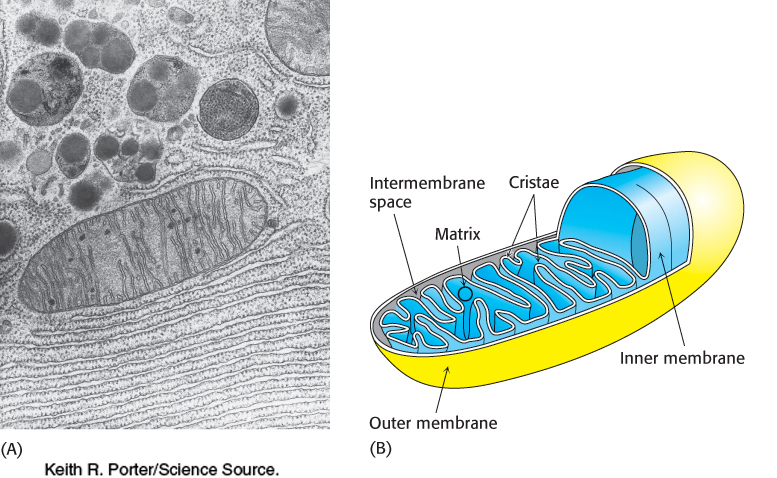
Electron microscopic studies revealed that mitochondria have two membrane systems: an outer membrane and an extensive, highly folded inner membrane. The inner membrane is folded into a series of internal ridges called cristae. Hence, there are two compartments in mitochondria: (1) the intermembrane space between the outer and the inner membranes and (2) the matrix, which is bounded by the inner membrane (Figure 18.2). The mitochondrial matrix is the site of most of the reactions of the citric acid cycle and fatty acid oxidation. In contrast, oxidative phosphorylation takes place in the inner mitochondrial membrane. The increase in surface area of the inner mitochondrial membrane provided by the cristae creates more sites for oxidative phosphorylation than would be the case with a simple, unfolded membrane. Humans contain an estimated 14,000 m2 of inner mitochondrial membrane, which is the approximate equivalent of three football fields in the United States.

Figure 18.2: Electron micrograph (A) and diagram (B) of a mitochondrion.
[(A) Keith R. Porter/Science Source. (B) Information from Wolfe, Biology of the Cell, 2e, © 1981 Brooks/Cole, a part of Cengage Learning, Inc. Reproduced by permission www.cengage.com/permission 3.]

The outer membrane is quite permeable to most small molecules and ions because it contains mitochondrial porin, a 30- to 35-kDa poreforming protein also known as VDAC, for voltage-dependent anion c hannel. VDAC, the most prevalent protein in the outer mitochondrial membrane, plays a role in the regulated flux of metabolites—usually anionic species such as phosphate, chloride, organic anions, and the adenine nucleotides—across the outer membrane. In contrast, the inner membrane is impermeable to nearly all ions and polar molecules. A large family of transporters shuttles metabolites such as ATP, pyruvate, and citrate across the inner mitochondrial membrane. The two faces of this membrane will be referred to as the matrix side and the cytoplasmic side (the latter because it is freely accessible to most small molecules in the cytoplasm). They are also called the N and P sides, respectively, because the membrane potential is negative on the matrix side and positive on the cytoplasmic side.

525

In prokaryotes, the electron-driven proton pumps and ATP-synthesizing complexes are located in the cytoplasmic membrane, the inner of two membranes. The outer membrane of bacteria, like that of mitochondria, is permeable to most small metabolites because of the presence of porins.

Mitochondria are the result of an endosymbiotic event

Figure 18.3: Sizes of mitochondrial genomes. The sizes of three mitochondrial genomes compared with the genome of Rickettsia, a relative of the presumed ancestor of all mitochondria. For genomes of more than 60 kbp, the DNA coding region for genes with known function is shown in red.

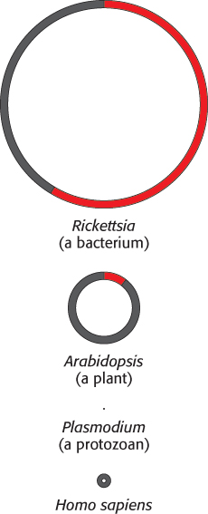
Mitochondria are semiautonomous organelles that live in an endosymbiotic relation with the host cell. These organelles contain their own DNA, which encodes a variety of different proteins and RNAs. Mitochondrial DNA is usually portrayed as being circular, but recent research suggests that the mitochondrial DNA of many organisms may be linear. The genomes of mitochondria range broadly in size across species. The mitochondrial genome of the protist Plasmodium falciparum consists of fewer than 6000 base pairs (bp), whereas those of some land plants comprise more than 200,000 bp (Figure 18.3). Human mitochondrial DNA comprises 16,569 bp and encodes 13 respiratory-chain proteins as well as the small and large ribosomal RNAs and enough tRNAs to translate all codons. However, mitochondria also contain many proteins encoded by nuclear DNA. Cells that contain mitochondria depend on these organelles for oxidative phosphorylation, and the mitochondria in turn depend on the cell for their very existence. How did this intimate symbiotic relation come to exist?

An endosymbiotic event is thought to have occurred whereby a free-living organism capable of oxidative phosphorylation was engulfed by another cell. The double-membrane, circular DNA (with exceptions) and the mitochondrial-specific transcription and translation machinery all point to this conclusion. Thanks to the rapid accumulation of sequence data for mitochondrial and bacterial genomes, speculation on the origin of the “original” mitochondrion with some authority is now possible. The most mitochondrial-like bacterial genome is that of Rickettsia prowazekii, the cause of louse-borne typhus. The genome for this organism is more than 1 million base pairs in size and contains 834 protein-encoding genes. Sequence data suggest that all extant mitochondria are derived from an ancestor of R. prowazekii as the result of a single endosymbiotic event.

526

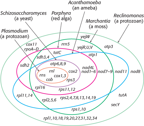
The evidence that modern mitochondria result from a single event comes from examination of the most bacteria-like mitochondrial genome, that of the protozoan Reclinomonas americana. Its genome contains 97 genes, of which 62 specify proteins. The genes encoding these proteins include all of the protein-coding genes found in all of the sequenced mitochondrial genomes (Figure 18.4). Yet, this genome encodes less than 2% of the protein-coding genes in the bacterium E. coli. In other words, a small fraction of bacterial genes—2%—is found in all examined mitochondria. How is it possible that all mitochondria have the same 2% of the bacterial genome? It seems unlikely that mitochondrial genomes resulting from several endosymbiotic events could have been independently reduced to the same set of genes found in R. americana. Thus, the simplest explanation is that the endosymbiotic event took place just once and all existing mitochondria are descendants of that ancestor.

Figure 18.4: Overlapping gene complements of mitochondria. The genes present within each oval are those present within the organism represented by the oval. Only rRNA- and protein-coding genes are shown. The genome of Reclinomonas contains all the protein-coding genes found in all the sequenced mitochondrial genomes.
[Data from M. W. Gray, G. Burger, and B. F. Lang, Science 283:1476–1481, 1999.]

Note that transient engulfment of prokaryotic cells by larger cells is not uncommon in the microbial world. In regard to mitochondria, such a transient relationship it incapable of independent living, and the host cell became dependent on the ATP generated by its tenant.