29.4
The Discovery of Catalytic RNA Was Revealing in Regard to Both Mechanism and Evolution
RNAs form a surprisingly versatile class of molecules. As we have seen, splicing is catalyzed largely by RNA molecules, with proteins playing a secondary role. Another enzyme that contains a key RNA component is ribonuclease P, which catalyzes the maturation of tRNA by endonucleolytic cleavage of nucleotides from the 5′ end of the precursor molecule. Finally, as we shall see in Chapter 30, the RNA component of ribosomes is the catalyst that carries out protein synthesis.
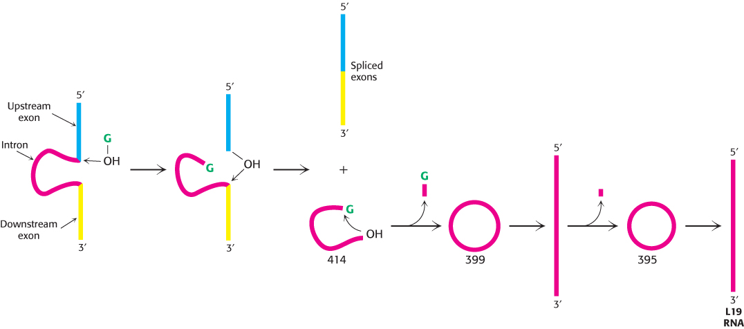
The versatility of RNA first became clear from observations of the processing of ribosomal RNA in a single-cell eukaryote. In Tetrahymena (a ciliated protozoan), a 414-nucleotide intron is removed from a 6.4-kb precursor to yield the mature 26S rRNA molecule (Figure 29.42). In an elegant series of studies of this splicing reaction, Thomas Cech and his coworkers established that the RNA spliced itself to precisely excise the intron. These remarkable experiments demonstrated that an RNA molecule can splice itself in the absence of protein. Indeed, the RNA alone is catalytic and, under certain conditions, is thus a ribozyme. More than 1500 similar introns have since been found in species as widely dispersed as bacteria and eukaryotes, though not in vertebrates. Collectively, they are referred to as group I introns.

FIGURE 29.42Self-splicing. A ribosomal RNA precursor from Tetrahymena, representative of the group I introns, splices itself in the presence of a guanosine cofactor (G, shown in green). A 414-nucleotide intron (red) is released in the first splicing reaction. This intron then splices itself twice again to produce a linear RNA that has lost a total of 19 nucleotides. This L19 RNA is catalytically active.
[Information from T. Cech. RNA as an enzyme. Copyright © 1986 by Scientific American, Inc. All rights reserved.]
The self-splicing reaction in the group I intron requires an added guanosine nucleotide. Nucleotides were originally included in the reaction mixture because it was thought that ATP or GTP might be needed as an energy source. Instead, the nucleotides were found to be necessary as cofactors. The required cofactor proved to be a guanosine unit, in the form of guanosine, GMP, GDP, or GTP. G (denoting any one of these species) serves not as an energy source but as an attacking group that becomes transiently incorporated into the RNA (Figure 29.42). G binds to the RNA and then attacks the 5′ splice site to form a phosphodiester bond with the 5′ end of the intron. This transesterification reaction generates a 3′ OH group at the end of the upstream exon. This newly attached 3′
 OH group then attacks the 3′ splice site. This second transesterification reaction joins the two exons and leads to the release of the 414-nucleotide intron.
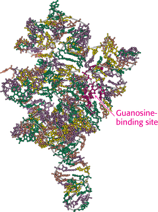
Self-splicing depends on the structural integrity of the RNA precursor. Much of the group I intron is needed for self-splicing. This molecule, like many RNAs, has a folded structure formed by many double-helical stems and loops (Figure 29.43), with a well-defined pocket for binding the guanosine. Examination of the three-dimensional structure of a catalytically active group I intron determined by x-ray crystallography reveals the coordination of magnesium ions in the active site analogous to that observed in protein enzymes such as DNA polymerase.

 FIGURE 29.43 Structure of a self-splicing intron. The structure of a large fragment of the self-splicing intron from Tetrahymena reveals a complex folding pattern of helices and loops. Bases are shown in green, A; yellow, C; purple, G; and orange, U.
[Drawn from 1GRZ.pdb].

FIGURE 29.44Self-splicing mechanism. The catalytic mechanism of the group I intron includes a series of transesterification reactions.
[Information from T. Cech. RNA as an enzyme. Copyright © 1986 by Scientific American, Inc. All rights reserved.]
Analysis of the base sequence of the rRNA precursor suggested that the splice sites are aligned with the catalytic residues by base-pairing between the internal guide sequence (IGS) in the intron and the 5′ and 3′ exons (Figure 29.44). The IGS first brings together the guanosine cofactor and the 5′ splice site so that the 3′ OH group of G can nucleophilically attack the phosphorus atom at this splice site. The IGS then holds the downstream exon in position for attack by the newly formed 3′
 OH group of the upstream exon. A phosphodiester bond is formed between the two exons, and the intron is released as a linear molecule. Like catalysis by protein enzymes, self-catalysis of bond formation and breakage in this rRNA precursor is highly specific.
The finding of enzymatic activity in the self-splicing intron and in the RNA component of RNase P has opened new areas of inquiry and changed the way in which we think about molecular evolution. As mentioned in an earlier chapter, the discovery that RNA can be a catalyst as well as an information carrier suggests that an RNA world may have existed early in the evolution of life, before the appearance of DNA and protein.
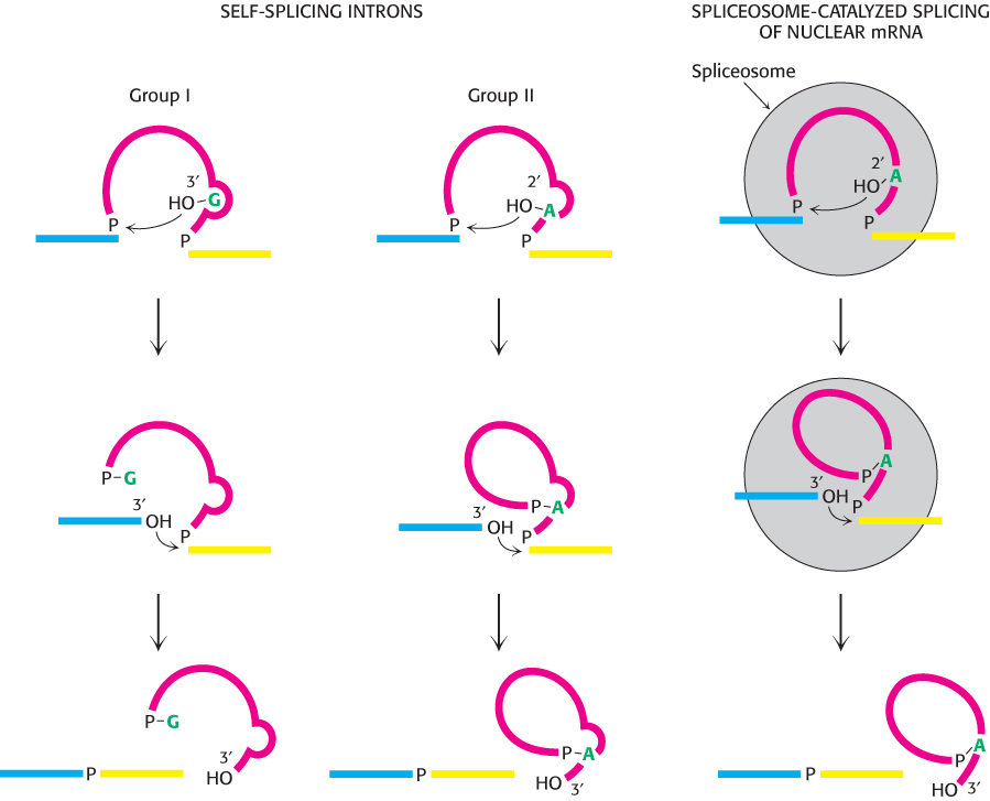
Messenger RNA precursors in the mitochondria of yeast and fungi also undergo self-splicing, as do some RNA precursors in the chloroplasts of unicellular organisms such as Chlamydomonas. Self-splicing reactions can be classified according to the nature of the unit that attacks the upstream splice site. Group I self-splicing is mediated by a guanosine cofactor, as in Tetrahymena. The attacking moiety in group II splicing is the 2′ OH group of a specific adenylate of the intron (Figure 29.45).

FIGURE 29.45Comparison of splicing pathways. The exons being joined are shown in blue and yellow and the attacking unit is shown in green. The catalytic site is formed by the intron itself (red) in group I and group II splicing. In contrast, the splicing of nuclear mRNA precursors is catalyzed by snRNAs and their associated proteins in the spliceosome.
[Information from P. A. Sharp, Science 235:766–771, 1987.]
Group I and group II self-splicing resembles spliceosome-catalyzed splicing in two respects. First, in the initial step, a ribose hydroxyl group attacks the 5′ splice site. The newly formed 3′-OH terminus of the upstream exon then attacks the 3′ splice site to form a phosphodiester bond with the downstream exon. Second, both reactions are transesterifications in which the phosphate moieties at each splice site are retained in the products. The number of phosphodiester bonds stays constant. Group II splicing is like the spliceosome-catalyzed splicing of mRNA precursors in several additional ways. The attack at the 5′ splice site is carried out by a part of the intron itself (the 2′-OH group of adenosine) rather than by an external cofactor (G). In both cases, the intron is released in the form of a lariat. Moreover, in some instances, the group II intron is transcribed in pieces that assemble through hydrogen bonding to the catalytic intron, in a manner analogous to the assembly of the snRNAs in the spliceosome.
 These similarities have led to the suggestion that the spliceosome-catalyzed splicing of mRNA precursors evolved from RNA-catalyzed self-splicing. Group II splicing may well be an intermediate between group I splicing and the splicing in the nuclei of higher eukaryotes. A major step in this transition was the transfer of catalytic power from the intron itself to other molecules. The formation of spliceosomes gave genes a new freedom because introns were no longer constrained to provide the catalytic center for splicing. Another advantage of external catalysts for splicing is that they can be more readily regulated. However, it is important to note that similarities do not establish ancestry. The similarities between group II introns and mRNA splicing may be a result of convergent evolution. Perhaps there are only a limited number of ways to carry out efficient, specific intron excision. The determination of whether these similarities stem from ancestry or from chemistry will require expanding our understanding of RNA biochemistry.

 OH group at the end of the upstream exon. This newly attached 3′
 OH group then attacks the 3′ splice site. This second transesterification reaction joins the two exons and leads to the release of the 414-
 FIGURE 29.43 Structure of a self-
 OH group of G can nucleophilically attack the phosphorus atom at this splice site. The IGS then holds the downstream exon in position for attack by the newly formed 3′
 OH group of the upstream exon. A phosphodiester bond is formed between the two exons, and the intron is released as a linear molecule. Like catalysis by protein enzymes, self-
 OH group of a specific adenylate of the intron (Figure 29.45).
 These similarities have led to the suggestion that the spliceosome-