30.2 Aminoacyl Transfer RNA Synthetases Read the Genetic Code

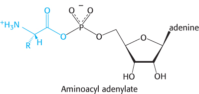
Before codon and anticodon meet, the amino acids required for protein synthesis must first be attached to specific tRNA molecules, linkages that are crucial for two reasons. First, the attachment of a given amino acid to a particular tRNA establishes the genetic code. When an amino acid has been linked to a tRNA, it will be incorporated into a growing polypeptide chain at a position dictated by the anticodon of the tRNA. Second, because the formation of a peptide bond between free amino acids is not thermodynamically favorable, the amino acid must first be activated in order for protein synthesis to proceed. The activated intermediates in protein synthesis are amino acid esters, in which the carboxyl group of an amino acid is linked to either the 2′- or the 3′-hydroxyl group of the ribose unit at the 3′ end of tRNA. An amino acid ester of tRNA is called an aminoacyl-tRNA or sometimes a charged tRNA (Figure 30.6). For a specific amino acid attached to its cognate tRNA—for instance, threonine—the charged tRNA is designated Thr-tRNAThr.

Amino acids are first activated by adenylation

The activation reaction is catalyzed by specific aminoacyl-tRNA synthetases. The first step is the formation of an aminoacyl adenylate from an amino acid and ATP.

899

This activated species is a mixed anhydride in which the carboxyl group of the amino acid is linked to the phosphoryl group of AMP; hence, it is also known as aminoacyl-AMP.

The next step is the transfer of the aminoacyl group of aminoacyl-AMP to a particular tRNA molecule to form aminoacyl-tRNA.

The sum of these activation and transfer steps is

The ΔG°′ of this reaction is close to 0, because the free energy of hydrolysis of the ester bond of aminoacyl-tRNA is similar to that for the hydrolysis of ATP to AMP and PPi. As we have seen many times, the reaction is driven by the hydrolysis of pyrophosphate. The sum of these three reactions is highly exergonic:

Thus, the equivalent of two molecules of ATP is consumed in the synthesis of each aminoacyl-tRNA. One of them is consumed in forming the ester linkage of aminoacyl-tRNA, whereas the other is consumed in driving the reaction forward.

The activation and transfer steps for a particular amino acid are catalyzed by the same aminoacyl-tRNA synthetase. Indeed, the aminoacyl-AMP intermediate does not dissociate from the synthetase. Rather, it is tightly bound to the active site of the enzyme by noncovalent interactions.

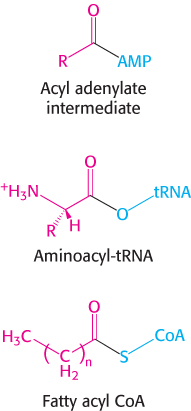
We have already encountered an acyl adenylate intermediate in fatty acid activation (Section 22.2). The major difference between these reactions is that the acceptor of the acyl group is CoA in fatty acid activation and tRNA in amino acid activation. The energetics of these biosyntheses are very similar: both are made irreversible by the hydrolysis of pyrophosphate.

Aminoacyl-tRNA synthetases have highly discriminating amino acid activation sites

Each aminoacyl-tRNA synthetase is highly specific for a given amino acid. Indeed, a synthetase will incorporate the incorrect amino acid only once in 104 or 105 reactions. How is this level of specificity achieved? Each aminoacyl-tRNA synthetase takes advantage of the properties of its amino acid substrate. Let us consider the challenge faced by threonyl-tRNA synthetase. Threonine is especially similar to two other amino acids—namely, valine and serine. Valine has almost exactly the same shape as that of threonine, except that valine has a methyl group in place of a hydroxyl group. Serine has a hydroxyl group, as does threonine, but lacks the methyl group. How can the threonyl-tRNA synthetase avoid coupling these incorrect amino acids to threonyl-tRNA?

900

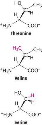
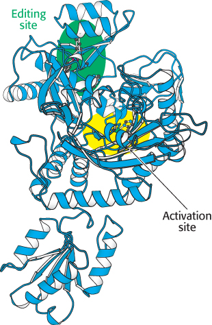
The structure of the amino acid-binding site of threonyl-tRNA synthetase reveals how valine is avoided (Figure 30.8). The synthetase contains a zinc ion, bound to the enzyme by two histidine residues and one cysteine residue. The remaining coordination sites are available for substrate binding. Threonine binds to the zinc ion through its amino group and its side-chain hydroxyl group. The side-chain hydroxyl group is further recognized by an aspartate residue that hydrogen bonds to it. The methyl group present in valine in place of this hydroxyl group cannot participate in these interactions; it is excluded from this active site and, hence, does not become adenylated and transferred to threonyl-tRNA (abbreviated tRNAThr). The use of a zinc ion appears to be unique to threonyl-tRNA synthetase; other aminoacyl-tRNA synthetases have different strategies for recognizing their cognate amino acids. The carboxylate group of the correctly positioned threonine is available to attack the α phosphoryl group of ATP to form the aminoacyl adenylate.

Figure 30.8: Active site of threonyl-tRNA synthetase. Notice that the amino acid-binding site includes a zinc ion (green ball) that binds threonine through its amino and hydroxyl groups.

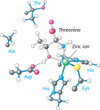
The zinc site is less able to discriminate against serine because this amino acid does have a hydroxyl group that can bind to the zinc ion. Indeed, with only this mechanism available, threonyl-tRNA synthetase does mistakenly couple serine to threonyl-tRNA at a rate 10−2 to 10−3 times that for threonine. This error rate is likely to lead to many translation errors. How is a higher level of specificity achieved?

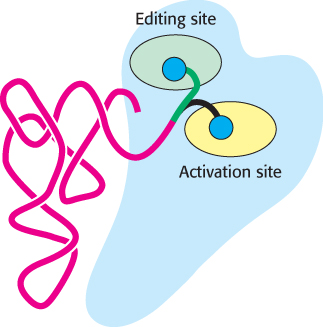
Proofreading by aminoacyl-tRNA synthetases increases the fidelity of protein synthesis

Figure 30.9: Editing site. Mutagenesis studies revealed the position of the editing site (shown in green) in threonyl-tRNA synthetase. The activation site is shown in yellow. Only one subunit of the dimeric enzyme is shown here and in subsequent illustrations.
[Drawn from 1QF6.pdb.]

Threonyl-tRNA synthetase can be incubated with tRNAThr that has been covalently linked with serine (Ser-tRNAThr); the tRNA has been “mpara-93ischarged.” The reaction is immediate: a rapid hydrolysis of the aminoacyl-tRNA forms serine and free tRNA. In contrast, incubation with correctly charged Thr-tRNAThr results in no reaction. Thus, threonyl-tRNA synthetase contains an additional functional site that hydrolyzes Ser-tRNAThr but not Thr-tRNAThr. This editing site provides an opportunity for the synthetase to correct its mistakes and improve its fidelity to less than one mistake in 104. The results of structural and mutagenesis studies revealed that the editing site is more than 20 Å from the activation site (Figure 30.9). This editing site readily accepts and cleaves Ser-tRNAThr but does not cleave Thr-tRNAThr. The discrimination of serine from threonine is easy because threonine contains an extra methyl group; a site that conforms to the structure of serine will sterically exclude threonine. The structure of the complex between threonyl-tRNA synthetase and its substrate reveals that the aminoacylated CCA can swing out of the activation site and into the editing site (Figure 30.10). Thus, the aminoacyl-tRNA can be edited without dissociating from the synthetase. This proofreading depends on the conformational flexibility of a short stretch of polynucleotide sequence.

Figure 30.10: Editing of aminoacyl-tRNA. The flexible CCA arm of an aminoacyl-tRNA can move the amino acid between the activation site and the editing site. If the amino acid fits well into the editing site, the amino acid is removed by hydrolysis.

901

Most aminoacyl-tRNA synthetases contain editing sites in addition to activation sites. These complementary pairs of sites function as a double sieve to ensure very high fidelity. In general, the acylation site rejects amino acids that are larger than the correct one because there is insufficient room for them, whereas the hydrolytic site cleaves activated species that are smaller than the correct one.

A few synthetases achieve high accuracy without editing. For example, tyrosyl-tRNA synthetase has no difficulty discriminating between tyrosine and phenylalanine; the hydroxyl group on the tyrosine ring enables tyrosine to bind to the enzyme 104 times as strongly as phenylalanine. Proofreading has been selected in evolution only when fidelity must be enhanced beyond what can be obtained through an initial binding interaction.

Synthetases recognize various features of transfer RNA molecules

Figure 30.11: Threonyl-tRNA synthetase complex. The structure shows the complex between threonyl-tRNA synthetase (blue) and tRNAThr (red). Notice that the synthetase binds to both the acceptor stem and the anticodon loop.
[Drawn from 1QF6.pdb.]

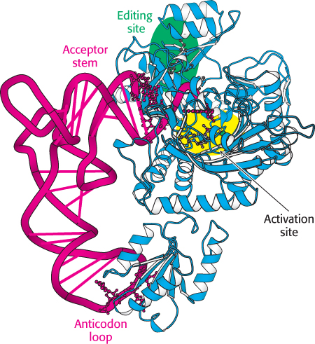
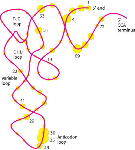
How do synthetases choose their tRNA partners? This enormously important step is the point at which “translation” takes place—at which the correlation between the amino acid and the nucleic acid worlds is made. In a sense, aminoacyl-tRNA synthetases are the only molecules in biology that “know” the genetic code. Their precise recognition of tRNAs is as important for high-fidelity protein synthesis as is the accurate selection of amino acids. In general, tRNA recognition by the synthetase is different for each synthetase and tRNA pair. Consequently, generalities are difficult to make.

Some synthetases recognize their tRNA partners primarily on the basis of their anticodons, although they may also recognize other aspects of tRNA structure that vary among different tRNAs. The most direct evidence comes from crystallographic studies of complexes formed between synthetases and their cognate tRNAs. Consider, for example, the structure of the complex between threonyl-tRNA synthetase and tRNAThr (Figure 30.11). As expected, the CCA arm extends into the zinc-containing activation site, where it is well positioned to accept threonine from threonyl adenylate. The enzyme interacts extensively not only with the acceptor stem of the tRNA, but also with the anticodon loop. The interactions with the anticodon loop are particularly revealing. Each base within the sequence 5′-CGU-3′ of the anticodon participates in hydrogen bonds with the enzyme; those with the second two bases (G and U) appear to be more important because the synthetase interacts just as efficiently with the anticodons GGU and UGU. Although interactions between the enzyme and the anticodon are often crucial for correct recognition,Figure 30.12 shows that many aspects of tRNA molecules are recognized by synthetases. Note that many of the recognition sites are loops rich in unusual bases that can provide structural identifiers.

Figure 30.12: Recognition sites on tRNA. Circles represent nucleotides, and the sizes of the circles are proportional to the frequency with which they are used as recognition sites by aminoacyl-tRNA synthetases. The numbers indicate the positions of the nucleotides in the base sequence, beginning from the 5′ end of the tRNA molecule.
[Information from M. Ibba, and D. Söll, Annu. Rev. Biochem. 69:617–650, 1981, p. 636.]

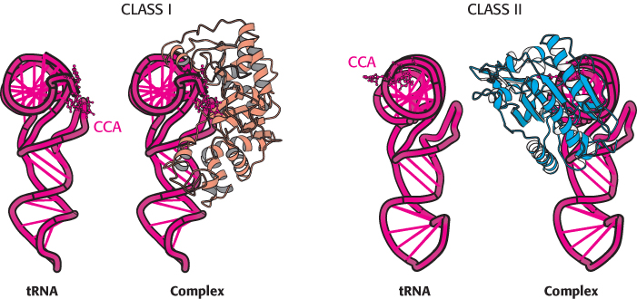
Aminoacyl-tRNA synthetases can be divided into two classes

Class I

Class II

Arg (α)

Ala (α4)

Cys (α)

Asn (α2)

Gln (α)

Asp (α2)

Glu (α)

Gly (α2β2)

IIe (α)

His (α2)

Leu (α)

Lys (α2)

Met (α)

Phe (α2β2)

Trp (α2)

Ser (α2)

Tyr (α2)

Pro (α2)

Val (α)

Thr (α2)

Table 30.3: Classification and subunit structure of aminoacyl-tRNA ­synthetases in E. coli

At least one aminoacyl-tRNA synthetase exists for each amino acid. The diverse sizes, subunit composition, and sequences of these enzymes were bewildering for many years. Could it be that essentially all synthetases evolved independently? The determination of the three-dimensional structures of several synthetases followed by more-refined sequence comparisons revealed that different synthetases are, in fact, related. Specifically, synthetases fall into two classes, termed class I and class II, each of which includes enzymes specific for 10 of the 20 amino acids (Table 30.3). Intriguingly, synthetases from the two classes bind to different faces of the tRNA molecule (Figure 30.13). The CCA arm of tRNA adopts different conformations to accommodate these interactions; the arm is in the helical conformation observed for free tRNA (Figures 30.4 and 30.5) for class II enzymes and in a hairpin conformation for class I enzymes. These two classes also differ in other ways.

Figure 30.13: Classes of aminoacyl-tRNA synthetases. Notice that class I and class II synthetases recognize different faces of the tRNA molecule. The CCA arm of tRNA adopts different conformations in complexes with the two classes of synthetase. Note that the CCA arm of the tRNA is turned toward the viewer (Figures 30.4 and 30.5).
[Drawn from 1EUY.pdb and 1QF6.pdb.]

902

1. Class I enzymes acylate the 2′-hydroxyl group of the terminal adenosine of tRNA, whereas class II enzymes (except the enzyme for Phe-tRNA) acylate the 3′-hydroxyl group.

2. The two classes bind ATP in different conformations.

3. Most class I enzymes are monomeric, whereas most class II enzymes are dimeric.

Why did two distinct classes of aminoacyl-tRNA synthetases evolve? The observation that the two classes bind to distinct faces of tRNA suggests a possibility. Recognition sites on both faces of tRNA may have been required to allow the recognition of 20 different tRNAs.