32.1 Eukaryotic DNA Is Organized into Chromatin

Eukaryotic DNA is tightly bound to a group of small basic proteins called histones. In fact, histones constitute half the mass of a eukaryotic chromosome. The entire complex of a cell’s DNA and associated protein is called chromatin. Chromatin compacts and organizes eukaryotic DNA and its presence has dramatic consequences for gene regulation.

Nucleosomes are complexes of DNA and histones

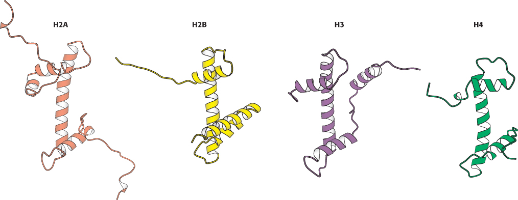
Chromatin is made up of repeating units, each containing 200 bp of DNA and two copies each of four histone proteins H2A, H2B, H3, and H4. Histones have strikingly basic properties because a quarter of the residues in each histone are either arginine or lysine, positively charged amino acids that strongly interact with the negatively charged DNA. The protein complex is called the histone octamer. The repeating units of the histone octamer and the associated DNA are known as nucleosomes. Chromatin viewed with the electron microscope has the appearance of beads on a string (Figure 32.2); each bead has a diameter of approximately 100 Å. Partial digestion of chromatin with DNase yields the isolated beads. These particles consist of fragments of DNA about 200 bp in length bound to the histone octamer. More-extensive digestion yields a shorter DNA fragment of 145 bp bound to the octamer. The smaller complex formed by the histone octamer and the 145-bp DNA fragment is the nucleosome core particle. The DNA connecting core particles in undigested chromatin is called linker DNA. Histone H1 binds, in part, to the linker DNA.

DNA wraps around histone octamers to form nucleosomes

The overall structure of the nucleosome was revealed through electron microscopic and x
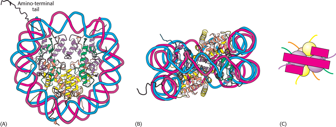
-ray crystallographic studies pioneered by Aaron Klug and his colleagues. More recently, the three-dimensional structure of a reconstituted nucleosome core particle (Figure 32.3) was determined to higher resolution by x-ray diffraction methods. The four types of histone that make up the protein core are homologous and similar in structure (Figure 32.4). The eight histones in the core are arranged into a (H3)2(H4)2 tetramer and a pair of H2A–H2B dimers. The tetramer and dimers come together to form a left-handed superhelical ramp around which the DNA wraps. In addition, each histone has an amino-terminal tail that extends out from the core structure. These tails are flexible and contain a number of lysine and arginine residues. As we shall see, covalent modifications of these tails play an essential role in regulating gene expression.

Figure 32.3: Nucleosome core particle. The structure consists of a core of eight histone proteins surrounded by DNA. (A) A view showing the DNA wrapping around the histone core. (B) A 90-degree rotation of the view in part (A). Notice that the DNA forms a left-handed superhelix as it wraps around the core. (C) A schematic view.
[Drawn from 1AOI.pdb.]
Figure 32.4: Homologous histones. Histones H2A, H2B, H3, and H4 adopt a similar three-dimensional structure as a consequence of common ancestry. Some parts of the tails at the termini of the proteins are not shown.
[Drawn from 1AOI.pdb.]
Figure 32.5: Higher-order chromatin structure. A proposed model for chromatin arranged in a helical array consisting of six nucleosomes per turn of helix. The DNA double helix (shown in red) is wound around each histone octamer (shown in blue). The actual structure in cells is likely more heterogeneous and dynamic than suggested by this model.
[Information from J. T. Finch and A. Klug, Proc. Natl. Acad. Sci. U. S. A. 73:1897–1901, 1976.]

944

The DNA forms a left-handed superhelix as it wraps around the outside of the histone octamer. The protein core forms contacts with the inner surface of the DNA superhelix at many points, particularly along the phosphodiester backbone and the minor groove. Nucleosomes will form on almost all DNA sites, although some sequences are preferred because the dinucleotide steps are properly spaced to favor bending around the histone core. A histone with a different structure from that of the others, called histone H1, seals off the nucleosome at the location at which the linker DNA enters and leaves. The amino acid sequences of histones, including their amino-terminal tails, are remarkably conserved from yeast to human beings.

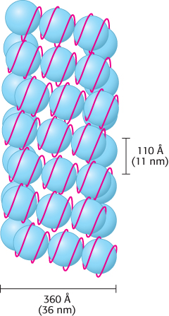
The winding of DNA around the nucleosome core contributes to the packing of DNA by decreasing its linear extent. An extended 200-bp stretch of DNA would have a length of about 680 Å. Wrapping this DNA around the histone octamer reduces the length to approximately 100 Å along the long dimension of the nucleosome. Thus the DNA is compacted by a factor of 7. However, human chromosomes in metaphase, which are highly condensed, are compacted by a factor of 104. Clearly, the nucleosome is just the first step in DNA compaction. What is the next step? The nucleosomes themselves are arranged in a helical array approximately 360 Å across, forming a series of stacked layers approximately 110 Å apart (Figure 32.5). The folding of these fibers of nucleosomes into loops further compacts DNA.

The wrapping of DNA around the histone octamer as a left-handed helix also stores negative supercoils; if the DNA in a nucleosome is straightened out, the DNA will be underwound. This underwinding is exactly what is needed to separate the two DNA strands during replication and transcription.

945