Prenatal Development
The most dramatic and extensive transformation of life occurs before birth. To make it easier to study, prenatal development is often divided into three main periods. The first two weeks are called the germinal period the third through the eighth week is the embryonic period from then until birth is the fetal period. (Alternative terms are presented in Table 4.1.)
Germinal: The First 14 Days
You learned in Chapter 3 that the one-
About a week after conception, the cell mass, now called a blastocyst, forms two distinct parts—
The first task of the outer cells is to achieve implantation—that is, to embed themselves in the nurturing lining of the uterus (see Figure 4.1). This process is far from automatic; about half of natural conceptions and an even larger proportion of in vitro conceptions never implant (see Table 4.2). Most new life ends before an embryo begins (Sadler, 2012).
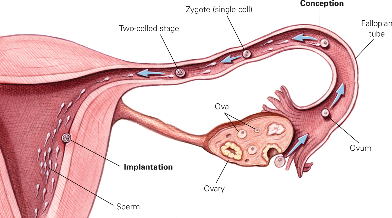
The Most Dangerous Journey In the first 10 days after conception, the organism does not increase in size because it is not yet nourished by the mother. However, the number of cells increases rapidly as the organism prepares for implantation, which occurs successfully not quite half of the time.
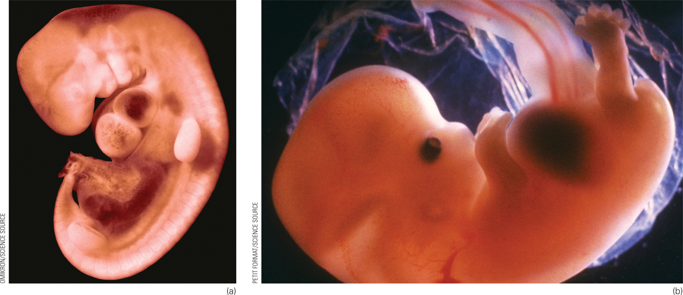
Embryo: From the Third Through the Eighth Week
The start of the third week after conception initiates the embryonic period, during which the formless mass of cells becomes a distinct being—
First, a thin line (called the primitive streak) appears down the middle of the embryo; it will become the neural tube between 20 and 27 days after conception and eventually develop into the central nervous system (the brain and spinal column) (Stiles & Jernigan, 2010). The head appears in the fourth week, as eyes, ears, nose, and mouth start to form. Also in the fourth week, a minuscule blood vessel that will become the heart begins to pulsate.
By the fifth week, buds that will become arms and legs emerge. The upper arms and then forearms, palms, and webbed fingers grow. Legs, knees, feet, and webbed toes, in that order, emerge a few days later, each having the beginning of a skeletal structure. Then, 52 and 54 days after conception, respectively, the fingers and toes separate (Sadler, 2012).
As you can see, prenatally, the head develops first, in a cephalo-
Fetus: From the Ninth Week Until Birth
The organism is called a fetus from the beginning of the ninth week after conception until birth. The fetal period encompasses dramatic change, from a tiny, sexless creature smaller than the final joint of your thumb to a boy or girl about 20 inches (51 centimeters) long.
The Third Month
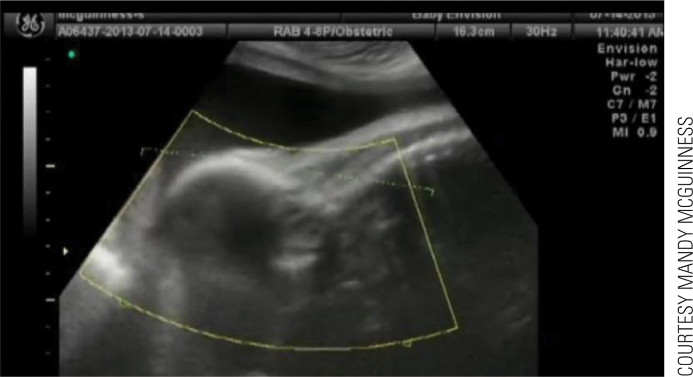

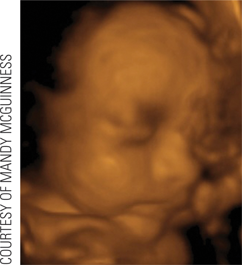
Video: Prenatal Period: 3D Ultrasound shows a real-
If the 23rd chromosomes are XY, the SRY gene on the Y triggers the development of male sexual organs. Otherwise, female organs develop. The male fetus experiences a rush of the hormone testosterone, affecting many structures and connections in the brain (Filová et al., 2013).
Of course, the range of brain and behavioral variations among males and among females is greater than the variations between the average man and woman. Nonetheless, neurological sex differences begin early in prenatal development.
By the end of the third month, the sex organs may be visible via ultrasound (in a sonogram), which is similar to an X-
The Middle Three Months
Especially for Biologists Many people believe that the differences between the sexes are sociocultural, not biological. Is there any prenatal support for that view?
Only one of the 46 human chromosomes determines sex, and the genitals develop last in the prenatal sequence, suggesting that dramatic male–
In the fourth, fifth, and sixth months, the heartbeat becomes stronger. Digestive and excretory systems develop. Fingernails, toenails, and buds for teeth form, and hair grows (including eyelashes).
The brain increases about six times in size and develops many new neurons (neurogenesis) and synapses (synaptogenesis). Indeed, mid-
Brain development occurs in every prenatal month, but these middle three months are especially crucial (Johnson, 2011). The entire central nervous system becomes responsive during mid-
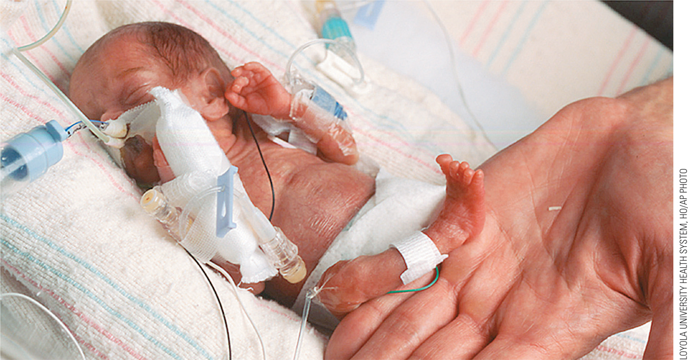
Survival is far from automatic; almost all 22-
In Japan, with excellent neonatal care, 20 percent of 22-
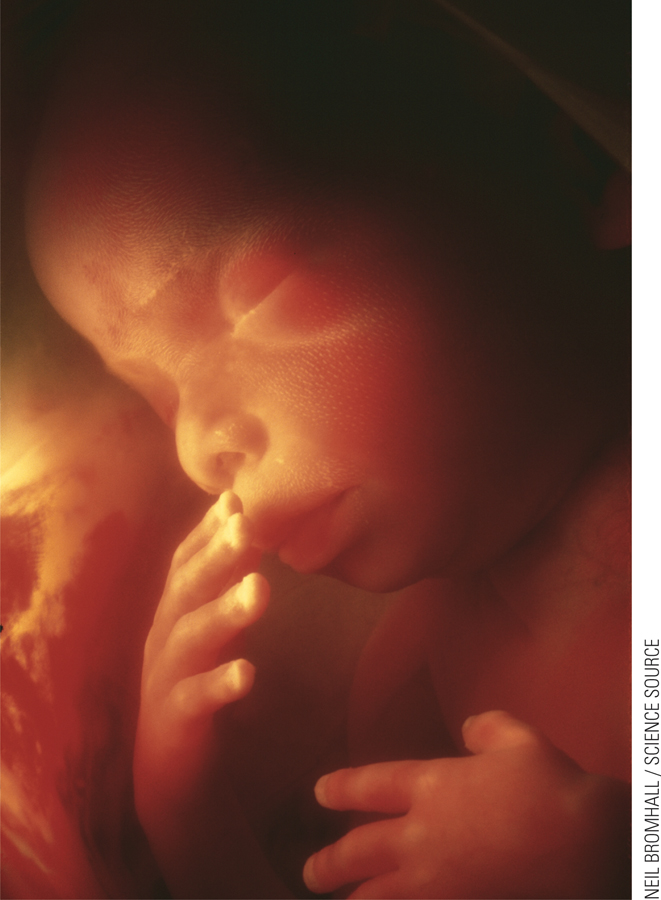
The Final Three Months
Reaching viability simply means that life outside the womb is possible. Many babies born between 22 and 24 weeks die, and survivors born before 27 weeks often develop slowly because they have missed some essential brain development in the uterus (Månsson & Stjernqvist, 2014). Each day of the final three months improves the odds not only of survival but also of a healthy life and normal cognition. (More on preterm birth appears later in this chapter.) Many aspects of prenatal life are awe-
Organ Maturation
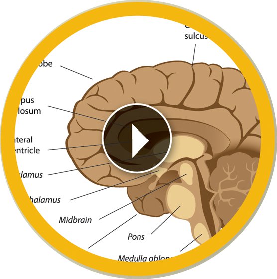
Video: Brain Development Animation: PrenatalALILA MEDICAL MEDIA/SHUTTERSTOCK
Even at 26 weeks, a preterm infant is a tiny creature requiring intensive care for each gram of nourishment and every shallow breath. Breathing tubes and feeding tubes are needed, and often other special equipment as well. By contrast, after 37 weeks or more, the typical full-
The critical difference between life and death, or between a fragile preterm newborn and a robust one, is maturation of the neurological, respiratory, and cardiovascular systems. As the brain matures, the organs of the body begin to work in harmony. The heart beats faster during activity; fetal movement as well as heart rate quiet down during rest (not necessarily when the mother wants to sleep).
In the final three months of prenatal life, the lungs begin to expand and contract, and breathing muscles are exercised as the fetus swallows (into the lungs, not the stomach) and spits out amniotic fluid. The valves of the heart go through a final maturation, as do the arteries and veins throughout the body.
In those final prenatal months, the brain not only develops more neurons, but also connects them to each other, destroying neurons that are not functioning. As the body matures, the skin of the skull thickens. That prevents “brain bleeds,” one of the hazards of preterm birth in which paper-
The fetus usually gains at least 4½ pounds (2.1 kilograms) in the third trimester, increasing to an average of about 7½ pounds (about 3.4 kilograms) at birth. By full term, human brain growth is so extensive that the cortex (the brain’s advanced outer layers) has formed several folds in order to fit into the skull (see Figure 4.2). Although some large mammals (whales, for instance) have bigger brains than humans, no other creature needs as many folds because, relative to size, the human cortex contains much more material than the brains of non-
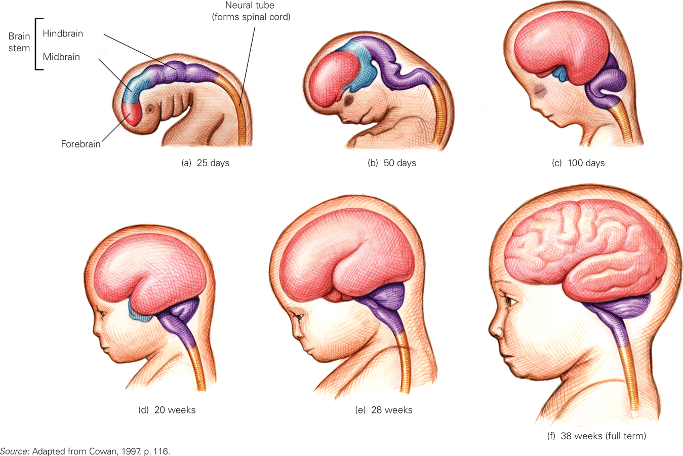
Prenatal Growth of the Brain Just 25 days after conception (a), the central nervous system is already evident. The brain looks distinctly human by day 100 (c). By 200 days after gestation (e), at the very time brain activity begins, the various sections of the brain are recognizable. When the fetus is full term (266 days) (f), all the parts of the brain, including the cortex (the outer layers), are formed, folding over one another and becoming more convoluted, or wrinkled, as the number of brain cells increases.
The Mother–Child Relationship
The relationship between mother and child intensifies during the final three months as the fetus’s size and movement make the pregnant woman very aware of it. In turn, her sounds, the tastes of her food (via amniotic fluid), and her behavior patterns become part of fetal consciousness. If she is up at dawn, her newborn infant is likely to be wide-
Auditory communication from mother to child begins at the 28th week and improves each week as fetal hearing (or newborn hearing if a baby is born early) becomes more acute (Bisiacchi et al., 2009). The fetus startles and kicks at loud noises, listens to the mother’s heartbeat and voice, and is comforted by rhythmic music and movement, such as when the mother sings as she walks. Other voices are also heard, and many men begin to relate to their future child by stroking the mother’s abdomen and talking to the fetus, thrilling to feel the kick.
If the mother is fearful or anxious, the fetal heart beats faster and body movements increase, and later, the infant is likely to startle at noises and unexpected sights. Maternal stress in pregnancy affects the fetus, infant, and child in many ways—
SUMMING UP In the first two weeks of rapid cell duplication, differentiation, and finally implantation, the newly conceived organism is transformed from a one-
WHAT HAVE YOU LEARNED?
Question 4.1
What are three major developments in the germinal period?
Within hours after conception, the zygote begins duplication and division. After about the 16–cell stage, duplication and division continue and a third process, differentiation, begins. Soon cells specialize, taking different forms and reproducing at various rates, depending on where they are located. Question 4.2
What body parts develop during the period of the embryo?
During the period of the embryo, the brain and spinal column, eyes, ears, nose, and mouth start to form, as do the heart, arms, and legs. This is followed by the upper arm, forearm, palms, webbed fingers, legs, knees, feet, and webbed toes.Question 4.3
How much weight does the fetus gain, and when?
The fetus usually gains at least 4½ pounds (2.1 kilograms) in the third trimester, increasing to an average of about 7½ pounds (about 3.4 kilograms) at birth.Question 4.4
How does brain development affect survival?
The critical difference between life and death, or between a fragile preterm newborn and a robust one, is maturation of the neurological, respiratory, and cardiovascular systems. As the brain matures, the organs of the body begin to work in harmony. The heart beats faster during activity; fetal movement as well as heart rate quiet down during rest (not necessarily when the mother wants to sleep). In the final prenatal months, the brain not only develops more neurons, but also connects them to each other, destroying neurons that are not functioning. As the body matures, the skin of the skull thickens. That prevents “brain bleeds,” one of the hazards of preterm birth in which paper–thin blood vessels in the skull collapse. Question 4.5
What occurs between age of viability and full-
term development? Reaching viability simply means that life outside the womb is possible. Many babies born between 22 and 24 weeks die, and survivors born before 27 weeks often develop slowly because they have missed some essential brain development in the uterus. Each day of the final three months improves the odds not only of survival but also of a healthy life and normal cognition. Many aspects of prenatal life are awe–inspiring; the fact that an ordinary woman provides a far better home for a fetus than the most advanced medical technology can attain is one of them.