 Prenatal Development
germinal period The first two weeks of prenatal development after conception, characterized by rapid cell division and the beginning of cell differentiation.
embryonic period The stage of prenatal development from approximately the third through the eighth week after conception, during which the basic forms of all body structures, including internal organs, develop.
fetal period The stage of prenatal development from the ninth week after conception until birth, during which the fetus gains about 7 pounds (more than 3,000 grams) and organs become more mature, gradually able to function on their own.
The most dramatic and extensive transformation of one’s entire life occurs before birth. To make it easier to study, prenatal development is often divided into three main periods. The first two weeks are called the germinal period; the third through the eighth week is the embryonic period; from then until birth is the fetal period. (Alternative terms are presented in Table 4.1.)
Popular and professional books use various phrases to segment the stages of pregnancy. The following comments may help to clarify the phrases used.
|
Germinal: The First 14 Days
You learned in Chapter 3 that the one-
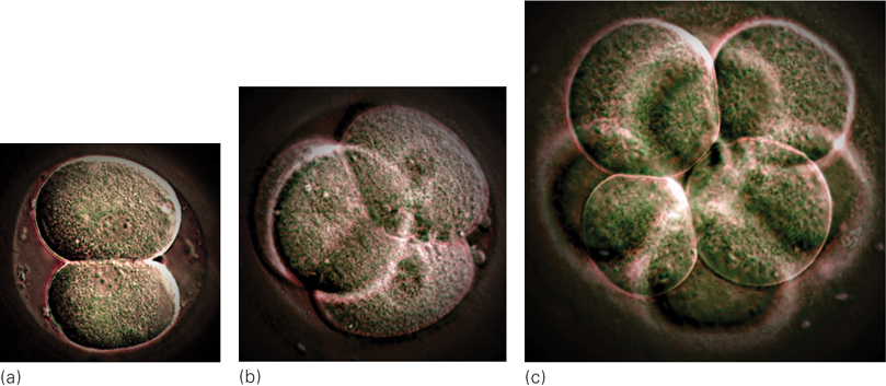
About a week after conception, the cell mass, now called a blastocyst, forms two distinct parts—
implantation The process, beginning about 10 days after conception, in which the developing organism burrows into the placenta that lines the uterus, where it can be nourished and protected as it continues to develop.
The first task of the outer cells is to achieve implantation—that is, to embed themselves in the nurturing lining of the uterus (see Figure 4.1). This process is far from automatic; about half of natural conceptions and an even larger proportion of in vitro conceptions never implant (see Table 4.2): Most new life ends before an embryo begins (Sadler, 2010).
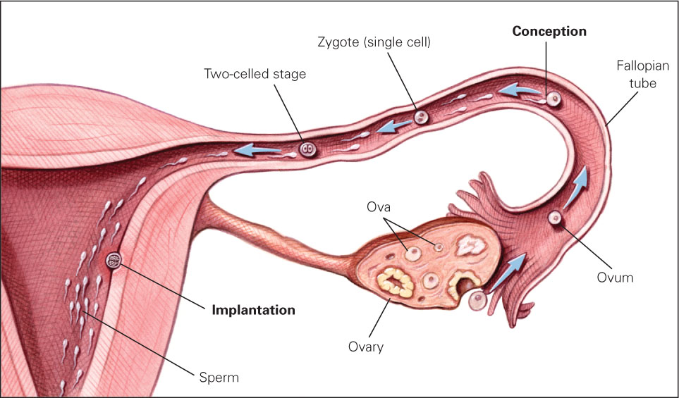
FIGURE 4.1
The Most Dangerous Journey In the first 10 days after conception, the organism does not increase in size because it is not yet nourished by the mother. However, the number of cells increases rapidly as the organism prepares for implantation, which occurs successfully not quite half of the time.| The Germinal PeriodAn estimated 60 percent of all zygotes do not grow or implant properly and thus do not survive the germinal period. Many of these organisms are abnormal; few women realize they were pregnant. |
| The Embryonic PeriodAbout 20 percent of all embryos are aborted spontaneously, most often because of chromosomal abnormalities. This is usually called an early miscarriage. |
| The Fetal PeriodAbout 5 percent of all fetuses are aborted spontaneously before viability at 22 weeks or are stillborn, defined as born dead after 22 weeks. This is much more common in poor nations. |
| BirthBecause of all these factors, only about 31 percent of all zygotes grow and survive to become living newborn babies. Age is crucial. One estimate is that less than 3 percent of all conceptions after age 40 result in live births. |
| Sources: Bentley & Mascie- |
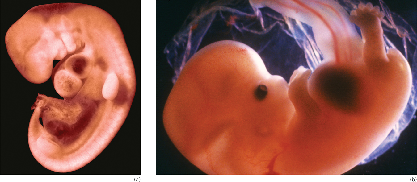
Embryo: From the Third Through the Eighth Week
embryo The name for a developing human organism from about the third through the eighth week after conception.
The start of the third week after conception initiates the embryonic period, during which the formless mass of cells becomes a distinct being—
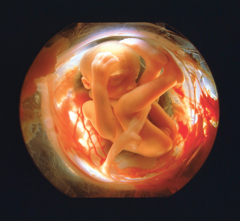
PETIT FORMAT/SCIENCE SOURCE
First, a thin line (called the primitive streak) appears down the middle of the embryo; it will become the neural tube 22 days after conception and eventually develop into the central nervous system (the brain and spinal column). The head appears in the fourth week, as eyes, ears, nose, and mouth start to form. Also in the fourth week, a minuscule blood vessel that will become the heart begins to pulsate.
By the fifth week, buds that will become arms and legs emerge. The upper arms and then forearms, palms, and webbed fingers grow. Legs, knees, feet, and webbed toes, in that order, are apparent a few days later, each having the beginning of a skeletal structure. Then, 52 and 54 days after conception, respectively, the fingers and toes separate (Sadler, 2010).
As you can see, prenatally, the head develops first, in a cephalocaudal (literally, “head-
Fetus: From the Ninth Week Until Birth
fetus The name for a developing human organism from the start of the ninth week after conception until birth.
The organism is called a fetus from the beginning of the ninth week after conception until birth. The fetal period encompasses dramatic change, from a tiny, sexless creature smaller than the final joint of your thumb to a boy or girl about 20 inches (51 centimeters) long.
The Third Month
If the 23rd chromosomes are XY, the SRY gene on the Y triggers the development of male sexual organs. Otherwise, female organs develop. The male fetus experiences a rush of the hormone testosterone, affecting many structures and connections in the brain (Filová et al., 2013). Of course, the range of brain and behavioral variations among males and among females is greater than the variations between the average man and woman. Nonetheless, some neurological sex differences begin early in prenatal development.
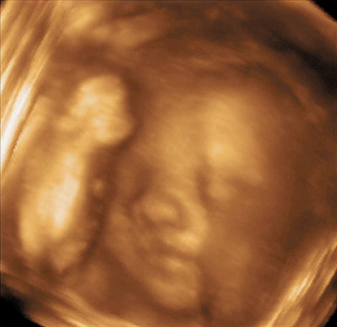
ultrasound An image of a fetus (or an internal organ) produced by using high-
By the end of the third month, the sex organs may be visible via ultrasound (in a sonogram), which is similar to an X-
Especially for Biologists Many people believe that the differences between the sexes are sociocultural, not biological. Is there any prenatal support for that view?
Response for Biologists: Only one of the 46 human chromosomes determines sex, and the genitals develop last in the prenatal sequence, suggesting that dramatic male-
The Middle Three Months
In the fourth, fifth, and sixth months, the heartbeat becomes stronger. Digestive and excretory systems develop. Fingernails, toenails, and buds for teeth form, and hair grows (including eyelashes). The brain increases about six times in size and develops many new neurons (neurogenesis) and synapses (synaptogenesis). Indeed, up to half a million brain cells per minute are created at peak growth during mid-
age of viability The age (about 22 weeks after conception) at which a fetus might survive outside the mother’s uterus if specialized medical care is available.
Brain development occurs in every prenatal month, but these middle three months may be the most crucial (Johnson, 2010). Brain growth is critical at this point because the entire central nervous system becomes responsive during mid-
With intensive medical care, some babies survive at 22 weeks past conception, although many hospitals worldwide do not routinely initiate intensive care unless the fetus is at least 25 weeks old. The age of viability decreased dramatically in the twentieth century, but it now seems stuck at about 22 weeks (Pignotti, 2010) because even the most advanced technology cannot maintain life without some brain response. (Reports of survivors born before 22 weeks are suspect because the date of conception is unknown.)
As the brain matures, the organs of the body begin to work in harmony. The heart beats faster during activity; fetal movement as well as heart rate quiet down during rest (not necessarily when the mother wants to sleep).
The Final Three Months
Reaching viability simply means that life outside the womb is possible. Each day of the final three months improves the odds, not only of survival but also of life without disability (Iacovidou et al., 2010). (More on viability is presented later in this chapter.)
A preterm infant born in the seventh month is a tiny creature requiring intensive care for each gram of nourishment and every shallow breath. By contrast, after nine months or so, the typical full-
The critical difference between life and death, or between a fragile preterm newborn and a robust one, is maturation of the neurological, respiratory, and cardiovascular systems. In the final three months of prenatal life, the lungs begin to expand and contract, and breathing muscles are exercised as the fetus swallows and spits out amniotic fluid. The valves of the heart go through a final maturation, as do the arteries and veins throughout the body. Among other things, this helps to prevent “brain bleeds,” one of the hazards of preterm birth in which paper-
The fetus usually gains at least 4½ pounds (2.1 kilograms) in the third trimester, increasing to almost 7½ pounds (about 3.4 kilograms) at birth. By full term, human brain growth is so extensive that the cortex (the brain’s advanced outer layers) forms several folds in order to fit into the skull (see Figure 4.2). Although some large mammals (whales, for instance) have bigger brains than humans, no other creature needs as many folds because, relative to size, the human cortex contains much more material than the brains of nonhumans.
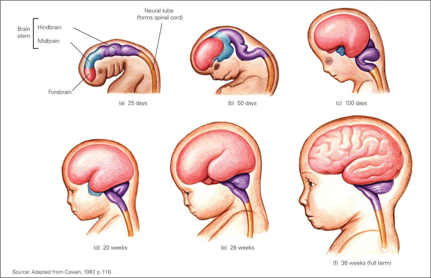
FIGURE 4.2
Prenatal Growth of the Brain Just 25 days after conception (a), the central nervous system is already evident. The brain looks distinctly human by day 100 (c). By the 28th week of gestation (e), at the very time brain activity begins, the various sections of the brain are recognizable. When the fetus is full term (f), all the parts of the brain, including the cortex (the outer layers), are formed, folding over one another and becoming more convoluted, or wrinkled, as the number of brain cells increases.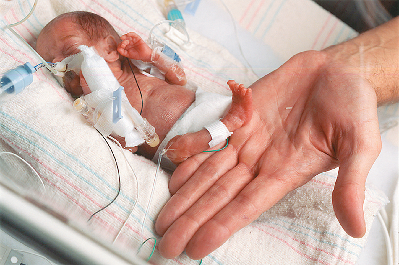

The relationship between mother and child intensifies during the final three months as the fetus’s size and movement make the pregnant woman very aware of it. In turn, her sounds, the tastes of her food (via amniotic fluid), and her behavior patterns become part of fetal consciousness.
Auditory communication from mother to child begins at the 28th week and improves each week as fetal hearing (or newborn hearing if a baby is born early) becomes more acute (Bisiacchi et al., 2009). The fetus startles and kicks at loud noises, listens to the mother’s heartbeat and voice, and is comforted by rhythmic music and movement, such as when the mother sings as she walks. If the mother is fearful or anxious, the fetal heart beats faster and body movements increase (DiPietro et al., 2002).
SUMMING UP
In two weeks of rapid cell duplication, differentiation, and finally implantation, the newly conceived organism is transformed from a one-