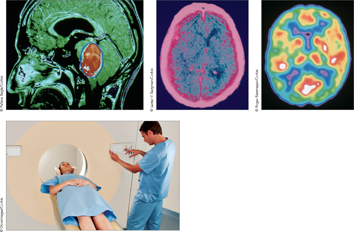
Traditional scanning The most widely used neuroimaging techniques in clinical practice—the MRI (lower left), CAT, and PET—take pictures of the living brain. Here, an MRI scan (above left) reveals a large tumor, colored in orange; a CAT scan (above center) reveals a mass of blood within the brain; and a PET scan (above right) shows which areas of the brain are active (those colored in red, orange, and yellow) when an individual is being stimulated. Images clockwise from top left: Pallava Bagla/Corbis; Lester V. Bergman/Corbis; Roger Ressmeyer/Corbis; Glowimages/Corbis.