Chapter 57. Touch, Movement, and Pain
Learning Objectives
Identify the receptor cells for the sensations generated within the skin and internal organs.
Identify the brain area that processes and perceives the information from body senses within the skin and internal organs.
Explain how touch signals can sometimes diminish the experience of pain.
Review
Review
Select the NEXT button to continue with the Review.

1. How many senses do humans have? If you say “five,” you are lumping several distinctively different senses under the label touch. It would be better to call the sense of touch something like “body-sense,” to include a group of systems that detect the following: a) external objects pressing or vibrating against our skin, b) temperature, c) pain signals indicating damage to our skin or internal organs, and d) changes to our body position.
Review
Review
Select the NEXT button to continue with the Review.
2. The body sensations we call touch originate in specialized sensory receptor cells buried in the skin. Most of these cells are mechanoreceptors, because they generate neural impulses when the skin around them is squeezed or moved. Some receptors respond primarily to light touch or vibration, while others are specialized for heavy, continuous pressure. Other cells called thermoreceptors detect warmth or cold.
Review
Review
Select the NEXT button to continue with the Review.
3. Scattered throughout the body, in both the skin and internal organs, are special “harm detectors” called nociceptors. These sensory receptors generate neural impulses when they detect potentially damaging events, such as extreme heat or a cut on the skin. The brain uses these signals to create the perception of pain, which helps us avoid further damage.
Review
Review
Select the NEXT button to continue with the Review.
4. In addition to detecting external objects, our “body-sense” detects the position and movement of the body. Mechanoreceptors in the muscles, tendons, and joints generate neural impulses when the arms or legs are moved—a sense called kinesthesia. Highly specialized cells in the semicircular canals of the inner ear are responsible for our vestibular sense, detecting rotation of the head or forward-backward movement of the head (and the whole body).
Review
Review
Select the NEXT button to continue with the Review.
5. All the neural impulses from these various “body-sense” receptors travel to the brain’s parietal lobe, where they are processed in the somatosensory cortex, creating perceptions of events happening outside or inside the body.
Practice 1: Exploring the Body Senses
Practice 1: Exploring the Body Senses
Roll over each label to view that component of somatosensory perception.
Special mechanoreceptors in the semicircular canals within the inner ear detect rotation of the head and straight-line movement of the head.
Nociceptors in the skin and internal organs detect injury or potential damage to body tissues.
Thermoreceptors in the skin and internal organs detect warmth and cold.
Mechanoreceptors embedded in the skin detect light touch, heavy pressure, vibration, and stretching of the skin.
Mechanoreceptors in muscles, tendons, and joints detect position and movement of arms and legs.
Practice 2: Interaction of Pain and Touch
Practice 2: Interaction of Pain and Touch
This person has an injured elbow. Select the "Pain pathway" button to view the signals that will be perceived as pain. Then select the "Block pain" button to view one way of reducing the experience of pain.
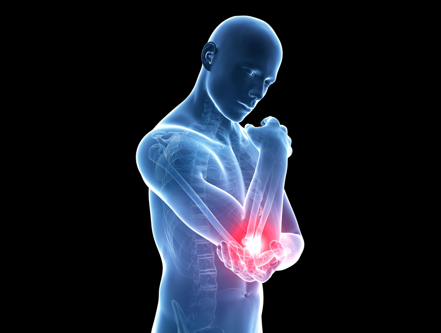
1. Nociceptors in elbow joint are stimulated by the damage and generate neural impulses.
2. These impulses travel to spinal cord on thin, unmyelinated axons.
3. Message is transferred to spinal neurons that carry the impulses to the thalamus.
4. Thalamus relays the impulses to the somatosensory cortex, where the message is perceived as dull, aching pain.
1. Massaging elbow with hand stimulates mechanoreceptors in skin around elbow.
2. Neural impulses travel from skin to spinal cord on thick, myelinated axons.
3. Touch message is transferred to spinal neurons, blocking some of the impulses from the nociceptors.
4. Touch message is relayed through thalamus to the somatosensory cortex, where the touch message is perceived strongly.
5. Weaker message from the nociceptors is perceived as diminished pain.

Quiz 1
Quiz 1
Match the terms to their descriptions by dragging each colored circle to the appropriate gray circle. When all the circles have been placed, select the CHECK ANSWER button.
Quiz 2
Quiz 2
Indicate whether each of the statements below is True or False. Then, select the CHECK ANSWER button.
| True | False | |
|---|---|---|
The sensory receptors that the body uses to detect touch and vibration are different from the receptors that detect tissue damage. |
||
Signals travel from nociceptors to the spinal cord on thick, myelinated axons. |
||
The experience of pain can be influenced by other simultaneous events detected by the somatosensory system. |
Conclusion