Chapter 1. Mirror Experiment Activity 36.20
Mirror Experiment Activity 36.20
The experiment described below explored the same concepts as the one described in Figure 36.20 in the textbook. Read the description of the experiment and answer the questions below the description to practice interpreting data and understanding experimental design.
Mirror Experiment activities practice skills described in the brief Experiment and Data Analysis Primers, which can be found by clicking on the “Resources” button on the upper right of your LaunchPad homepage. Certain questions in this activity draw on concepts described in the Experimental Design and Data and data Presentation primers. Click on the “Key Terms” buttons to see definitions of terms used in the question, and click on the “Primer Section” button to pull up a relevant section from the primer.
Experiment
Background
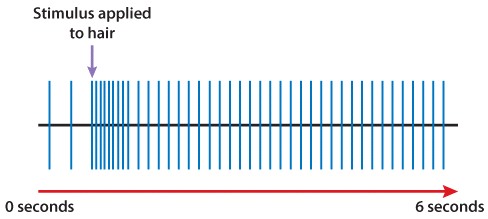
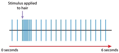
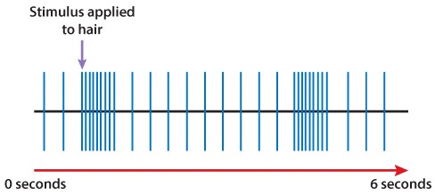
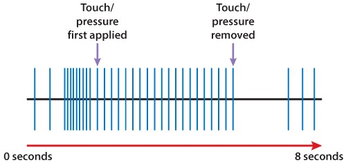
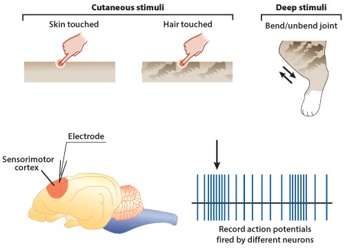
As you have learned, the somatosensory cortex is responsible for processing “touch” stimuli. If someone were tickling the bottom of your foot, mechanoreceptors in the skin of your foot would fire action potentials. These signals would (ultimately) be relayed to the somatosensory cortex portion of your brain, and then your motor cortex. Following this chain of events, you might jerk your foot away from the tickler.
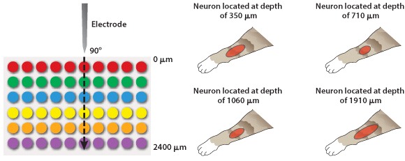
If you were to take a cross section of the somatosensory cortex, you would find that neurons are arranged in six distinct layers; the first layer would be composed of superficial neurons located near the brain surface, and the sixth layer would be composed of the “deepest” neurons (that is, those closest to the white matter). How are neurons that respond to touch stimuli organized in the somatosensory cortex? Do neurons in the six different layers of the somatosensory cortex respond to different types of stimuli?
Hypothesis
Vernon Mountcastle hypothesized that researchers could create a diagram of the somatosensory cortex by tracking which neurons responded to different types of touch stimuli.
Experiment
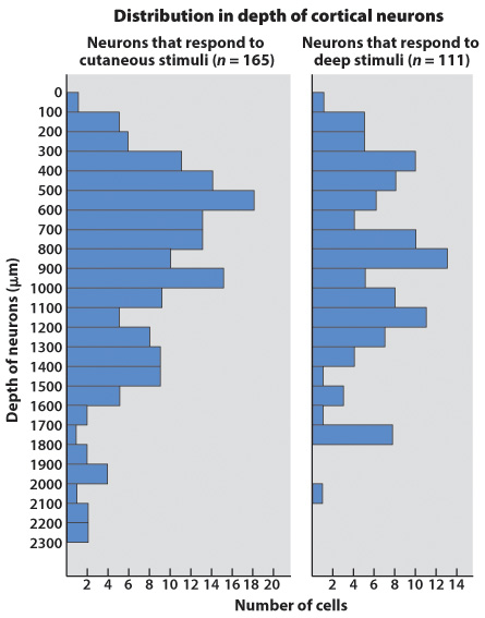
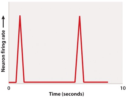
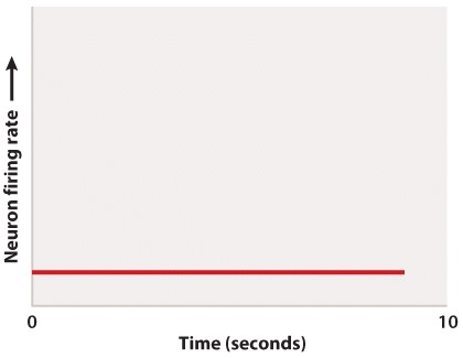
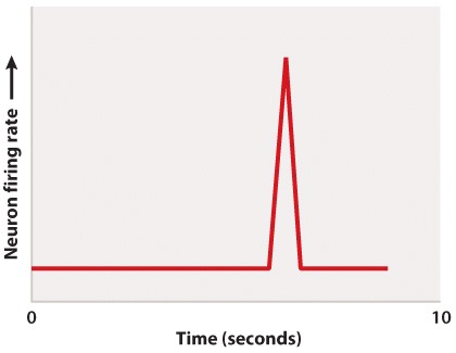
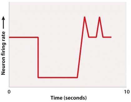
Mountcastle exposed cats to two types of stimuli: (1) cutaneous or superficial stimuli, which included touching hairs or touching the skin; and (2) deep stimuli, which included bending and extending joints or touching the connective tissue surrounding muscles. He was able to track which neurons in the somatosensory cortex fired action potentials in response to these two types of stimuli, and measured their firing rates (Figure 1).
Results
Mountcastle determined that neurons involved in processing the same type of stimuli are organized in “vertical columns.” These columns are composed of cells belonging to different layers of the somatosensory cortex stacked one on top of another. These results demonstrated that just because a neuron responds to deep stimuli does not mean that this neuron will be found deep within the brain; similarly, a neuron that responds to cutaneous stimuli will not necessarily be located near the brain surface. In addition to identifying vertical columns of neurons, Mountcastle also determined that these columns are arranged in a “mosaic” pattern in the somatosensory cortex. Columns composed of neurons responding to deep stimuli occur side-by-side with columns composed of neurons responding to cutaneous stimuli.
Source
Mountcastle, V. B., 1957. Modality and topographic properties of single neurons of cat's somatic sensory cortex. J Neurophysiol. 20, 408-34.
Question
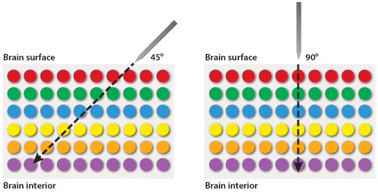
Mountcastle noted that within the somatosensory cortex, neurons that respond to the same type of stimulus are arranged in vertical columns. One way Mountcastle discovered this vertical arrangement was by inserting a measuring device (to record action potentials) into the somatosensory cortex at different angles. This approach is depicted in Figure 2 below. Which of the following statements is true regarding measuring devices that were inserted into the brain at angles of 45° and 90°?
| A. |
| B. |
| C. |
| D. |
| E. |
Data from Mountcastle, V. B., 1957. Modality and topographic properties of single neurons of cat's somatic sensory cortex. J Neurophysiol. 20, 408-34.