Chapter 1. Mirror Experiment Activity 9.5
Mirror Experiment Activity 9.5
The experiment described below explored the same concepts as the one described in Figure 9.5 in the textbook. Read the description of the experiment and answer the questions below the description to practice interpreting data and understanding experimental design.
Mirror Experiment activities practice skills described in the brief Experiment and Data Analysis Primers, which can be found by clicking on the “Resources” button on the upper right of your LaunchPad homepage. Certain questions in this activity draw on concepts described in the Experimental Design and Data and Data Presentation primers. Click on the “Key Terms” buttons to see definitions of terms used in the question, and click on the “Primer Section” button to pull up a relevant section from the primer.
Experiment
Background
As you have learned in Fig. 9.5, growth-promoting factors produced by platelets in the blood can cause fibroblasts to grow and divide. In Chapter 10, you will learn that fibroblasts are cells that contribute to skin structure; they generate a scaffold to which cells can attach and grow. Are the effects of platelet-produced factors specific only to fibroblasts? Can other types of cells, such as muscle cells, proliferate in response to signals produced by platelets?
Hypothesis
Russell Ross and his colleagues were interested in atherosclerosis, a disease that you may know better as hardening of the arteries. Although atherosclerosis is typically associated with cholesterol and diet, it also involves the proliferation of smooth muscle cells (SMCs) in the artery walls themselves. Ross and colleagues speculated that, as a result of artery damage (i.e., a plaque beginning to form), platelets in the blood may accumulate at a specific location in an artery and secrete growth-promoting factors. Researchers hypothesized that these factors might cause SMCs to proliferate, ultimately contributing to atherosclerosis. Ross and colleagues predicted that if SMCs were treated with serum or with plasma containing platelets (or their associated growth factors), SMCs would rapidly grow and divide.
Experiment
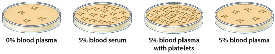
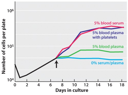
Ross and colleagues adopted a similar protocol to that discussed in Fig. 9.5. Researchers isolated SMCs from the arteries of monkeys and grew these cells in petri dishes. SMCs were then exposed to a variety of treatments generated from monkey blood: blood serum (derived from clotted blood in which platelets had presumably released growth factors), blood plasma (derived from unclotted blood that did not contain platelets or their associated growth factors), and blood plasma supplemented with monkey platelets. Researchers then determined which treatments (if any) resulted in the proliferation of SMCs by counting the number of cells in petri dishes after a 9-11 day culture period (Figure 1).
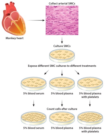
Photo credit: SPL/Science Source
Results
Much like the experiments of Kohler and Lipton discussed in Fig. 9.5, Ross and colleagues determined that SMCs treated with blood serum grew and rapidly divided, whereas cells treated only with blood plasma did not proliferate (or did so only slightly). When supplemented with platelets, blood plasma could cause SMCs to proliferate. This observation provided evidence that growth factors produced by platelets could cause other cells – aside from fibroblasts – to grow and divide.
Source
Ross, R., et al., 1974. A platelet-dependent serum factor that stimulates the proliferation of arterial smooth muscle cells in vitro. Proc Natl Acad Sci U S A. 71, 1207-10.
Question
As you may recall from Fig. 9.5, blood plasma is derived from unclotted blood that (presumably) does not contain any platelet-produced factors. How could Ross and colleagues have collected blood plasma from freshly drawn monkey blood that met this criteria?
| A. |
| B. |
| C. |
| D. |
| E. |