Chapter 47. The Cerebral Cortex

Learning Objectives

angular gyrus
area in the parietal lobe that converts written language into sounds so that Wernicke’s area can understand the meaning
motor cortex
area in the frontal lobes specialized for initiating muscle movements
association areas
regions of the brain’s cerebral cortex that receive inputs from more than one sensory system; involved in higher-level thinking and memory
neuron
a single nerve cell, forming the basic unit of the nervous system
auditory cortex
area in the temporal lobes that receives and processes information from the ears
occipital lobe
lower back part of each hemisphere; processes sensory input for vision
Broca’s area
area in the frontal lobe involved in the production of language; usually in left hemisphere
parietal lobe
upper back part of each hemisphere; processes sensory input for touch and body position
cerebellum
brain structure that coordinates body movement and balance; involved in simple forms of learning
pons
part of brainstem that relays messages between the cerebellum and the cerebral cortex to help coordinate body movements; plays a role in sleep
cerebral cortex
six thin layers of interconnected neurons that cover the outer surface of the brain and do most of the brain's processing
somatosensory cortex
area within the parietal lobes that receives and processes information from the body senses
cerebral hemispheres
the two halves of the brain
temporal lobe
lower side of each hemisphere; processes sensory input for hearing
frontal lobe
front part of each hemisphere; initiates speech and muscle movements; involved in planning and decision making
visual cortex
area in the occipital lobes that receives and processes information from the eyes
hemisphere
one half of the brain; divided into four lobes
Wernicke’s area
area in the temporal lobe involved in understanding language sounds; usually in left hemisphere
The Cerebral Cortex
true
true
true
asset/activities/cerebral_cortex/images/intro_image_b.svg
Chris Parsons/Getty Images
Learning Objectives:

Identify the four lobes of the cerebral cortex.

Describe the main functional areas of each lobe of the cerebral cortex.

Review

concept_review

Review

Select the NEXT button to continue with the Review.

asset/activities/cerebral_cortex/images/review_1.svg

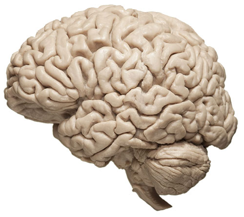
1. The cerebral cortex is a layer of tightly packed neurons that covers the outer surface of the brain’s two cerebral hemispheres. If you opened a person's skull, the cortex is what you would see.

Review

concept_review

Review

Select the NEXT button to continue with the Review.

asset/activities/cerebral_cortex/images/review_2.svg

2. The cerebral cortex is structured in six thin layers, with each layer containing billions of interconnected neurons that do most of the brain's complicated processing.

Review

concept_review

Review

Select the NEXT button to continue with the Review.

asset/activities/cerebral_cortex/images/review_3.svg

3. Each of the two cerebral hemispheres is divided into four lobes named for their location within the skull.

Review

concept_review

Review

Select the NEXT button to continue with the Review.

asset/activities/cerebral_cortex/images/review_4-fixed.svg

4. Three of the cortex lobes contain regions specialized for the processing of sensory information from the outside world and from the body's internal organs. The occipital lobe contains the primary visual cortex, which processes information from the eyes.

Review

concept_review

Review

Select the NEXT button to continue with the Review.

asset/activities/cerebral_cortex/images/review_5.svg

5. The temporal lobe contains the primary auditory cortex, which processes information from the ears.

Review

concept_review

Review

Select the NEXT button to continue with the Review.

asset/activities/cerebral_cortex/images/review_6-fixed-2.svg

6. The parietal lobe contains a long vertical strip called the primary somatosensory cortex, which processes information from the outside world through the skin (such as touch, vibration, temperature, and pain). This area also processes internal body signals, such as position of the head and movement of the arms and legs, as well as pain in internal organs.

Review

concept_review

Review

Select the NEXT button to continue with the Review.

asset/activities/cerebral_cortex/images/review_7.svg

7. The areas that process sensations of taste and smell are not visible on the surface of the brain. They are hidden deep in the fissure, or crack, that separates the temporal lobe from the frontal lobe.

Review

concept_review

Review

Select the NEXT button to continue with the Review.

asset/activities/cerebral_cortex/images/review_8.svg

8. The frontal lobe contains an area that controls voluntary muscle movements. This primary motor cortex initiates the movement, but lower brain areas, including the cerebellum and pons, coordinate the muscle groups that actually perform the movement.

Review

concept_review

Review

Select the NEXT button to continue with the Review.

The figure is a side view of the brain with the four lobes labeled.  The frontal lobe is located in the front of the brain, behind the forehead and eyes, up to the midpoint on the top of the head.  The parietal lobe is located on the top of the brain from the midpoint behind the frontal lobe to where the head starts to slope down in the back.  The temporal lobe is located under the parietal lobe where the ears are located on the side of the head. The occipital lobe is located in the back of the head, under the parietal lobe and behind the temporal lobe. The Broca's area is located in the back of the frontal lobe, above the temporal lobe.  The Wernicke's area is located at the top of the temporal lobe, under the parietal lobe.  The Angular gyrus is located in the parietal lobe, behind the temporal lobe.

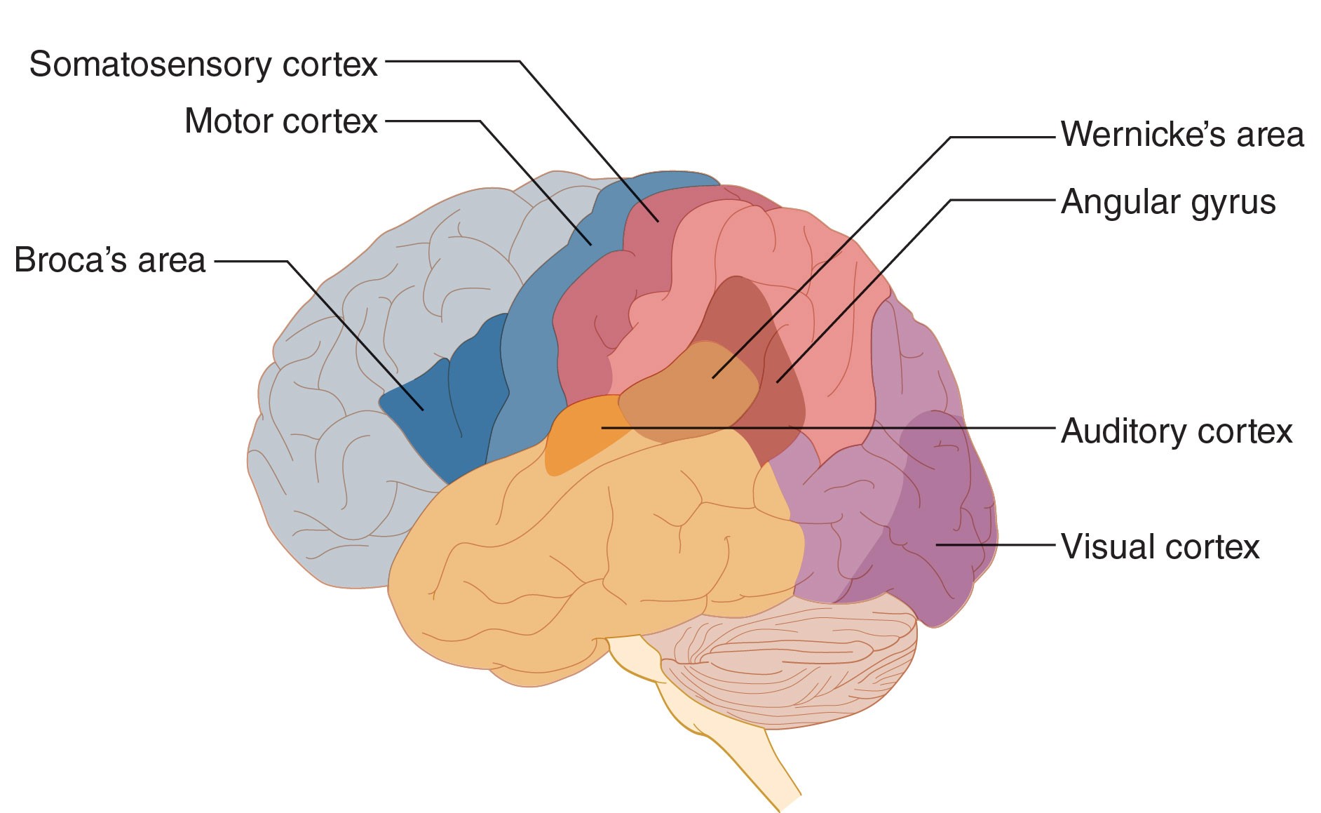
9. The specialized areas of the cortex that produce and understand language are usually located in the left hemisphere only. Broca’s area is mainly involved in the production of language. Wernicke’s area is involved in the understanding of language. With spoken language, Wernicke’s area gets input from the auditory cortex. With written language, the visual cortex must first send the words to the angular gyrus, which converts the words to sounds that Wernicke’s area can interpret.

Review

concept_review

Review

Select the NEXT button to continue with the Review.

asset/activities/cerebral_cortex/images/review_10.svg

10. Finally, the cortex contains association areas that integrate information from many brain regions to interpret the meaning of events, make judgments and decisions, and plan future actions.

Practice 1: Lobes of the Cortex

hover_review
true

Practice 1: Lobes of the Cortex

Roll over each lobe to see a brief description of its role.

The cerebral cortex is the outer surface of the upper part of the brain. Its four lobes contain the body's ultimate control centers and information-processing areas.

Temporal lobe
Occipital lobe
Parietal lobe
Frontal lobe
Function of the selected language area of the cerebral cortex:

receives and processes auditory information from the ears

receives and processes visual information from the eyes

receives and processes sensory information from the skin and body senses

controls muscle movement and production of speech; is involved in making plans and judgments

asset/activities/cerebral_cortex/images/n1-cerebral-cortex-step12-v4.svg

Practice 2: Sensory and Motor Areas

hover_review
true

Practice 2: Sensory and Motor Areas

Roll over each area to see a brief description of its role in information processing or muscle control.

These special areas within the four lobes control voluntary muscle movement and process information from the senses.

Motor cortex
Somatosensory cortex
Auditory cortex
Visual cortex
Function of the selected area of the cerebral cortex:

controls voluntary muscle movements on the opposite side of the body (that is, the right hemisphere's motor cortex controls movement on the left side of the body)

processes sensations from the skin and internal organs on the opposite side of the body (that is, the right hemisphere's sensory cortex processes skin/organ information from the body's left side)

processes information from the ears, primarily from the opposite side of the body (that is, the right hemisphere's auditory cortex processes sounds entering the left ear)

processes information from the eyes coming from the opposite visual field (that is, the right hemisphere's visual cortex processes information from the left visual field)

asset/activities/cerebral_cortex/images/n1-practice_2.svg

Practice 3: Language Areas

hover_review
true

Practice 3: Language Areas

Roll over each area to see a brief description of its role in language.

These special areas, usually in the left hemisphere, control production and comprehension of spoken and written language.

Broca’s area
Wernicke’s area
Angular gyrus
Function of the selected area of the cerebral cortex:

generates spoken and written language and controls mouth muscles by way of the motor cortex

processes the information from speech or writing and comprehends its meaning

is involved in reading (turns written words into an auditory code to be interpreted by Wernicke's area)

asset/activities/cerebral_cortex/images/n1-practice_3.svg

Quiz 1

dnd_test

Quiz 1

Drag each label to the gray area connected to the appropriate lobe of the cerebral cortex. When all the labels have been placed, select the CHECK ANSWER button.

The figure is a lateral image of the brain with the various elements unlabeled.  The labels listed on the page should be placed in their appropriate location.  The labels include the following: temporal lobe, frontal lobe, occipital lobe, and parietal lobe.
Perhaps you should go back to review the lobes of the cerebral cortex.
Select the NEXT button and move to Quiz 2.
Frontal lobe
Parietal lobe
Occipital lobe
Temporal lobe

Quiz 2

dnd_test

Quiz 2

Drag each label to the gray area connected to the appropriate area of the cerebral cortex. When all the labels have been placed, select the CHECK ANSWER button.

 The figure is a lateral image of the brain with the various elements unlabeled.  The labels listed on the page should be placed in their appropriate location.  The labels include the following: visual cortex, somatosensory cortex, auditory cortex, motor cortex, Broca's area, Wernicke's area, and angular gyrus.
Perhaps you should go back to review the areas of the cerebral cortex.
Select the NEXT button and move to Quiz 3.
Visual cortex
Auditory cortex
Somatosensory cortex
Motor cortex
Broca’s area
Wernicke’s area
Angular gyrus

Quiz 3

matching_test

Quiz 3

Match the brain structures to their functions by dragging each colored circle to the appropriate gray circle. When all the circles have been placed, select the CHECK ANSWER button.

Select the NEXT button and move to the Conclusion.
Perhaps you should go back to review the areas of the cerebral cortex.
visual cortex
auditory cortex
somatosensory cortex
motor cortex
Broca’s area
Wernicke’s area
angular gyrus
in temporal lobe; processes information from the ears
generates spoken and written language
in occipital lobe; processes information from the eyes
in parietal lobe; processes skin and body sensations
in frontal lobe; controls voluntary muscle movements
comprehends spoken and written language
involved in reading; converts writing into auditory code

Conclusion

end_slide
asset/activities/cerebral_cortex/images/intro_image_b.svg
Chris Parsons/Getty Images
Congratulations!
You have completed the activity Title