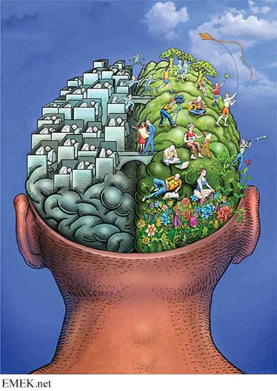
Specialization in the Cerebral Hemispheres
KEY THEME
Although they have many functions in common, the two hemispheres of the cerebral cortex are specialized for different tasks.
KEY QUESTIONS
How did Broca, Wernicke, and Sperry contribute to our knowledge of the brain?
Why would the corpus callosum be surgically severed, and what effects would that produce?
How do the functions of the right and left cerebral hemispheres differ?
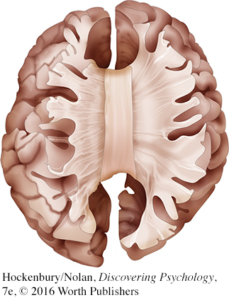
If you hold a human brain in your hand, the two cerebral hemispheres would appear to be symmetrical. Although the left and right hemispheres are very similar in appearance, they are not identical. Anatomically, one hemisphere may be slightly larger than the other. There are also subtle differences in the sizes of particular structures, in the distribution of gray matter and white matter, and in the patterns of folds, bulges, and grooves that make up the surface of the cerebral cortex (Ocklenburg & Güntürkun, 2012).
What about differences in the functions of the two hemispheres? In many cases, the functioning of the left and right hemispheres is symmetrical, meaning that the same functions are located in roughly the same places on each hemisphere. Examples of such functional symmetry include the primary motor cortex and the somatosensory cortex, which we discussed in the previous section. With regard to other important processes, however, the left and right cerebral hemispheres do differ—
Here’s a rough analogy. Imagine two computers that are linked through a network. One computer is optimized for handling word processing, the other for handling graphic design. Although specialized for different functions, the two computers actively share information and can communicate with each other across the network. In this analogy, the two computers correspond to the left and right cerebral hemispheres, and the network that links them is the corpus callosum.
CRITICAL THINKING
“His” and “Her” Brains?
Do men and women have fundamentally different brains? Some popular authors claim men’s and women’s brains are “hardwired” by hormones and genes to produce “separate realities” (Brizendine, 2006, 2010). According to this view, there are innate differences between the brains of males and females, differences that cause gender differences in behaviors, attitudes, personality traits, and skills (Fine, 2012, 2014). But what are these differences? Do they cause men and women to “think, feel, and behave differently,” as some headlines and popular books claim? Let’s look at the scientific research.
MYTH SCIENCE
Is it true that because their brains are wired differently, men and women think, feel, and behave differently?
A case in point is a recent study that used diffusion tensor imaging (see p. 62) to compare the neural pathways, or connectomes, of over 900 children and young adults. Madhura Ingalhalikar and her colleagues (2014) concluded that the scans of the adolescent and young adult participants showed “fundamental sex differences in the structural architecture in the human brain.” On average, males had significantly more neural connections within the left and right hemispheres than females. And, females had significantly more neural connections between the left and right hemispheres than males.
The media jumped on the findings as evidence that sex differences were “hardwired” in the brain. Going well beyond the study’s findings, print articles and online blogs claimed that the findings explained many supposed sex differences, such as why men were better at map-
But just how definitive were the study’s results? First, while statistically significant, the differences identified in the study were still quite small (Cossins, 2015; Joel & Tarrasch, 2014). Second, the differences were quantitative rather than qualitative: that is, the differences were a matter of degree, not kind. Although the average number of connections differed, the general pattern of connections was the same in male and female participants (Fine, 2014).
And, critics noted that male brains are, on average, about 10% larger than female brains. Thus, the differences could have been associated with brain size rather than brain sex. A later study found that larger brains tend to have more interhemispheric connectivity than smaller brains, regardless of sex. In men’s and women’s brains that were the same size, there were no differences in the pattern of connections: the apparent sex effect disappeared (Hänggi & others, 2014).
Finally, the analysis did not take into account the participants’ past experience, such as participation in sports or hobbies (Fine, 2014). Yet, as we’ve shown in this chapter, experience changes the brain.
Thinking Critically About Brain Differences
How should you interpret media sound bites about profound sex differences in the brain? Or claims that certain personality traits or behaviors are “hardwired in the brain”?
First, it’s important to think critically about media claims. Unlike reporters, scientists are usually careful to qualify their conclusions and describe the limitations of their research. These limitations are rarely mentioned in media reports. For example, brain studies are typically based on small groups of participants, who may or may not be representative of the wider population of men and women. And, while findings of sex differences in brain structure or function tend to be widely reported in the media, findings of no difference go unreported. So, often, does the failure of studies to be replicated by other researchers (Eliot, 2011; Fine, 2013a, 2013b). In other words, the findings of individual research studies are rarely as earth-
Second, most sex differences amount to minor variations in the size of a particular brain region or to statistical differences in the average level of activation of particular brain regions. Brain structures and functioning are essentially the same in men and women—
The hardwiring paradigm erases the effect of the social world in producing sex/gender differences, so that sex/gender hierarchies appear natural. Neuroscientific explanations of sex/gender differences have added a new allure to an old fashioned sexism.
—Rebecca Jordan-
Finally, it’s important to remember that even differences that are biological in origin are not necessarily fixed, permanent, or inevitable (Fine, 2014; Hyde, 2014). Brain development and function are affected by biological influences, such as exposure to the sex hormones produced both before birth and throughout your life (Lombardo & others, 2012). However, these biological factors themselves are strongly influenced by environmental factors, ranging from the food we eat to the stressful circumstances we experience. As we’ve emphasized throughout this chapter, both brain function and structure are highly responsive to environmental influences (Fine & others, 2013). Thus, sex differences in structures or function might well be the result of the different life experiences of men and women, rather than the cause (Rippon & others, 2014).
CRITICAL THINKING QUESTIONS
Why are sweeping claims about fundamental sex differences in the human brain misleading?
What is wrong with the statement that certain behaviors or personality traits are “hardwired” in the male or female brain?
How might claims that sex differences are “hardwired” or innate affect attitudes or behavior?
Why is the notion that sex differences might be due to brain differences so appealing to many people?
As you’ll see in this section, the first discoveries about the differing abilities of the two brain hemispheres were made more than a hundred years ago by two important pioneers in brain research, Pierre Paul Broca and Karl Wernicke.
Language and the Left Hemisphere
THE EARLY WORK OF BROCA AND WERNICKE
By the end of the 1700s it had already been well established that injury to one side of the brain could produce muscle paralysis or loss of sensation on the opposite side of the body. By the early 1800s, animal experiments had shown that specific functions would be lost if particular brain areas were destroyed. And, as discussed in the Science Versus Pseudoscience box on page 61, phrenology triggered scientific debates about cortical localization, or localization of function—the idea that particular brain areas are associated with specific functions.
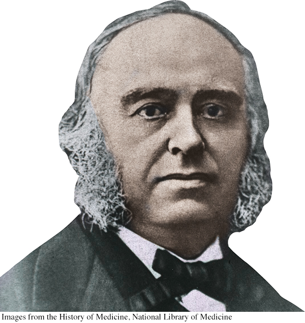
In the 1860s, more compelling evidence for cortical localization was presented by a French surgeon and neuroanatomist named Pierre Paul Broca. Broca treated a series of patients who had great difficulty speaking but could comprehend written or spoken language. Subsequent autopsies of these patients revealed a consistent finding—
About a decade after Broca’s discovery, a young German neurologist named Karl Wernicke discovered another area in the left hemisphere that, when damaged, produced a different type of language disturbance. Unlike Broca’s patients, Wernicke’s patients had great difficulty understanding spoken or written communications. They could speak quickly and easily, but their speech sometimes made no sense. They sometimes used meaningless words or even nonsense syllables, though their sentences seemed to be grammatical. In response to the question “How are you feeling?” a patient might say something like, “Don’t glow glover. Yes, uh, ummm, bick, bo chipickers the dallydoe mick more work mittle.” Autopsies of these patients’ brains revealed consistent damage to an area on the left temporal lobe that today is called Wernicke’s area (see Figure 2.21).

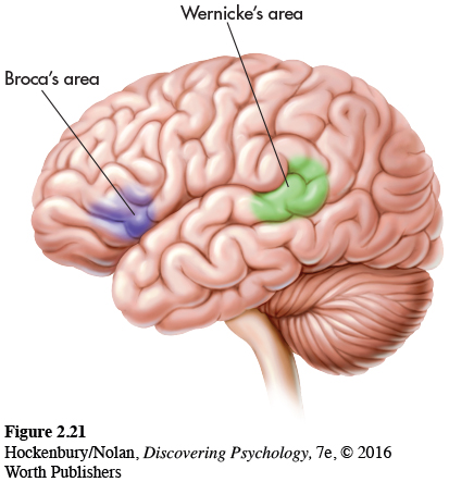
The discoveries of Broca and Wernicke provided the first compelling clinical evidence that language and speech functions are performed primarily by the left cerebral hemisphere. If similar brain damage occurs in the exact same locations on the right hemisphere, these severe disruptions in language and speech are usually not seen.
The notion that one hemisphere exerts more control over or is more involved in the processing of a particular psychological function is termed lateralization of function. Speech and language functions are lateralized on the left hemisphere. Generally, the left hemisphere exerts greater control over speech and language abilities in virtually all right-
The language disruptions demonstrated by Broca’s and Wernicke’s patients represent different types of aphasia. Aphasia refers to the partial or complete inability to articulate ideas or understand spoken or written language because of brain injury or damage. There are many different types of aphasia.
People with Broca’s aphasia find it difficult or impossible to produce speech, which is why it is often referred to as expressive aphasia. Despite their impairments in speaking, their comprehension of verbal or written words is relatively unaffected.

People with Wernicke’s aphasia have great difficulty comprehending written or spoken communication, which is why it is often referred to as receptive aphasia. Although they can speak, they often have trouble finding the correct words.
At the beginning of this chapter, we described the symptoms experienced by our friend Asha in the weeks before and the months following her stroke. Asha, who is right-
Asha also discovered that she had lost the ability to read. She could see the words on the page, but they seemed to have no meaning. Her husband, Paul, brought some of their Christmas cards to the hospital. Asha recalls, “When I realized I couldn’t read the Christmas cards, I thought my life was over. I just lost it. I remember crying and telling the nurse, ‘I have a doctorate and I can’t read, write, or talk!’”
When we visited Asha in the hospital, we brought her a Christmas present: a portable music player with headphones and some albums of relaxing instrumental music. Little did we realize how helpful the music would be for her. One album was a recording of Native American flute music called Sky of Dreams. The music was beautiful and rather unusual, with intricate melodies and unexpected, complex harmonies. Although it was very difficult for Asha to follow normal speech, listening to Sky of Dreams was an entirely different experience. As Asha explained:
I tried cranking up the music very high and it soothed me. I could sleep. At the time, the flute music seemed to be just perfectly timed with the way my brain was working. It was tuning out all the other noises so I could focus on just one thing and sleep. So I would play the music over and over again at a very high level. I did that for a long time because my mind was so active and jumbled that I couldn’t think.
Asha’s language functions were severely disrupted, yet she was able to listen to and appreciate instrumental music—
Cutting the Corpus Callosum
THE SPLIT BRAIN
Since the discoveries by Broca and Wernicke, the most dramatic evidence illustrating the independent functions of the two cerebral hemispheres has come from a surgical procedure called the split-
What was the logic behind cutting the corpus callosum? An epileptic seizure typically occurs when neurons begin firing in a disorganized fashion in one region of the brain. The disorganized neuronal firing quickly spreads from one hemisphere to the other via the corpus callosum. If the corpus callosum is cut, seizures should be contained in just one hemisphere, reducing their severity or eliminating them altogether. This is exactly what happened when the split-
Surprisingly, cutting the corpus callosum initially seemed to produce no noticeable effect on the patients, other than reducing their epileptic seizures. Their ability to engage in routine conversations and tasks seemed to be unaffected. On the basis of these early observations, some brain researchers speculated that the corpus callosum served no function whatsoever. One famous psychologist, Karl Lashley, joked that the primary function of the corpus callosum seemed to be to keep the two hemispheres from sagging (Hoptman & Davidson, 1994).
In the 1960s, however, psychologist and neuroscientist Roger Sperry and his colleagues began unraveling the puzzle of the left and right hemispheres. Sperry and his colleagues used the apparatus shown in Figure 2.22 to test the abilities of split-
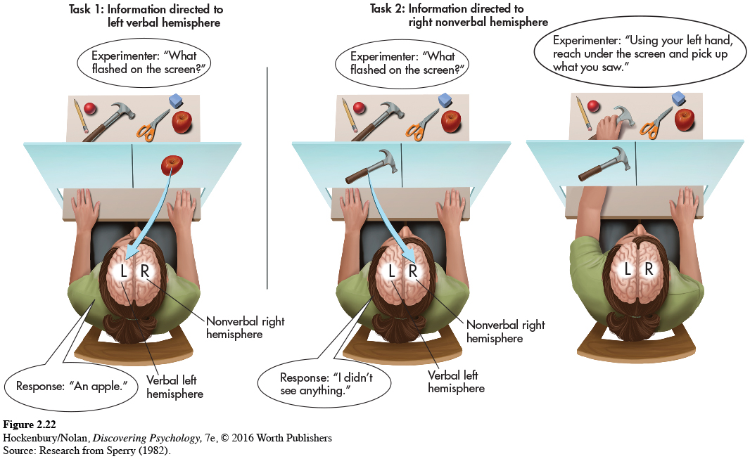
In this procedure, visual information to the right of the midpoint is projected to the person’s left hemisphere, and visual information to the left of the midpoint is projected to the right hemisphere. Behind the screen several objects were hidden from the split-
In a typical experiment, Sperry projected the image of an object concealed behind the screen, such as a hammer, to the left of the midpoint. This is shown in Task 2, Figure 2.22. Thus, the image of the hammer was sent to the right, nonverbal hemisphere. If a split-
Sperry’s experiments reconfirmed the specialized language abilities of the left hemisphere that Broca and Wernicke had discovered more than a hundred years earlier. But notice, even though the split-
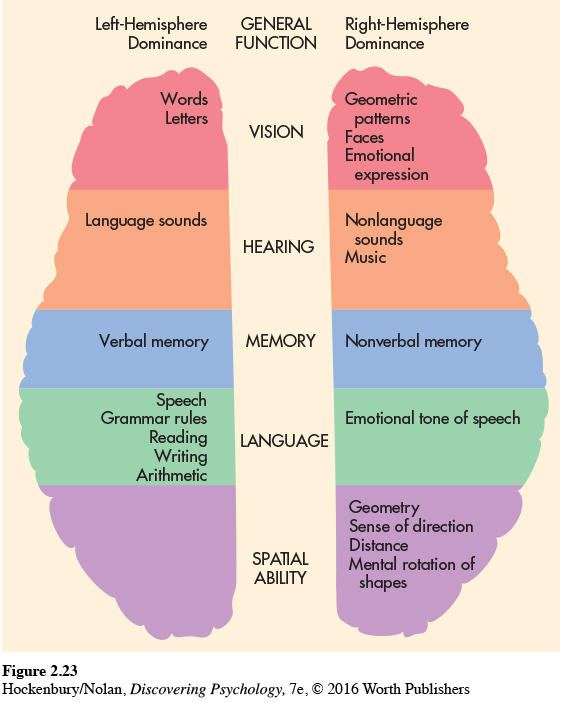
Over the past decades, researchers have gained numerous insights about the brain’s lateralization of functions by studying split-
In contrast, the right hemisphere is more involved in nonverbal emotional expression and visual-
Figure 2.23 summarizes the research findings for the different specialized abilities of the two hemispheres for right-
Think Like a SCIENTIST
Can you be classified as right-

SCIENCE VERSUS PSEUDOSCIENCE
Brain Myths
Is it true that some people are “right-
To investigate this question, researchers compared more than a thousand fMRI scans taken while participants rested. There was no evidence that participants preferentially relied on networks in the left or right hemisphere, as you would expect if some people were “left-
It certainly seems as if some people are more logical, analytical, or detail-
What about left-
Only about 10 to 13 percent of the population identify themselves as left-
It’s a myth that left-
Is the right brain responsible for creativity and intuition? Can you train your right brain?
MYTH SCIENCE
Is it true that the right brain is creative and intuitive, and the left brain is analytic and logical, but that left-
Although the right hemisphere is specialized for holistic processing, there is no evidence that the right hemisphere is any more “intuitive” or “creative” than the left hemisphere (Gazzaniga, 2005). In fact, a recent study found that a task requiring a creative solution involved greater left hemisphere activation than a task that required a noncreative solution (Aziz-
Given the basic findings on the laterality of different functions in the two hemispheres, can you speculate about why Asha was unable to read or follow a simple conversation but could easily concentrate on a complex piece of music? Why were her language abilities so disrupted, while her ability to focus on and appreciate music remained intact after her stroke?
A plausible explanation has to do with the location of the stroke’s damage on Asha’s left temporal lobe. Because language functions are usually localized on the left hemisphere, the stroke produced serious disruptions in Asha’s language abilities. However, her right cerebral hemisphere sustained no detectable damage. Because one of the right hemisphere’s abilities is the appreciation of musical sounds, Asha retained the ability to concentrate on and appreciate music.
Test your understanding of The Brain with  .
.