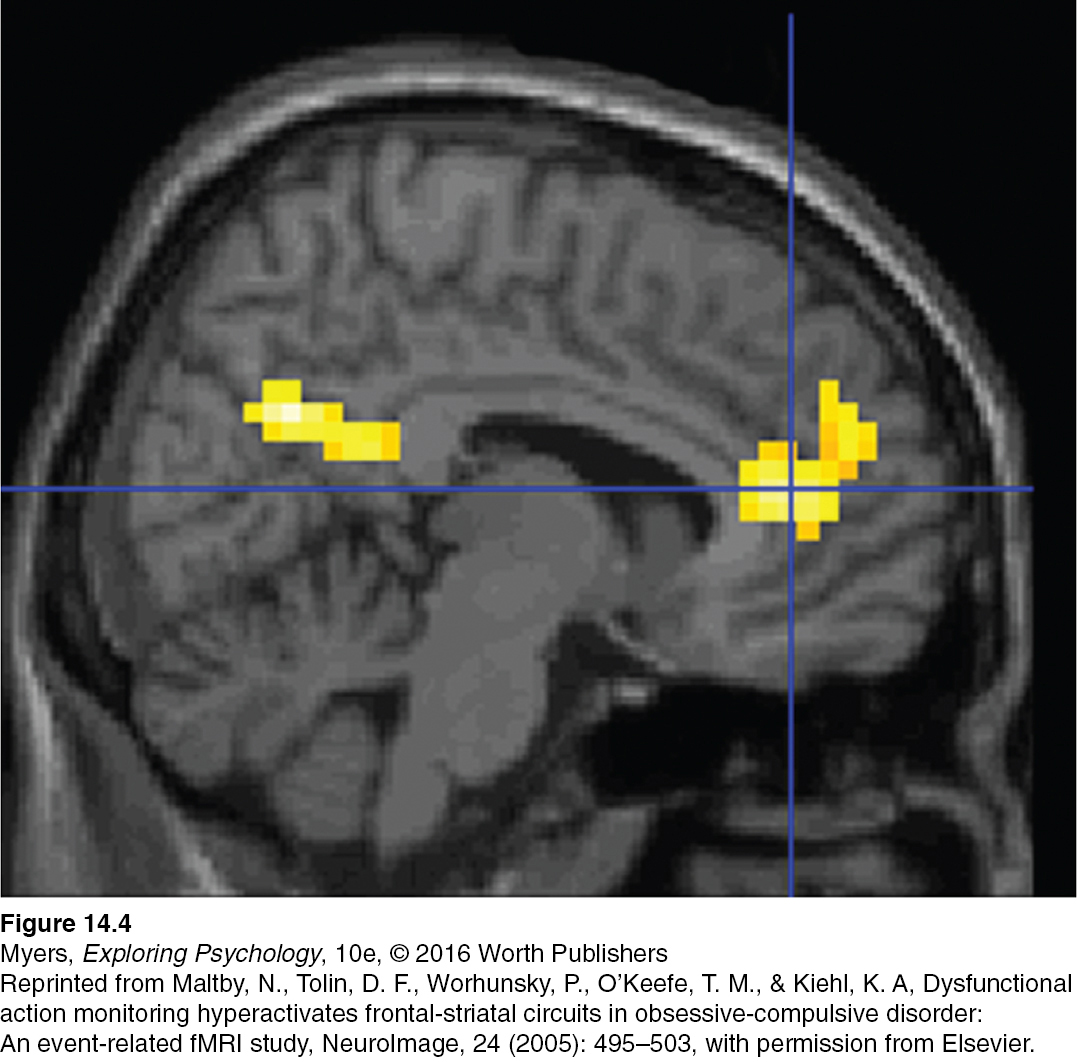
FIGURE 14.4 An obsessive- compulsive brain Neuroscientists Nicholas Maltby, David Tolin, and their colleagues (2005) used functional MRI scans to compare the brains of those with and without OCD as they engaged in a challenging cognitive task. The scans of those with OCD showed elevated activity in the anterior cingulate cortex in the brain’s frontal area (indicated by the yellow area on the far right).
Reprinted from Maltby, N., Tolin, D. F., Worhunsky, P., O’Keefe, T. M., & Kiehl, K. A, Dysfunctional action monitoring hyperactivates frontal- striatal circuits in obsessive- compulsive disorder: An event- related fMRI study, NeuroImage, 24 (2005): 495– 503, with permission from Elsevier.
[Leave] [Close]