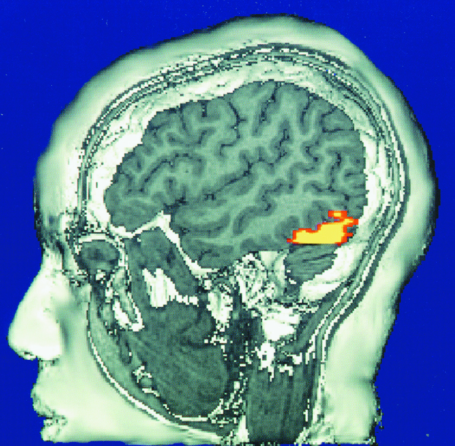
FIGURE 2.20 The brain in action This fMRI (functional MRI) scan shows the visual cortex in the occipital lobes activated (color represents increased bloodflow) as a research participant looks at a photo. When the person stops looking, the region instantly calms down.
NeuroImage, Vol. 4, V.P. Clark, K. Keill, J. Ma. Maisog, S. Courtney, L. G. Ungerleider, and J. V. Haxby, Functional Magnetic Resonance Imaging of Human Visual Cortex during Face Matching: A Comparison with Positron Emission Tomography, August 1996, with permission from Elsevier.
[Leave] [Close]