2.2 Tools of Discovery and Older Brain Structures
“I am a brain, Watson. The rest of me is a mere appendix.”
Sherlock Holmes, in Arthur Conan Doyle’s “The Adventure of the Mazarin Stone”
The mind seeking to understand the brain—
When you think about your brain, you’re thinking with your brain—
The Tools of Discovery: Having Our Head Examined
2-
A century ago, scientists had no tools high powered yet gentle enough to explore the living human brain. Early case studies helped localize some brain functions. Damage to one side of the brain often caused numbness or paralysis on the opposite side, suggesting that the body’s right side is wired to the brain’s left side, and vice versa. Damage to the back of the brain disrupted vision, and damage to the left-
lesion [LEE-
Now, within a lifetime, a new generation of neural mapmakers is charting the known universe’s most amazing organ. Whether in the interests of science or medicine, they can selectively lesion (destroy) tiny clusters of normal or defective brain cells, leaving the surrounding tissue unharmed. Today’s scientists can snoop on the messages of individual neurons, using modern microelectrodes with tips small enough to detect the electrical pulse in a single neuron. For example, they can now detect exactly where the information goes in a cat’s brain when someone strokes its whisker. They can also stimulate various brain parts and note the effect, eavesdrop on the chatter of billions of neurons, and see color representations of the brain’s energy-
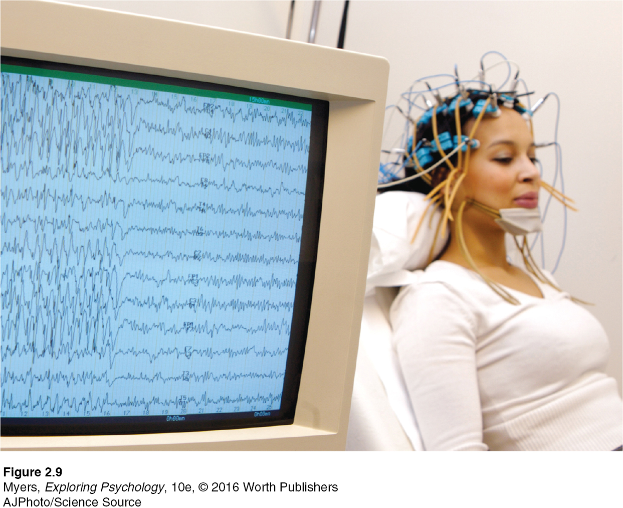
electroencephalogram (EEG) an amplified recording of the waves of electrical activity sweeping across the brain’s surface. These waves are measured by electrodes placed on the scalp.
Right now, your mental activity is emitting telltale electrical, metabolic, and magnetic signals that would enable neuroscientists to observe your brain at work. Electrical activity in your brain’s billions of neurons sweeps in regular waves across its surface. An electroencephalogram (EEG) is an amplified readout of such waves (FIGURE 2.9). Researchers record the brain waves through a shower-
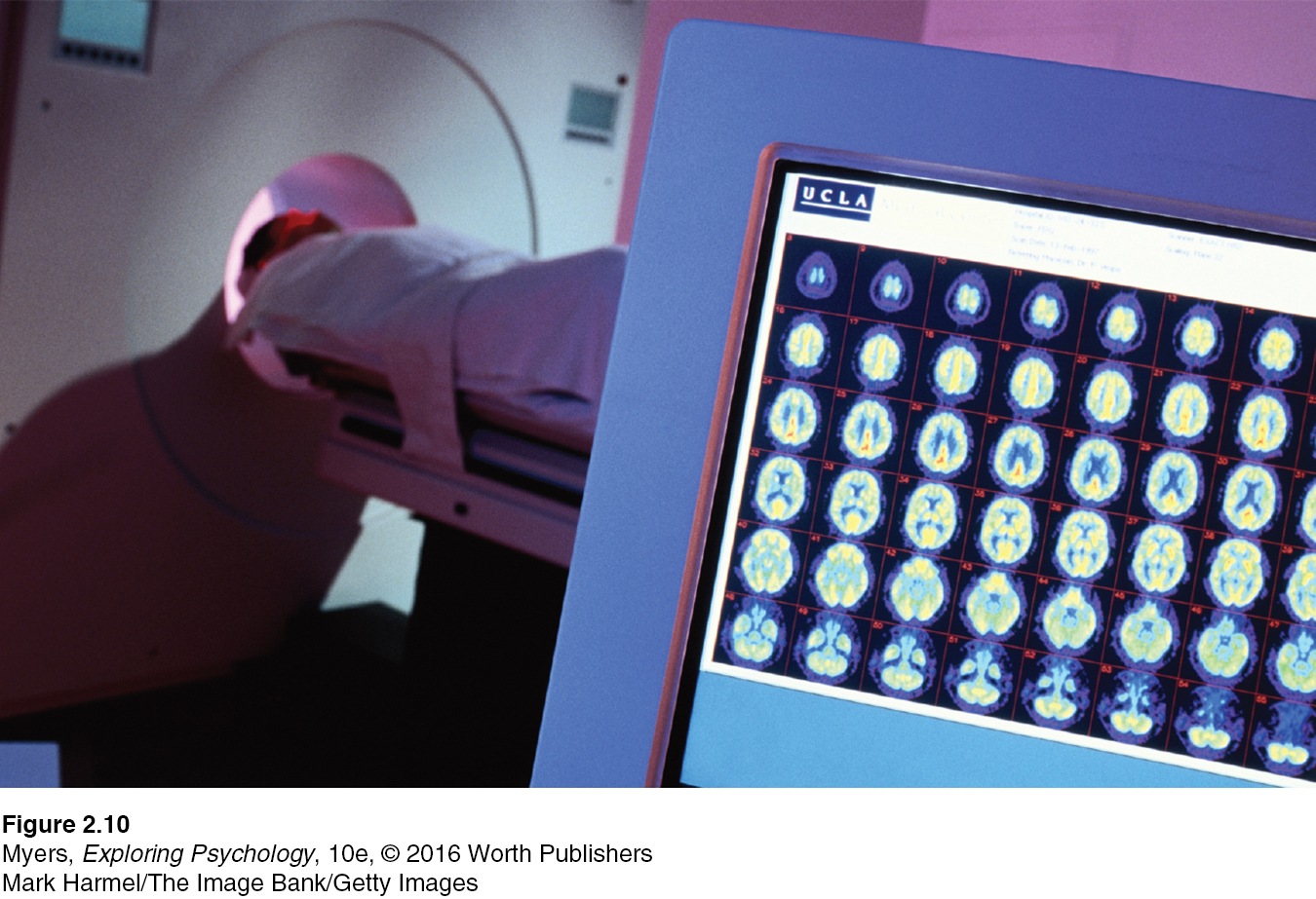
PET (positron emission tomography) scan a visual display of brain activity that detects where a radioactive form of glucose goes while the brain performs a given task.
“You must look into people, as well as at them,” advised Lord Chesterfield in a 1746 letter to his son. Unlike EEGs, newer neuroimaging techniques give us that Superman-
MRI (magnetic resonance imaging) a technique that uses magnetic fields and radio waves to produce computer-
In MRI (magnetic resonance imaging) brain scans, the person’s head is put in a strong magnetic field, which aligns the spinning atoms of brain molecules. Then, a radio-
fMRI (functional MRI) a technique for revealing bloodflow and, therefore, brain activity by comparing successive MRI scans. fMRI scans show brain function as well as structure.
A special application of MRI—
Such snapshots of the brain’s activity provide new insights into how the brain divides its labor. A mountain of recent fMRI studies suggests which brain areas are most active when people feel pain or rejection, listen to angry voices, think about scary things, feel happy, or become sexually excited. The technology enables a very crude sort of mind reading. One neuroscience team scanned 129 people’s brains as they did eight different mental tasks (such as reading, gambling, or rhyming). Later, they were able, with 80 percent accuracy, to predict which of these mental activities their participants had been doing (Poldrack et al., 2009).
You’ve seen the pictures—
Nevertheless, to learn about the neurosciences now is like studying world geography when Magellan explored the seas. The $40 million Human Connectome Project (2013; Gorman, 2014), for example, seeks “neural pathways [that] will reveal much about what makes us uniquely human and what makes every person different from all others.” It harnesses the power of diffusion spectrum imaging, a type of MRI technology that maps long-
RETRIEVE IT
Match the scanning technique with the correct description.
Question
fMRI scan PET scan MRI scan | uses magnetic fields and radio waves to show brain anatomy. tracks successive images of brain tissue to show brain function. tracks radioactive glucose to reveal brain activity. |
Older Brain Structures

2-
 For an introductory 12.5-
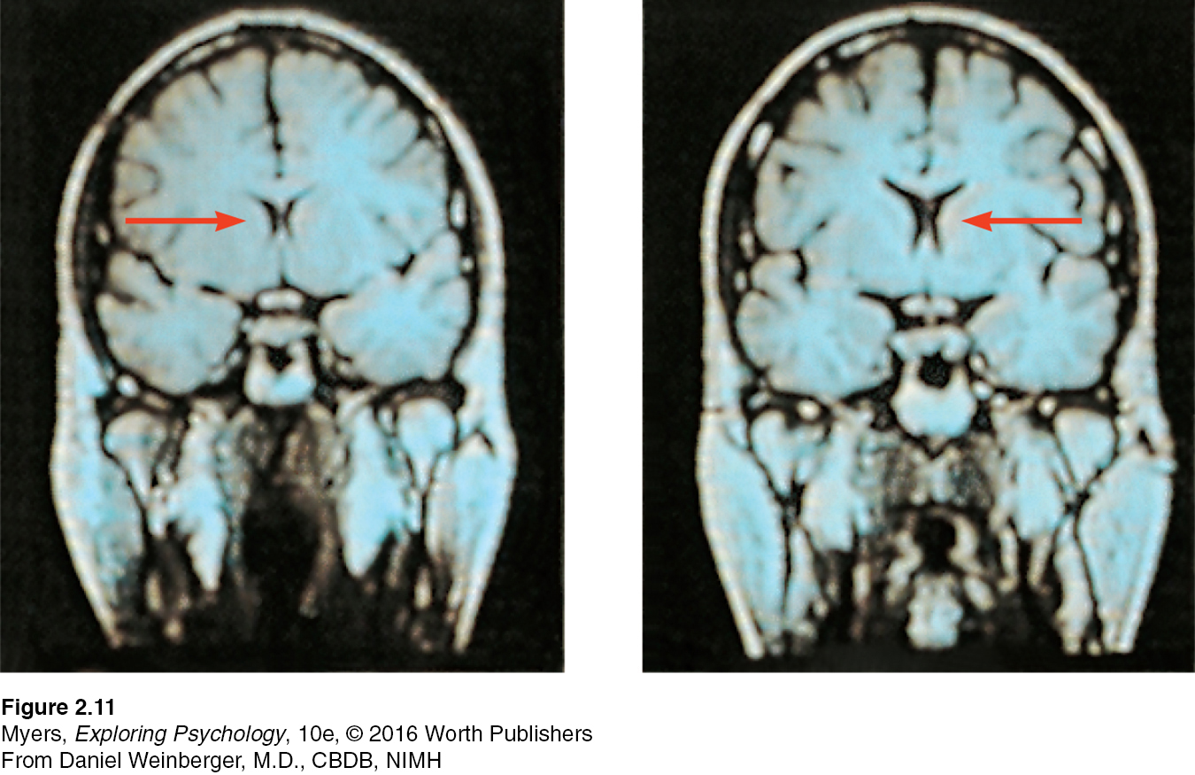
An animal’s capacities come from its brain structures. In primitive animals, such as sharks, a not-
This increasing complexity arises from new brain systems built on top of the old, much as Earth’s landscape covers the old with the new. Digging down, one discovers the fossil remnants of the past—
The Brainstem
brainstem the oldest part and central core of the brain, beginning where the spinal cord swells as it enters the skull; the brainstem is responsible for automatic survival functions.
medulla [muh-
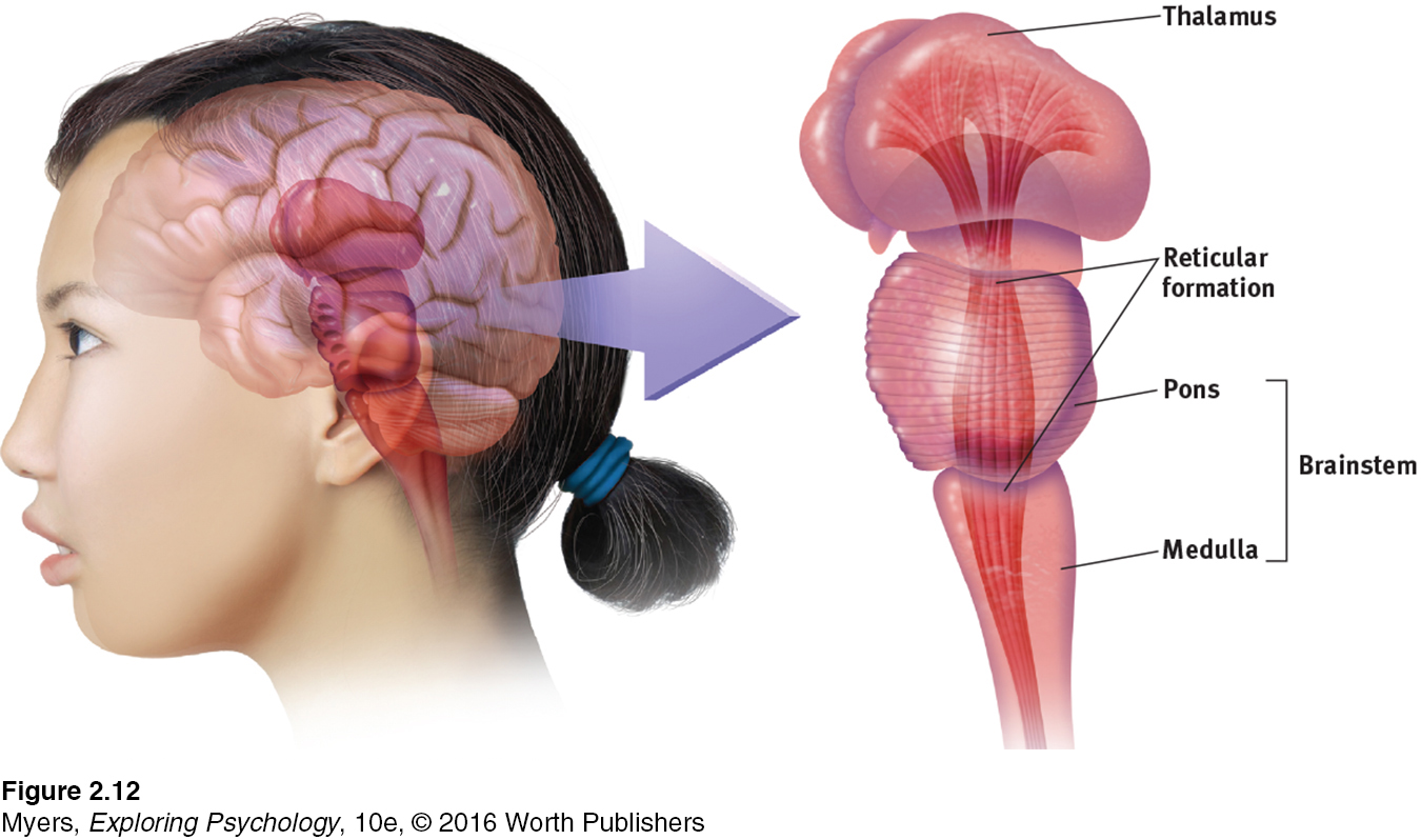
The brain’s oldest and innermost region is the brainstem. It begins where the spinal cord swells slightly after entering the skull. This slight swelling is the medulla (FIGURE 2.12). Here lie the controls for your heartbeat and breathing. As brain-
If a cat’s brainstem is severed from the rest of the brain above it, the animal will still breathe and live—
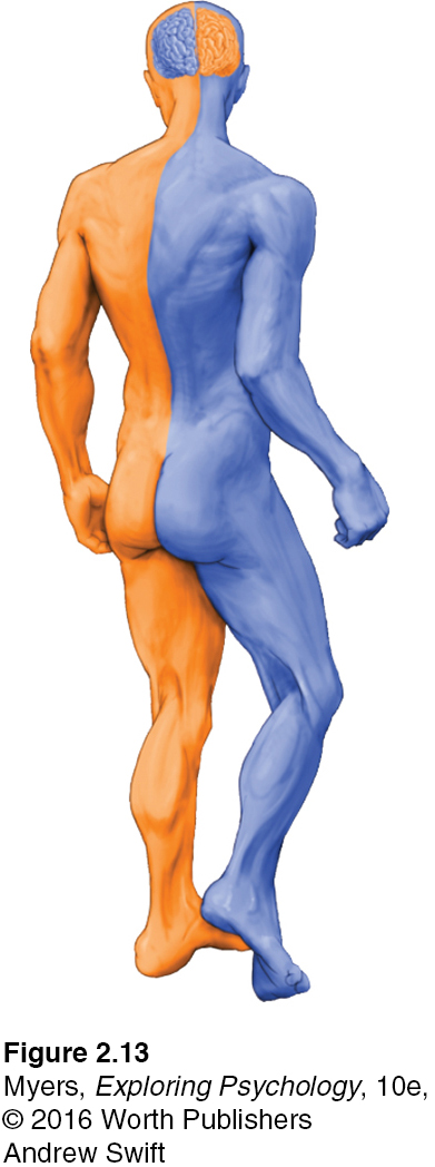
The brainstem is a crossover point, where most nerves to and from each side of the brain connect with the body’s opposite side (FIGURE 2.13). This peculiar cross-
RETRIEVE IT
Question
Nerves from the left side of the brain are mostly linked to the side of the body, and vice versa.
The Thalamus
thalamus [THAL-uh-muss] the brain’s sensory control center, located on top of the brainstem; it directs messages to the sensory receiving areas in the cortex and transmits replies to the cerebellum and medulla.
Sitting atop the brainstem is the thalamus, a pair of egg-shaped structures that act as the brain’s sensory control center (FIGURE 2.12). The thalamus receives information from all the senses except smell, and routes that information to higher brain regions that deal with seeing, hearing, tasting, and touching. The thalamus also receives some of the higher brain’s replies, which it then directs to the medulla and to the cerebellum. Think of the thalamus as being to sensory information what London is to England’s trains: a hub through which traffic passes en route to various destinations.
The Reticular Formation
reticular formation a nerve network that travels through the brainstem into the thalamus and plays an important role in controlling arousal.
Inside the brainstem, between your ears, lies the reticular (“netlike”) formation, a neuron network extending from the spinal cord right up through the thalamus. As the spinal cord’s sensory input flows up to the thalamus, some of it travels through the reticular formation, which filters incoming stimuli, relays important information to other brain areas, and controls arousal.
In 1949, Giuseppe Moruzzi and Horace Magoun discovered that electrically stimulating a sleeping cat’s reticular formation almost instantly produced an awake, alert animal. When Magoun severed a cat’s reticular formation without damaging nearby sensory pathways, the effect was equally dramatic: The cat lapsed into a coma from which it never awakened.
The Cerebellum
cerebellum [sehr-
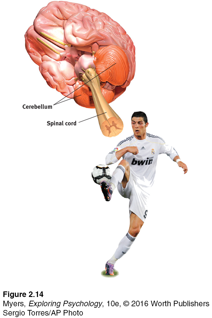
Extending from the rear of the brainstem is the baseball-
* * *
Note: These older brain functions all occur without any conscious effort. This illustrates another of our recurring themes: Our brain processes most information outside of our awareness. We are aware of the results of our brain’s labor—
RETRIEVE IT
Question
In what brain region would damage be most likely to (1) disrupt your ability to skip rope? (2) disrupt your ability to hear and taste? (3) perhaps leave you in a coma? (4) cut off the very breath and heartbeat of life?
The Limbic System
limbic system neural system (including the amygdala, hypothalamus, and hippocampus) located below the cerebral hemispheres; associated with emotions and drives.
2-
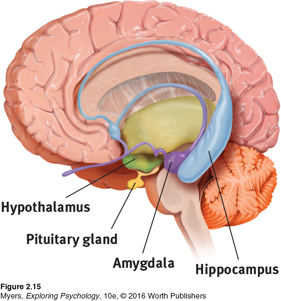
We’ve considered the brain’s oldest parts, but we’ve not yet reached its newest and highest regions, the cerebral hemispheres (the two halves of the brain). Between the oldest and newest brain areas lies the limbic system (limbus means “border”). This system contains the amygdala, the hypothalamus, and the hippocampus (FIGURE 2.15).
amygdala [uh-
THE AMYGDALA Research has linked the amygdala, two lima-

What then might happen if we electrically stimulated the amygdala of a normally placid domestic animal, such as a cat? Do so in one spot and the cat prepares to attack, hissing with its back arched, its pupils dilated, its hair on end. Move the electrode only slightly within the amygdala, cage the cat with a small mouse, and now it cowers in terror.
These and other experiments have confirmed the amygdala’s role in fear and rage. One study found math anxiety associated with hyperactivity in the right amygdala (Young et al., 2012). Other studies link criminal behavior with amygdala dysfunction (Boccardi et al., 2011; Ermer et al., 2012). But we must be careful. The brain is not neatly organized into structures that correspond to our behavior categories. When we feel or act in aggressive or fearful ways, there is neural activity in many areas of our brain—
RETRIEVE IT
Question
Electrical stimulation of a cat's amygdala provokes angry reactions. Which autonomic nervous system division is activated by such stimulation?
hypothalamus [hi-
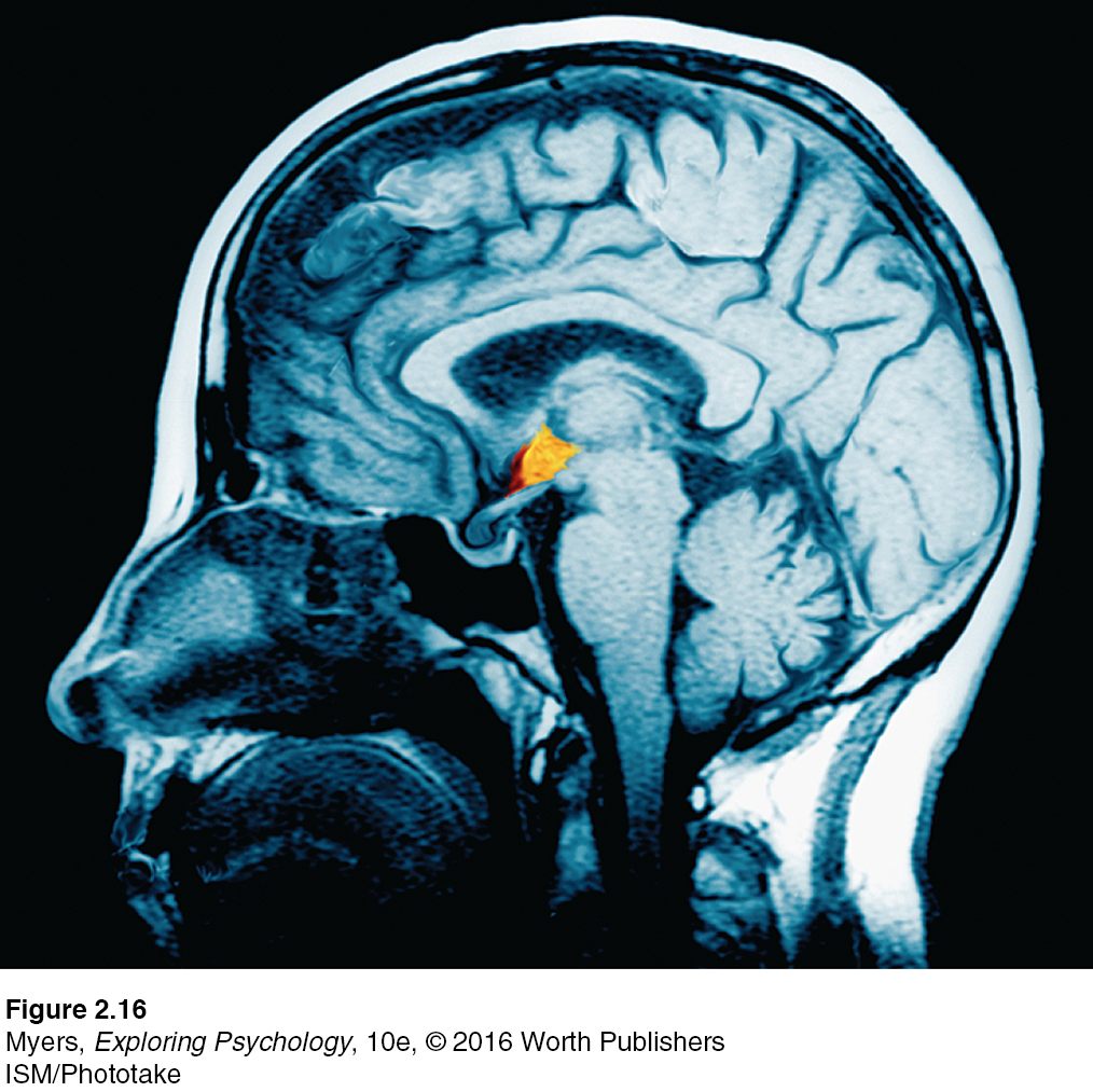
THE HYPOTHALAMUS Just below (hypo) the thalamus is the hypothalamus (FIGURE 2.16), an important link in the command chain governing bodily maintenance. Some neural clusters in the hypothalamus influence hunger; others regulate thirst, body temperature, and sexual behavior. Together, they help maintain a steady (homeostatic) internal state.
As the hypothalamus monitors the state of your body, it tunes into your blood chemistry and any incoming orders from other brain parts. For example, picking up signals from your brain’s cerebral cortex that you are thinking about sex, your hypothalamus will secrete hormones. These hormones will in turn trigger the adjacent “master gland” of the endocrine system, your pituitary (see FIGURE 2.15), to influence your sex glands to release their hormones. These will intensify the thoughts of sex in your cerebral cortex. (Once again, we see the interplay between the nervous and endocrine systems: The brain influences the endocrine system, which in turn influences the brain.)
A remarkable discovery about the hypothalamus illustrates how progress in science often occurs—
Later experiments located other “pleasure centers” (Olds, 1958). (What the rats actually experience only they know, and they aren’t telling. Rather than attribute human feelings to rats, today’s scientists refer to reward centers, not “pleasure centers.”) Just how rewarding are these reward centers? Enough to cause rats to self-
Do humans have limbic centers for pleasure? To calm violent patients, one neurosurgeon implanted electrodes in such areas. Stimulated patients reported mild pleasure; unlike Olds’ rats, however, they were not driven to a frenzy (Deutsch, 1972; Hooper & Teresi, 1986). Moreover, newer research reveals that stimulating the brain’s “hedonic hotspots” (its reward circuits) produces more desire than pure enjoyment (Kringelbach & Berridge, 2012).
“If you were designing a robot vehicle to walk into the future and survive, . . . you’d wire it up so that behavior that ensured the survival of the self or the species—
Candace Pert (1986)
Some researchers believe that substance use disorders may stem from malfunctions in natural brain systems for pleasure and well-
hippocampus a neural center located in the limbic system; helps process explicit memories for storage.
THE HIPPOCAMPUS The hippocampus processes conscious, explicit memories and decreases in size and function as we grow older. Animals or humans who lose their hippocampus to surgery or injury lose their ability to form new memories of facts and events. Those who survive a hippocampal brain tumor in childhood struggle to remember new information in adulthood (Jayakar et al., 2015). Chapter 4 discusses how hippocampus size and function decrease as we grow older. Chapter 8 explains how our two-
* * *
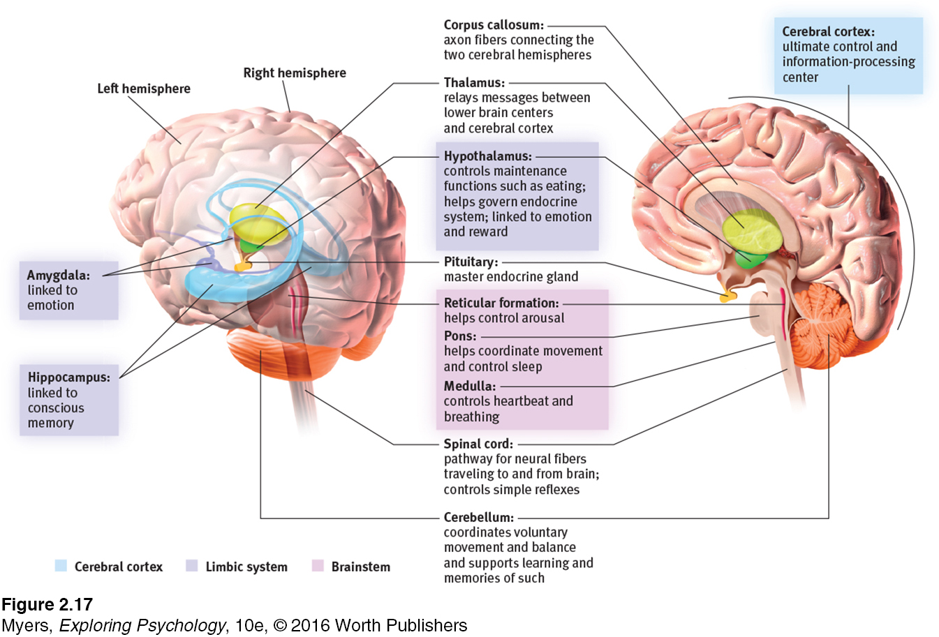
FIGURE 2.17 locates the brain areas we’ve discussed, as well as the cerebral cortex, our next topic.
To review and assess your understanding, visit LaunchPad’s Concept Practice: The Limbic System.
RETRIEVE IT
Question
What are the three key structures of the limbic system, and what functions do they serve?
REVIEW Tools of Discovery and Older Brain Structures
Learning Objectives
Test Yourself by taking a moment to answer each of these Learning Objective Questions (repeated here from within the chapter). Research suggests that trying to answer these questions on your own will improve your long-
Question
2-
Question
2-
Question
2-
Terms and Concepts to Remember
Test yourself on these terms.
Question
lesion (LEE- electroencephalogram (EEG) (p. 48) PET (positron emission tomography) scan (p. 48) MRI (magnetic resonance imaging) (p. 49) fMRI (functional MRI) (p. 49) brainstem (p. 50) medulla (muh- thalamus (THAL- reticular formation (p. 52) cerebellum (sehr- limbic system (p. 52) amygdala (uh- hypothalamus (hi- hippocampus (p. 54) | the brain's sensory control center, located on top of the brainstem; it directs messages to the sensory receiving areas in the cortex and transmits replies to the cerebellum and medulla. a neural structure lying below (hypo) the thalamus; it directs several maintenance activities (eating, drinking, body temperature), helps govern the endocrine system via the pituitary gland, and is linked to emotion and reward. a technique for revealing bloodflow and, therefore, brain activity by comparing successive MRI scans. fMRI scans show brain function as well as structure. a neural center located in the limbic system; helps process explicit memories for storage. a technique that uses magnetic fields and radio waves to produce computer- tissue destruction. A brain lesion is a naturally or experimentally caused destruction of brain tissue. a nerve network that travels through the brainstem into the thalamus and plays an important role in controlling arousal. the “little brain” at the rear of the brainstem; functions include processing sensory input, coordinating movement output and balance, and enabling nonverbal learning and memory. an amplified recording of the waves of electrical activity sweeping across the brain's surface. These waves are measured by electrodes placed on the scalp. neural system (including the amygdala, hypothalamus, and hippocampus) located below the cerebral hemispheres; associated with emotions and drives. two lima- a visual display of brain activity that detects where a radioactive form of glucose goes while the brain performs a given task. the oldest part and central core of the brain, beginning where the spinal cord swells as it enters the skull; the brainstem is responsible for automatic survival functions. the base of the brainstem; controls heartbeat and breathing. |
Experience the Testing Effect
Test yourself repeatedly throughout your studies. This will not only help you figure out what you know and don’t know; the testing itself will help you learn and remember the information more effectively thanks to the testing effect.
Question 2.11
1. The part of the brainstem that controls heartbeat and breathing is the
| A. |
| B. |
| C. |
| D. |
Question 2.12
2. The thalamus functions as a
| A. |
| B. |
| C. |
| D. |
Question 2.13
3. The lower brain structure that governs arousal is the
| A. |
| B. |
| C. |
| D. |
Question 2.14
4. The part of the brain that coordinates voluntary movement and enables nonverbal learning and memory is the .
Question 2.15
5. Two parts of the limbic system are the amygdala and the
| A. |
| B. |
| C. |
| D. |
Question 2.16
6. A cat's ferocious response to electrical brain stimulation would lead you to suppose the electrode had touched the .
Question 2.17
7. The neural structure that most directly regulates eating, drinking, and body temperature is the
| A. |
| B. |
| C. |
| D. |
Question 2.18
8. The initial reward center discovered by Olds and Milner was located in the .
Use  to create your personalized study plan, which will direct you to the resources that will help you most in
.
to create your personalized study plan, which will direct you to the resources that will help you most in
.