2.3 The Cerebral Cortex and Our Divided Brain
The Cerebral Cortex
2-
cerebral [seh-
Older brain networks sustain basic life functions and enable memory, emotions, and basic drives. Newer neural networks within the cerebrum—
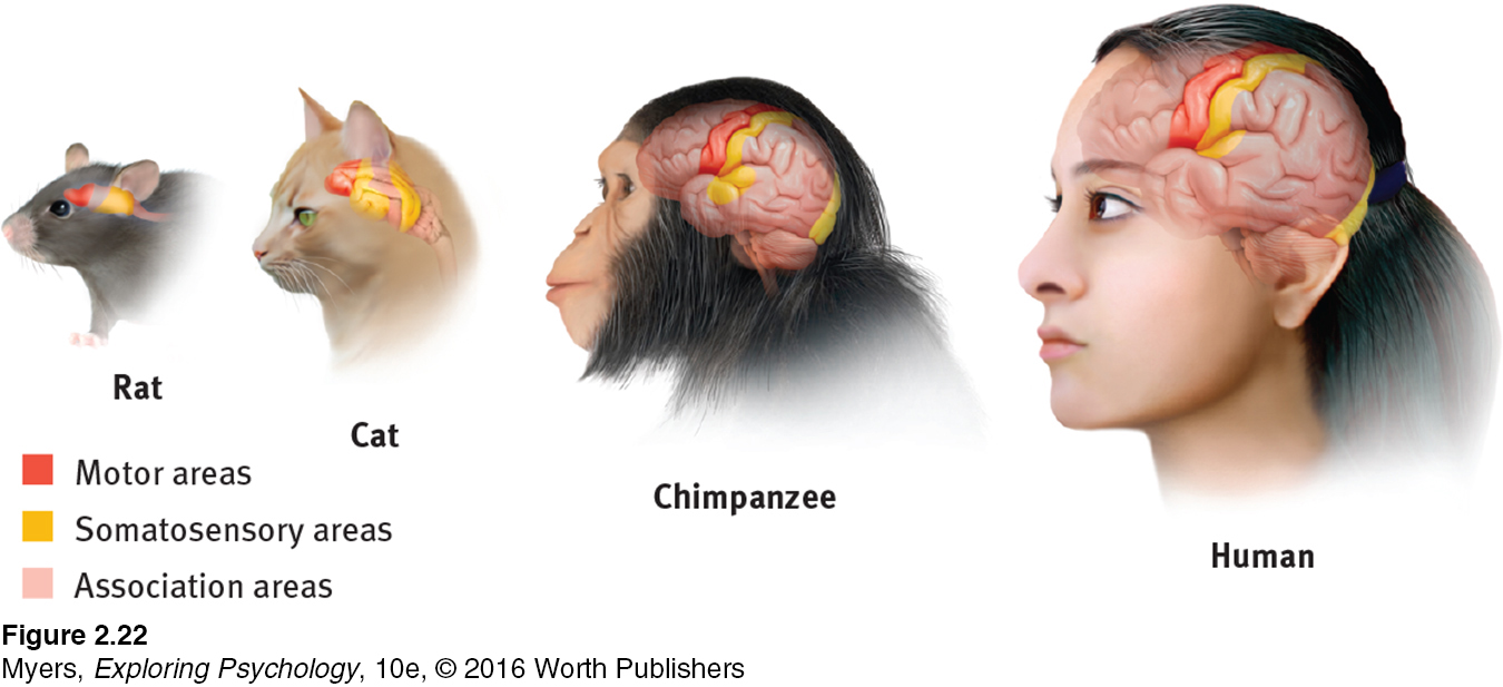
As we move up the ladder of animal life, the cerebral cortex expands, tight genetic controls relax, and the organism’s adaptability increases. Frogs and other small-
The people who first dissected and labeled the brain used the language of scholars—
RETRIEVE IT
Question
Which area of the human brain is most similar to that of less complex animals? Which part of the human brain distinguishes us most from less complex animals?
Structure of the Cortex
If you opened a human skull, exposing the brain, you would see a wrinkled organ, shaped somewhat like an oversized walnut. Without these wrinkles, a flattened cerebral cortex would require triple the area—
frontal lobes portion of the cerebral cortex lying just behind the forehead; involved in speaking and muscle movements and in making plans and judgments.
parietal [puh-
occipital [ahk-
temporal lobes portion of the cerebral cortex lying roughly above the ears; includes the auditory areas, each receiving information primarily from the opposite ear.
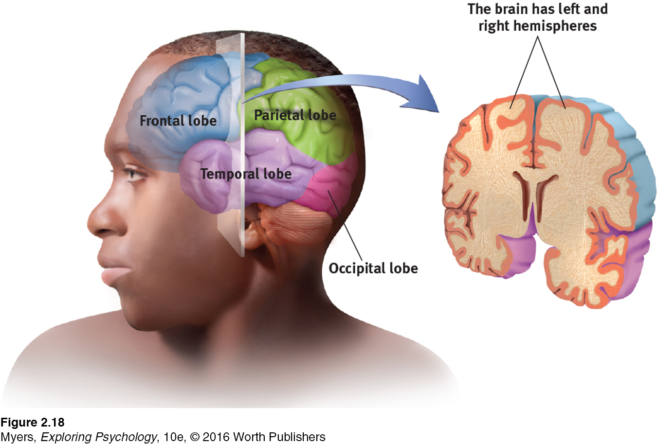
Each hemisphere’s cortex is subdivided into four lobes, separated by prominent fissures, or folds (FIGURE 2.18). Starting at the front of your brain and moving over the top, there are the frontal lobes (behind your forehead), the parietal lobes (at the top and to the rear), and the occipital lobes (at the back of your head). Reversing direction and moving forward, just above your ears, you find the temporal lobes. Each of the four lobes carries out many functions, and many functions require the interplay of several lobes.
Functions of the Cortex
More than a century ago, surgeons found damaged cortical areas during autopsies of people who had been partially paralyzed or speechless. This rather crude evidence did not prove that specific parts of the cortex control complex functions like movement or speech. After all, if the entire cortex controlled speech and movement, damage to almost any area might produce the same effect. A TV with its power cord cut would go dead, but we would be fooling ourselves if we thought we had “localized” the picture in the cord.
motor cortex an area at the rear of the frontal lobes that controls voluntary movements.
MOTOR FUNCTIONS Scientists had better luck in localizing simpler brain functions. For example, in 1870, German physicians Gustav Fritsch and Eduard Hitzig made an important discovery: Mild electrical stimulation to parts of an animal’s cortex made parts of its body move. The effects were selective: Stimulation caused movement only when applied to an arch-
MAPPING THE MOTOR CORTEX Lucky for brain surgeons and their patients, the brain has no sensory receptors. Knowing this, in the 1930s, Otfrid Foerster and Wilder Penfield were able to map the motor cortex in hundreds of wide-
RETRIEVE IT
Question
Try moving your right hand in a circular motion, as if cleaning a table. Then start your right foot doing the same motion, synchronized with your hand. Now reverse the right foot's motion, but not the hand's. Finally, try moving the left foot opposite to the right hand.
1. Why is reversing the right foot's motion so hard?
2. Why is it easier to move the left foot opposite to the right hand?
More recently, scientists were able to predict a monkey’s arm motion a tenth of a second before it moved—
What might happen, some researchers are asking, if we implant a device to detect motor cortex activity in humans? Could such devices help severely paralyzed people learn to command a cursor to write e-
somatosensory cortex area at the front of the parietal lobes that registers and processes body touch and movement sensations.
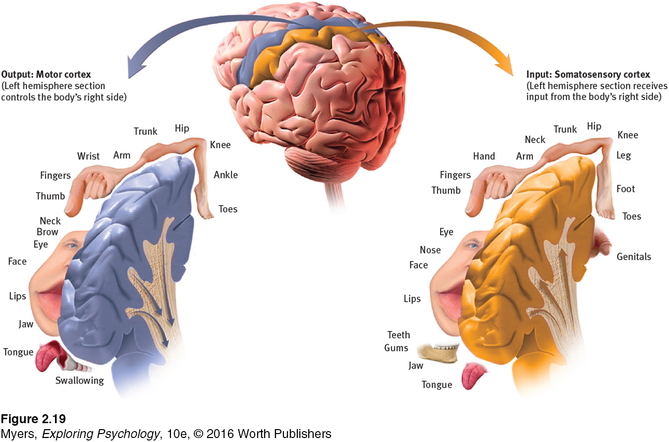
SENSORY FUNCTIONS If the motor cortex sends messages out to the body, where does the cortex receive incoming messages? Penfield identified a cortical area—
The more sensitive the body region, the larger the somatosensory cortex area devoted to it (FIGURE 2.19). Your supersensitive lips project to a larger brain area than do your toes, which is one reason we kiss rather than touch toes. Rats have a large area of the brain devoted to their whisker sensations, and owls to their hearing sensations.
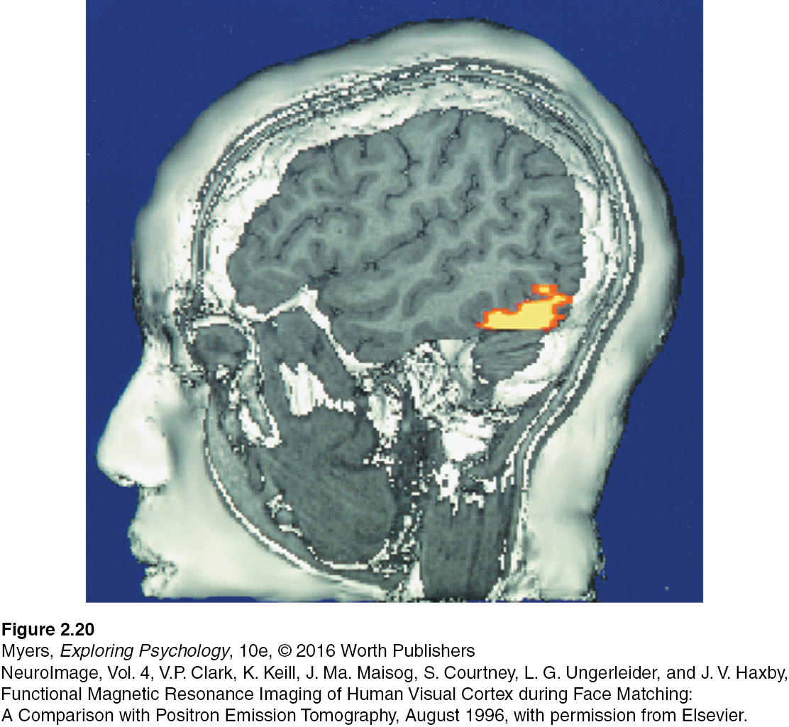
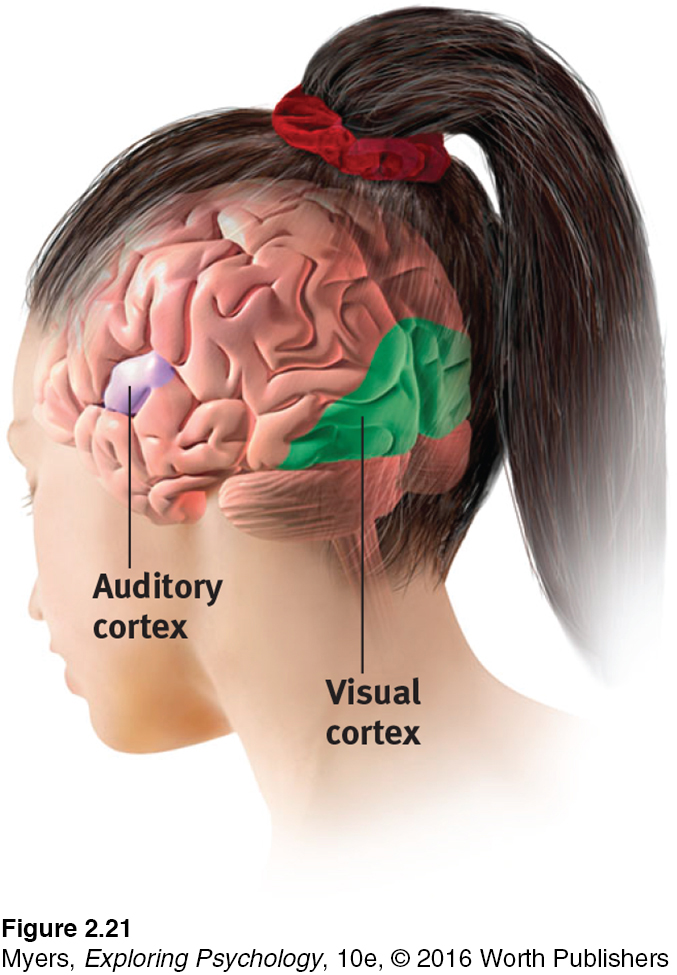
Scientists have identified additional areas where the cortex receives input from senses other than touch. Any visual information you are receiving now is going to the visual cortex in your occipital lobes, at the back of your brain (FIGURES 2.20 and 2.21). Stimulated in the occipital lobes, you might see flashes of light or dashes of color. (In a sense, we do have eyes in the back of our head!) Having lost much of his right occipital lobe to a tumor removal, a friend of mine [DM’s] was blind to the left half of his field of vision. Visual information travels from the occipital lobes to other areas that specialize in tasks such as identifying words, detecting emotions, and recognizing faces.
Any sound you now hear is processed by your auditory cortex in your temporal lobes (just above your ears; see FIGURE 2.21). Most of this auditory information travels a circuitous route from one ear to the auditory receiving area above your opposite ear. If stimulated in your auditory cortex, you might hear a sound. MRI scans of people with schizophrenia have revealed active auditory areas in the temporal lobes during the false sensory experience of auditory hallucinations (Lennox et al., 1999). Even the phantom ringing sound experienced by people with hearing loss is—
RETRIEVE IT
Question
Our brain's cortex registers and processes body touch and movement sensations. The cortex controls our voluntary movements.
association areas areas of the cerebral cortex that are not involved in primary motor or sensory functions; rather, they are involved in higher mental functions such as learning, remembering, thinking, and speaking
ASSOCIATION AREAS So far, we have pointed out small cortical areas that either receive sensory input or direct muscular output. Together, these occupy about one-fourth of the human brain’s thin, wrinkled cover. What, then, goes on in the remaining vast regions of the cortex? In these association areas (the peach-colored areas in FIGURE 2.22), neurons are busy with higher mental functions—
Electrically probing an association area won’t trigger any observable response. So, unlike the somatosensory and motor areas, association area functions cannot be neatly mapped. Their silence has led to what Donald McBurney (1996, p. 44) called “one of the hardiest weeds in the garden of psychology”: the claim that we ordinarily use only 10 percent of our brain. (If true, wouldn’t this imply a 90 percent chance that a bullet to your brain would land in an unused area?) Surgically lesioned animals and brain-damaged humans bear witness that association areas are not dormant. Rather, these areas interpret, integrate, and act on sensory information and link it with stored memories—
Association areas are found in all four lobes. The prefrontal cortex in the forward part of the frontal lobes enables judgment, planning, and processing of new memories. People with damaged frontal lobes may have intact memories, high scores on intelligence tests, and great cake-
 See LaunchPad’s Video: Case Studies for a helpful tutorial animation.
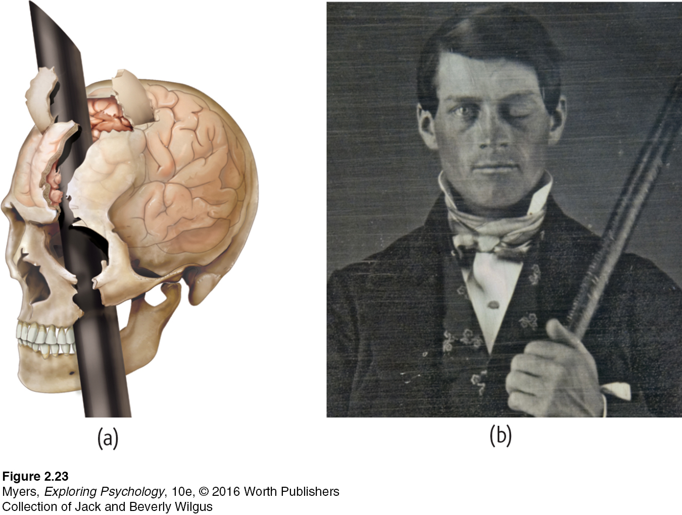
Frontal lobe damage also can alter personality and remove a person’s inhibitions. Consider the classic case study of railroad worker Phineas Gage. One afternoon in 1848, Gage, then 25 years old, was using a tamping iron to pack gunpowder into a rock. A spark ignited the gunpowder, shooting the rod up through his left cheek and out the top of his skull, leaving his frontal lobes damaged (FIGURE 2.23). To everyone’s amazement, Gage was immediately able to sit up and speak, and after the wound healed he returned to work. But having lost some of the neural tracts that enabled his frontal lobes to control his emotions (Van Horn et al., 2012), the affable, soft-
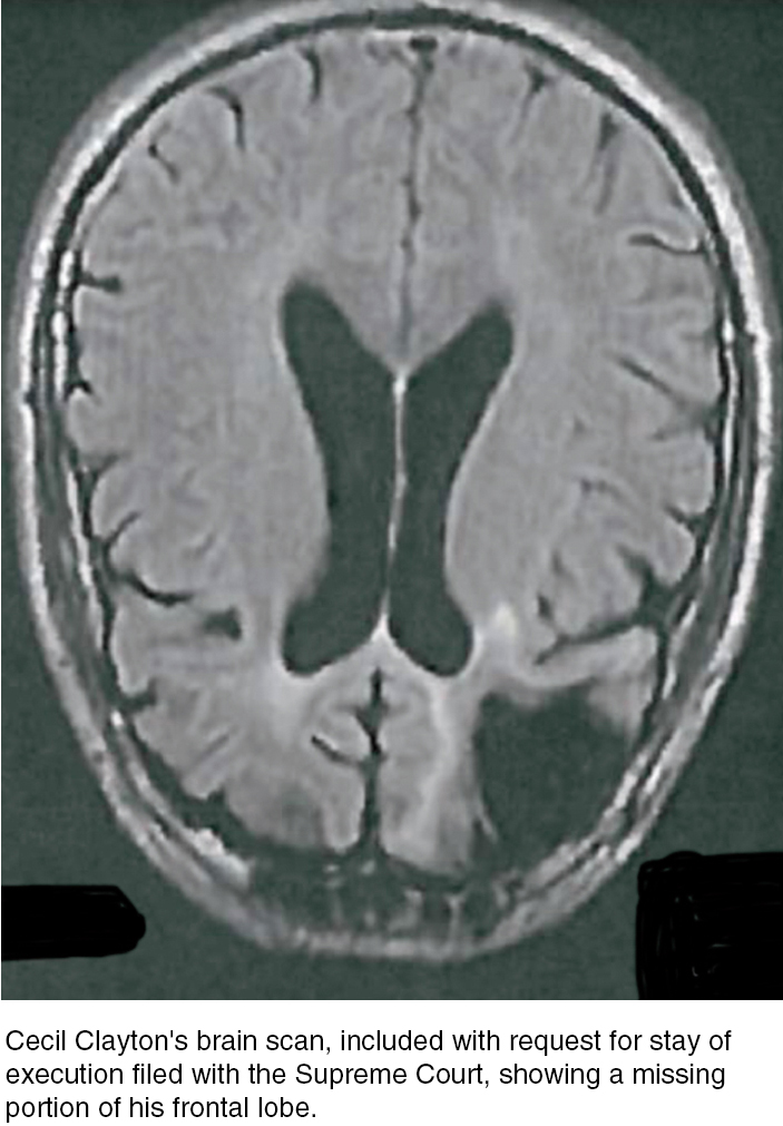
Studies of others with damaged frontal lobes have revealed similar impairments. Not only may they become less inhibited (without the frontal lobe brakes on their impulses), but their moral judgments may seem unrestrained. Cecil Clayton lost 20 percent of his left frontal lobe in a 1972 sawmill accident. Thereafter, his intelligence test score dropped to an elementary school level and he displayed increased impulsivity. In 1996, he fatally shot a deputy sheriff. In 2015, when he was 74, the State of Missouri executed him (Williams, 2015).
Would you advocate pushing one person in front of a runaway trolley to save five others? Most people would not, but those with damage to a brain area behind the eyes are often untroubled by such ethical dilemmas (Koenigs et al., 2007). The frontal lobes help steer us away from violent actions (Molenberghs et al., 2015; Yang & Raine, 2009).
Association areas also perform other mental functions. The parietal lobes, parts of which were large and unusually shaped in Einstein’s normal-
Nevertheless, complex mental functions don’t reside in any one place. There is no one spot in a rat’s small association cortex that, when damaged, will obliterate its ability to learn or remember a maze. Your memory, language, and attention result from synchronized activity among distinct brain areas and neural networks (Knight, 2007). Ditto for religious experience. More than 40 distinct brain regions become active in different religious states, such as prayer and meditation, indicating that there is no simple “God spot” (Fingelkurts & Fingelkurts, 2009). The point to remember: Our mental experiences arise from coordinated brain activity.
RETRIEVE IT
Question
Why are association areas important?
The Brain’s Plasticity
2-
plasticity the brain’s ability to change, especially during childhood, by reorganizing after damage or by building new pathways based on experience.
Our brains are sculpted not only by our genes but also by our experiences. In Chapter 4, we’ll focus more on how experience molds the brain. For now, let’s turn to another aspect of the brain’s plasticity: its ability to modify itself after damage.
Some brain-
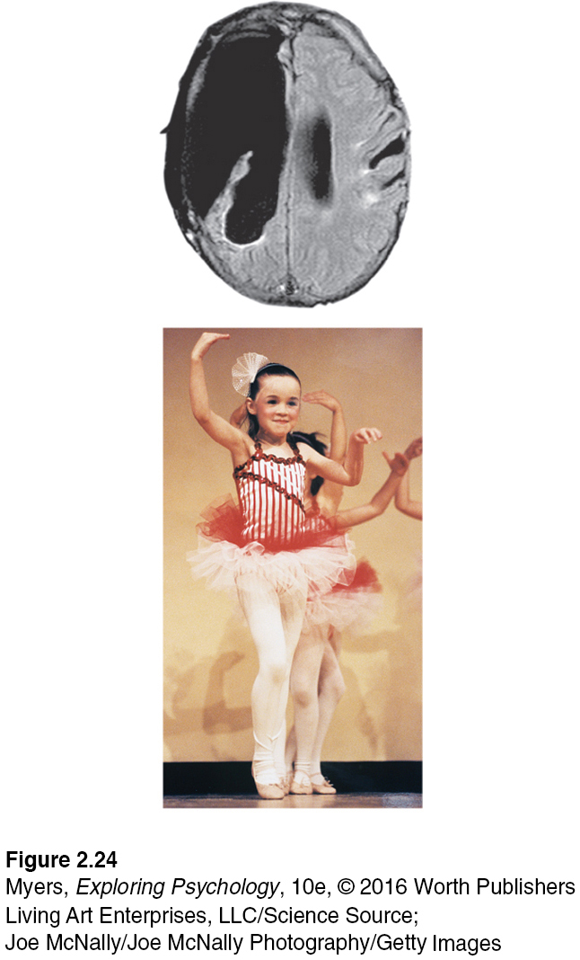
Plasticity may also occur after serious damage, especially in young children (Kolb, 1989; see also FIGURE 2.24). The brain’s plasticity is good news for those with vision or hearing loss. Blindness or deafness makes unused brain areas available for other uses (Amedi et al., 2005). If a blind person uses one finger to read Braille, the brain area dedicated to that finger expands as the sense of touch invades the visual cortex that normally helps people see (Barinaga, 1992a; Sadato et al., 1996).
Plasticity also helps explain why some studies have found that deaf people have enhanced peripheral and motion-
Similar reassignment may occur when disease or damage frees up other brain areas normally dedicated to specific functions. If a slow-
neurogenesis the formation of new neurons.
Although the brain often attempts self-
Master stem cells that can develop into any type of brain cell have also been discovered in the human embryo. If mass-
Our Divided Brain
2-
Our brain’s look-
Splitting the Brain
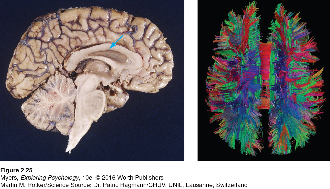
corpus callosum [KOR-
In 1961, Los Angeles neurosurgeons Philip Vogel and Joseph Bogen speculated that major epileptic seizures were caused by an amplification of abnormal brain activity bouncing back and forth between the two cerebral hemispheres, which work together as a whole system. If so, they wondered, could they end this biological tennis match by severing the corpus callosum, the wide band of axon fibers connecting the two hemispheres and carrying messages between them (FIGURE 2.25)? Vogel and Bogen knew that psychologists Roger Sperry, Ronald Myers, and Michael Gazzaniga had divided cats’ and monkeys’ brains in this manner, with no serious ill effects.
split brain a condition resulting from surgery that isolates the brain’s two hemispheres by cutting the fibers (mainly those of the corpus callosum) connecting them.
So the surgeons operated. The result? The seizures all but disappeared. The patients with these split brains were surprisingly normal, their personality and intellect hardly affected. Waking from surgery, one even joked that he had a “splitting headache” (Gazzaniga, 1967). By sharing their experiences, these patients have greatly expanded our understanding of interactions between the intact brain’s two hemispheres.
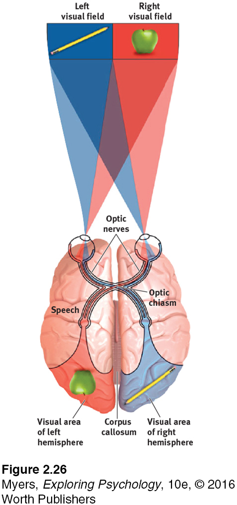
To appreciate these findings, we need to focus for a minute on the peculiar nature of our visual wiring, illustrated in FIGURE 2.26. Note that each eye receives sensory information from the entire visual field. But in each eye, information from the left half of your field of vision goes to your right hemisphere, and information from the right half of your visual field goes to your left hemisphere, which usually controls speech. Information received by either hemisphere is quickly transmitted to the other across the corpus callosum. In a person with a severed corpus callosum, this information-
Knowing these facts, Sperry and Gazzaniga could send information to a patient’s left or right hemisphere. As the person stared at a spot, they flashed a stimulus to its right or left. They could do this with you, too, but in your intact brain, the hemisphere receiving the information would instantly pass the news to the other side. Because the split-
In an early experiment, Gazzaniga (1967) asked split-
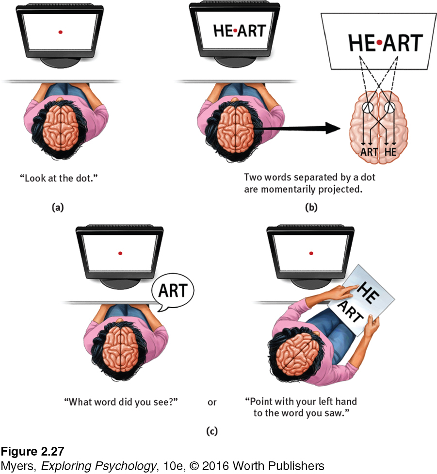
When a picture of a spoon was flashed to their right hemisphere, the patients could not say what they had viewed. But when asked to identify what they had viewed by feeling an assortment of hidden objects with their left hand, they readily selected the spoon. If the experimenter said, “Correct!” the patient might reply, “What? Correct? How could I possibly pick out the correct object when I don’t know what I saw?” It is, of course, the left hemisphere doing the talking here, bewildered by what the nonverbal right hemisphere knows.
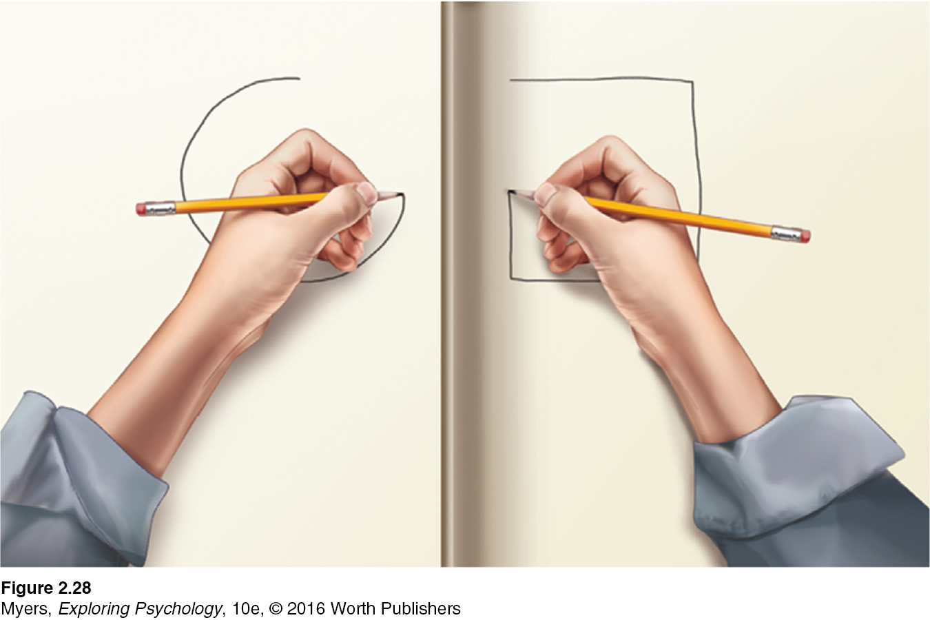
A few people who have had split-
“Do not let your left hand know what your right hand is doing.”
Matthew 6:3
IMMERSIVE LEARNING Have you ever been asked if you are “left-
When the “two minds” are at odds, the left hemisphere does mental gymnastics to rationalize reactions it does not understand. If a patient follows an order (“Walk”) sent to the right hemisphere, a strange thing happens. The left hemisphere, unaware of the order, doesn’t know why the patient begins walking. If asked why, the patient doesn’t reply, “I don’t know.” Instead, the left hemisphere improvises—
RETRIEVE IT
Question
(1) If we flash a red light to the right hemisphere of a person with a split brain, and flash a green light to the left hemisphere, will each observe its own color? (2) Will the person be aware that the colors differ? (3) What will the person verbally report seeing?
Right-Left Differences in the Intact Brain
So, what about the 99.99+ percent of us with undivided brains? Does each of our hemispheres also perform distinct functions? Several different types of studies indicate they do. When a person performs a perceptual task, for example, brain waves, bloodflow, and glucose consumption reveal increased activity in the right hemisphere. When the person speaks or calculates, activity usually increases in the left hemisphere.
A dramatic demonstration of hemispheric specialization happens before some types of brain surgery. To locate the patient’s language centers, the surgeon injects a sedative into the neck artery feeding blood to the left hemisphere, which usually controls speech. Before the injection, the patient is lying down, arms in the air, chatting with the doctor. Can you predict what probably happens when the drug puts the left hemisphere to sleep? Within seconds, the person’s right arm falls limp. If the left hemisphere is controlling language, the patient will be speechless until the drug wears off. If the drug is injected into the artery to the right hemisphere, the left arm will fall limp, but the person will still be able to speak.
To the brain, language is language, whether spoken or signed. Just as hearing people usually use the left hemisphere to process spoken language, deaf people use the left hemisphere to process sign language (Corina et al., 1992; Hickok et al., 2001). Thus, a left hemisphere stroke disrupts a deaf person’s signing, much as it would disrupt a hearing person’s speaking (Corina, 1998).
Although the left hemisphere is skilled at making quick, literal interpretations of language, the right hemisphere excels at making inferences (Beeman & Chiarello, 1998; Bowden & Beeman, 1998; Mason & Just, 2004). It also helps us modulate our speech to make meaning clear—
For a helpful animated review of split-
Simply looking at the two hemispheres, so alike to the naked eye, who would suppose they contribute uniquely to the harmony of the whole? Yet a variety of observations—
How does the brain’s intricate networking emerge? How does our heredity—
REVIEW The Cerebral Cortex and Our Divided Brain
Learning Objectives
Test Yourself by taking a moment to answer each of these Learning Objective Questions (repeated here from within the chapter). Research suggests that trying to answer these questions on your own will improve your long-
Question
2-
Question
2-
Question
2-
Terms and Concepts to Remember
Test yourself on these terms.
Question
cerebral (seh- frontal lobes (p. 56) parietal (puh- occipital (ahk- temporal lobes (p. 56) motor cortex (p. 57) somatosensory cortex (p. 58) association areas (p. 59) plasticity (p. 60) neurogenesis (p. 61) corpus callosum (KOR- split brain (p. 62) | portion of the cerebral cortex lying just behind the forehead; involved in speaking and muscle movements and in making plans and judgments. portion of the cerebral cortex lying at the back of the head; includes areas that receive information from the visual fields. portion of the cerebral cortex lying at the top of the head and toward the rear; receives sensory input for touch and body position. portion of the cerebral cortex lying roughly above the ears; includes the auditory areas, each receiving information primarily from the opposite ear. area at the front of the parietal lobes that registers and processes body touch and movement sensations. a condition resulting from surgery that isolates the brain's two hemispheres by cutting the fibers (mainly those of the corpus callosum) connecting them. areas of the cerebral cortex that are not involved in primary motor or sensory functions; rather, they are involved in higher mental functions such as learning, remembering, thinking, and speaking the intricate fabric of interconnected neural cells covering the cerebral hemispheres; the body's ultimate control and information- the large band of neural fibers connecting the two brain hemispheres and carrying messages between them. the brain's ability to change, especially during childhood, by reorganizing after damage or by building new pathways based on experience. the formation of new neurons. an area at the rear of the frontal lobes that controls voluntary movements. |
Experience the Testing Effect
Test yourself repeatedly throughout your studies. This will not only help you figure out what you know and don’t know; the testing itself will help you learn and remember the information more effectively thanks to the testing effect.
Question 2.19
1. If a neurosurgeon stimulated your right motor cortex, you would most likely
| A. |
| B. |
| C. |
| D. |
Question 2.20
2. How do different neural networks communicate with one another to let you respond when a friend greets you at a party?
Question 2.21
3. Which of the following body regions has the greatest representation in the somatosensory cortex?
| A. |
| B. |
| C. |
| D. |
Question 2.22
4. Judging and planning are enabled by the lobes.
Question 2.23
5. What would it be like to talk on the phone if you didn't have temporal lobe association areas? What would you hear? What would you understand?
Question 2.24
6. The “uncommitted” areas that make up about three-fourths of the cerebral cortex are called .
Question 2.25
7. Plasticity is especially evident in the brains of
| A. |
| B. |
| C. |
| D. |
Question 2.26
8. An experimenter flashes the word HERON across the visual field of a man whose corpus callosum has been severed. HER is transmitted to his right hemisphere and ON to his left hemisphere. When asked to indicate what he saw, the man says he saw but points to .
Question 2.27
9. Studies of people with split brains and brain scans of those with undivided brains indicate that the left hemisphere excels in
| A. |
| B. |
| C. |
| D. |
Question 2.28
10. Damage to the brain's right hemisphere is most likely to reduce a person's ability to
| A. |
| B. |
| C. |
| D. |
Use  to create your personalized study plan, which will direct you to the resources that will help you most in
.
to create your personalized study plan, which will direct you to the resources that will help you most in
.