5.1 The Cerebral Cortex

5-1 What are the functions of the various cerebral cortex regions?

cerebral [seh-REE-bruhl] cortex the intricate fabric of interconnected neural cells covering the cerebral hemispheres; the body’s ultimate control and information-processing center.

Older brain networks sustain basic life functions and enable memory, emotions, and basic drives. Newer neural networks within the cerebrum—the two cerebral hemispheres contributing 85 percent of the brain’s weight—form specialized work teams that enable our perceiving, thinking, and speaking. Like other structures above the brainstem (including the thalamus, hippocampus, and amygdala), the cerebral hemispheres come as a pair. Covering those hemispheres, like bark on a tree, is the cerebral cortex, a thin surface layer of interconnected neural cells. It is your brain’s thinking crown, your body’s ultimate control and information-processing center.

As we move up the ladder of animal life, the cerebral cortex expands, tight genetic controls relax, and the organism’s adaptability increases. Frogs and other small-cortex amphibians operate extensively on preprogrammed genetic instructions. The larger cortex of mammals offers increased capacities for learning and thinking, enabling them to be more adaptable. What makes us distinctively human mostly arises from the complex functions of our cerebral cortex.

The people who first dissected and labeled the brain used the language of scholars—Latin and Greek. Their words are actually attempts at graphic description: For example, cortex means “bark,” cerebellum is “little brain,” and thalamus is “inner chamber.”

RETRIEVE IT

Question

KfAkQZnbBMQOp2rGHpLXzeS/g9dXv4Lt6lQYYufhGNCvHeLw+YSqN3TSMWEqrOHchgXPUcHKSdrGwUfl4XMjiK2IhfYD+C75tu3Qb3sRz7MUiRWkzCDLJ/SWZj2NoupNBbsppMVeFrItta5UThAVL7c67PyFsg0wqPhz6Cswoxu1MlfpygIySOTUOMpCBCIeNczSTQCczURieqj7/EBN2nNcs3o=
ANSWERS: The brainstem; the cerebral cortex

Structure of the Cortex

If you opened a human skull, exposing the brain, you would see a wrinkled organ, shaped somewhat like an oversized walnut. Without these wrinkles, a flattened cerebral cortex would require triple the area—roughly that of a large pizza. The brain’s left and right hemispheres are filled mainly with axons connecting the cortex to the brain’s other regions. The cerebral cortex—that thin surface layer—contains some 20 to 23 billion of the brain’s nerve cells and 300 trillion synaptic connections (de Courten-Myers, 2005). Being human takes a lot of nerve.

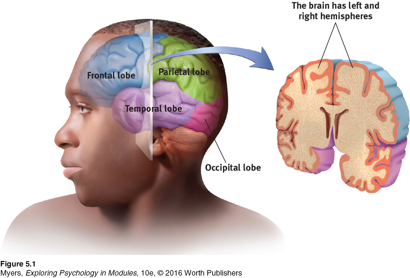
frontal lobes portion of the cerebral cortex lying just behind the forehead; involved in speaking and muscle movements and in making plans and judgments.

parietal [puh-RYE-uh-tuhl] lobes portion of the cerebral cortex lying at the top of the head and toward the rear; receives sensory input for touch and body position.

occipital [ahk-SIP-uh-tuhl] lobes portion of the cerebral cortex lying at the back of the head; includes areas that receive information from the visual fields.

temporal lobes portion of the cerebral cortex lying roughly above the ears; includes the auditory areas, each receiving information primarily from the opposite ear.

Each hemisphere’s cortex is subdivided into four lobes, separated by prominent fissures, or folds (FIGURE 5.1). Starting at the front of your brain and moving over the top, there are the frontal lobes (behind your forehead), the parietal lobes (at the top and to the rear), and the occipital lobes (at the back of your head). Reversing direction and moving forward, just above your ears, you find the temporal lobes. Each of the four lobes carries out many functions, and many functions require the interplay of several lobes.

image
Figure 2.18: FIGURE 5.1 The cortex and its basic subdivisions

Functions of the Cortex

57

More than a century ago, surgeons found damaged cortical areas during autopsies of people who had been partially paralyzed or speechless. This rather crude evidence did not prove that specific parts of the cortex control complex functions like movement or speech. After all, if the entire cortex controlled speech and movement, damage to almost any area might produce the same effect. A TV with its power cord cut would go dead, but we would be fooling ourselves if we thought we had “localized” the picture in the cord.

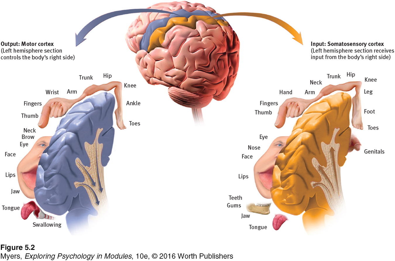
motor cortex an area at the rear of the frontal lobes that controls voluntary movements.

MOTOR FUNCTIONS Scientists had better luck in localizing simpler brain functions. For example, in 1870, German physicians Gustav Fritsch and Eduard Hitzig made an important discovery: Mild electrical stimulation to parts of an animal’s cortex made parts of its body move. The effects were selective: Stimulation caused movement only when applied to an arch-shaped region at the back of the frontal lobe, running roughly ear-to-ear across the top of the brain. Moreover, stimulating parts of this region in the left or right hemisphere caused movements of specific body parts on the opposite side of the body. Fritsch and Hitzig had discovered what is now called the motor cortex.

MAPPING THE MOTOR CORTEX Lucky for brain surgeons and their patients, the brain has no sensory receptors. Knowing this, in the 1930s, Otfrid Foerster and Wilder Penfield were able to map the motor cortex in hundreds of wide-awake patients by stimulating different cortical areas and observing the body’s responses. They discovered that body areas requiring precise control, such as the fingers and mouth, occupy the greatest amount of cortical space (FIGURE 5.2). In one of his many demonstrations of motor behavior mechanics, Spanish neuroscientist José Delgado stimulated a spot on a patient’s left motor cortex, triggering the right hand to make a fist. Asked to keep the fingers open during the next stimulation, the patient, whose fingers closed despite his best efforts, remarked, “I guess, Doctor, that your electricity is stronger than my will” (Delgado, 1969, p. 114).

image
Figure 2.19: FIGURE 5.2 Left hemisphere tissue devoted to each body part in the motor cortex and the somatosensory cortex As you can see from this classic though inexact representation, the amount of cortex devoted to a body part in the motor cortex (in the frontal lobes) or in the somatosensory cortex (in the parietal lobes) is not proportional to that body part’s size. Rather, the brain devotes more tissue to sensitive areas and to areas requiring precise control. Thus, the fingers have a greater representation in the cortex than does the upper arm.

58

RETRIEVE IT

Question

h8v42edQdzMxF3+5CoGkEEbF2pfc4zUg853bmZbrlV942xAVlq1s7dxajCYavcxr69T82aFqfsrFVaprmX0xVzxofuAcXzSNbDmY25lAG25KYexgyAewttDmyua7dN04eee3J+P6G3G0RvWNA+H4UfwLKwt3NVtxGWqgW7vS/A8HQouK8rEL37gNn3yY5aioEFsuF9vskogrZWbbWnnxT38eiHy3RmZDoEHQa+Fvug/wCg9KCka0UAj/EhC65gfR6QYlYKQND4rtbvhyfdttHLIoOlIBdNArhyBUVh/ugAOdnIdDFYTaTgpT2Tb0rBzdXJXPoGekis6bzHZZ7lpiVK02vw6ObB5a+khYQd0mPpzhEI+VTHYZ8Jqf8TW8L/uizdAxBn22AYMA6Lh12Is1Eoxj2/ghwVIR77endbk1ygmF7bcwrxHZQFz69Yh9eqHSn+EiJBHQQpp7Gi41a4cz3hxMFNNpFTOhngO6nxWQv/k6lylwh8zF30+Z7ITDllV6jctDPzwBCrlg+W/qMG9G8lJ0/6bwELjhRtmSU5N3qYaDYgx/
ANSWERS: 1. The right limbs' opposed activities interfere with each other because both are controlled by the same (left) side of your brain. 2. Opposite sides of your brain control your left and right limbs, so the reversed motion causes less interference.

More recently, scientists were able to predict a monkey’s arm motion a tenth of a second before it moved—by repeatedly measuring motor cortex activity preceding specific arm movements (Gibbs, 1996). Such findings have opened the door to research on brain-controlled computers.

What might happen, some researchers are asking, if we implant a device to detect motor cortex activity in humans? Could such devices help severely paralyzed people learn to command a cursor to write e-mail or work online? Clinical trials are now under way with people who have suffered paralysis or amputation (Andersen et al., 2010; Nurmikko et al., 2010). The first patient, a paralyzed 25-year-old man, was able to mentally control a TV, draw shapes on a computer screen, and play video games—all thanks to an aspirin-sized chip with 100 microelectrodes recording activity in his motor cortex (Hochberg et al., 2006). Since then, others with paralysis who have been given implants have learned to direct robotic arms with their thoughts (Collinger et al., 2013; Hochberg et al., 2012).

somatosensory cortex area at the front of the parietal lobes that registers and processes body touch and movement sensations.

image
Figure 2.20: FIGURE 5.3 The brain in action This fMRI (functional MRI) scan shows the visual cortex in the occipital lobes activated (color represents increased bloodflow) as a research participant looks at a photo. When the person stops looking, the region instantly calms down.
NeuroImage, Vol. 4, V.P. Clark, K. Keill, J. Ma. Maisog, S. Courtney, L. G. Ungerleider, and J. V. Haxby, Functional Magnetic Resonance Imaging of Human Visual Cortex during Face Matching: A Comparison with Positron Emission Tomography, August 1996, with permission from Elsevier.
image
Figure 2.21: FIGURE 5.4 The visual cortex and auditory cortex The visual cortex in the occipital lobes at the rear of your brain receives input from your eyes. The auditory cortex, in your temporal lobes—above your ears—receives information from your ears.

SENSORY FUNCTIONS If the motor cortex sends messages out to the body, where does the cortex receive incoming messages? Penfield identified a cortical area—at the front of the parietal lobes, parallel to and just behind the motor cortex—that specializes in receiving information from the skin senses and from the movement of body parts. We now call this area the somatosensory cortex (FIGURE 5.2). Stimulate a point on the top of this band of tissue and a person may report being touched on the shoulder; stimulate some point on the side and the person may feel something on the face.

The more sensitive the body region, the larger the somatosensory cortex area devoted to it (FIGURE 5.2). Your supersensitive lips project to a larger brain area than do your toes, which is one reason we kiss rather than touch toes. Rats have a large area of the brain devoted to their whisker sensations, and owls to their hearing sensations.

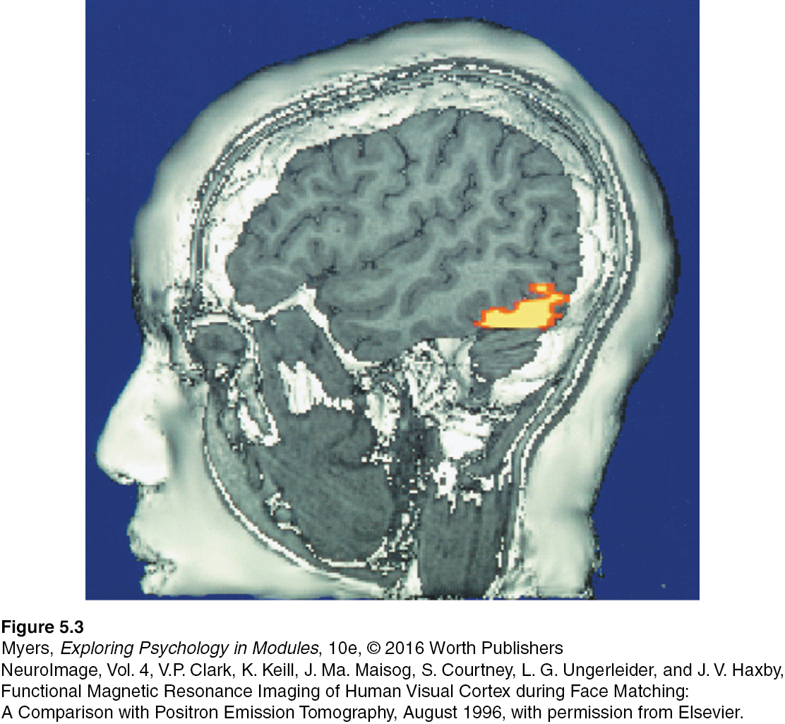
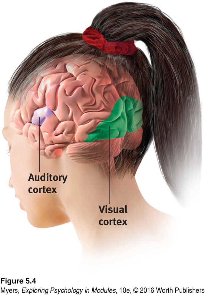
Scientists have identified additional areas where the cortex receives input from senses other than touch. Any visual information you are receiving now is going to the visual cortex in your occipital lobes, at the back of your brain (FIGURES 5.3 and 5.4). Stimulated in the occipital lobes, you might see flashes of light or dashes of color. (In a sense, we do have eyes in the back of our head!) Having lost much of his right occipital lobe to a tumor removal, a friend of mine [DM’s] was blind to the left half of his field of vision. Visual information travels from the occipital lobes to other areas that specialize in tasks such as identifying words, detecting emotions, and recognizing faces.

Any sound you now hear is processed by your auditory cortex in your temporal lobes (just above your ears; see FIGURE 5.4). Most of this auditory information travels a circuitous route from one ear to the auditory receiving area above your opposite ear. If stimulated in your auditory cortex, you might hear a sound. MRI scans of people with schizophrenia have revealed active auditory areas in the temporal lobes during the false sensory experience of auditory hallucinations (Lennox et al., 1999). Even the phantom ringing sound experienced by people with hearing loss is—if heard in one ear—associated with activity in the temporal lobe on the brain’s opposite side (Muhlnickel, 1998).

59

RETRIEVE IT

Question

Our brain's R4qmLlFH1vCsIXGvMq7h7xQVi7Q= cortex registers and processes body touch and movement sensations. The g87dwysVOp/+0qnv cortex controls our voluntary movements.

association areas areas of the cerebral cortex that are not involved in primary motor or sensory functions; rather, they are involved in higher mental functions such as learning, remembering, thinking, and speaking

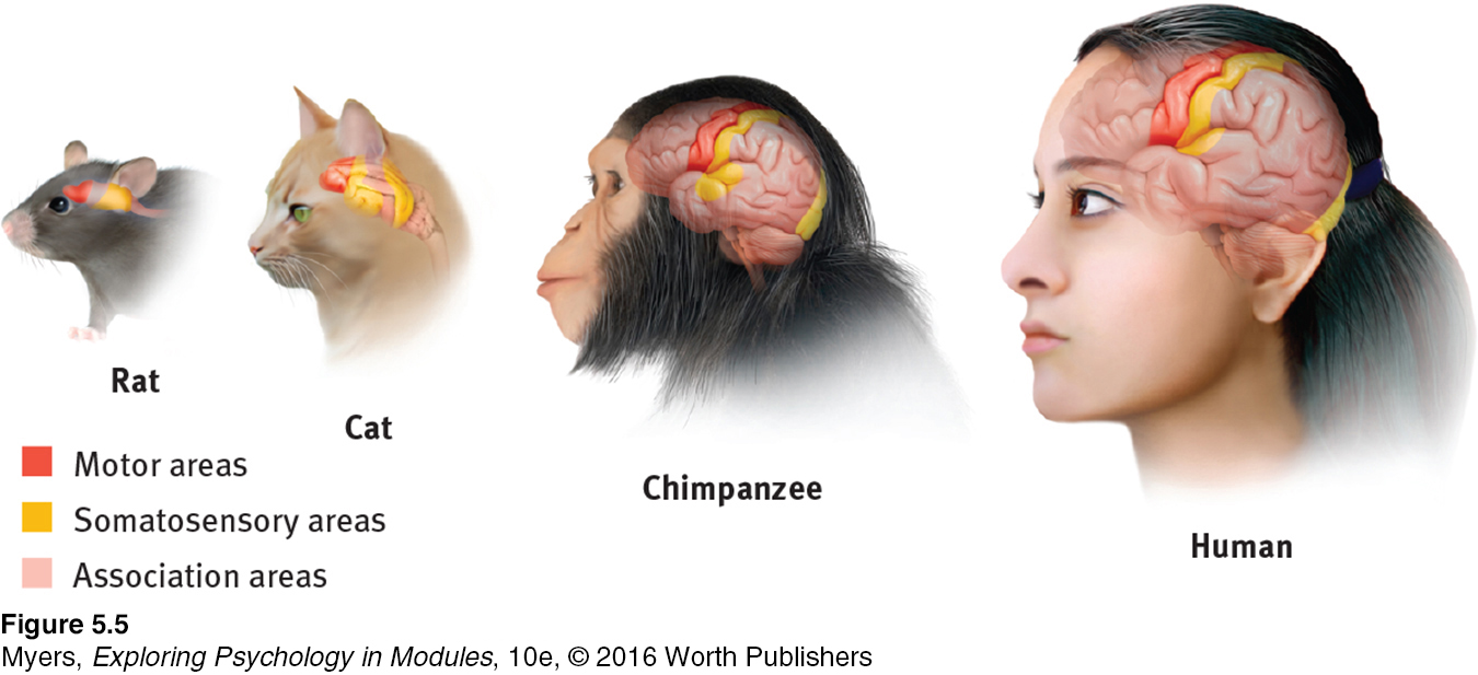
ASSOCIATION AREAS So far, we have pointed out small cortical areas that either receive sensory input or direct muscular output. Together, these occupy about one-fourth of the human brain’s thin, wrinkled cover. What, then, goes on in the remaining vast regions of the cortex? In these association areas (the peach-colored areas in FIGURE 5.5), neurons are busy with higher mental functions—many of the tasks that make us human.

image
Figure 2.22: FIGURE 5.5 Areas of the cortex in four mammals More intelligent animals have increased “uncommitted” or association areas of the cortex. These vast areas of the brain are responsible for interpreting, integrating, and acting on sensory information and linking it with stored memories.

Electrically probing an association area won’t trigger any observable response. So, unlike the somatosensory and motor areas, association area functions cannot be neatly mapped. Their silence has led to what Donald McBurney (1996, p. 44) called “one of the hardiest weeds in the garden of psychology”: the claim that we ordinarily use only 10 percent of our brain. (If true, wouldn’t this imply a 90 percent chance that a bullet to your brain would land in an unused area?) Surgically lesioned animals and brain-damaged humans bear witness that association areas are not dormant. Rather, these areas interpret, integrate, and act on sensory information and link it with stored memories—a very important part of thinking. Simple tasks often increase activity in small brain patches, involving far less than 10 percent of the brain. Yet complex tasks integrate many islands of brain activity, some performing automatic tasks and others requiring conscious control (Chein & Schneider, 2012). The brain is a whole system, with no dead spot for a stray bullet.

Association areas are found in all four lobes. The prefrontal cortex in the forward part of the frontal lobes enables judgment, planning, and processing of new memories. People with damaged frontal lobes may have intact memories, high scores on intelligence tests, and great cake-baking skills. Yet they would not be able to plan ahead to begin baking a cake for a birthday party (Huey et al., 2006).

image See LaunchPad’s Video: Case Studies for a helpful tutorial animation.

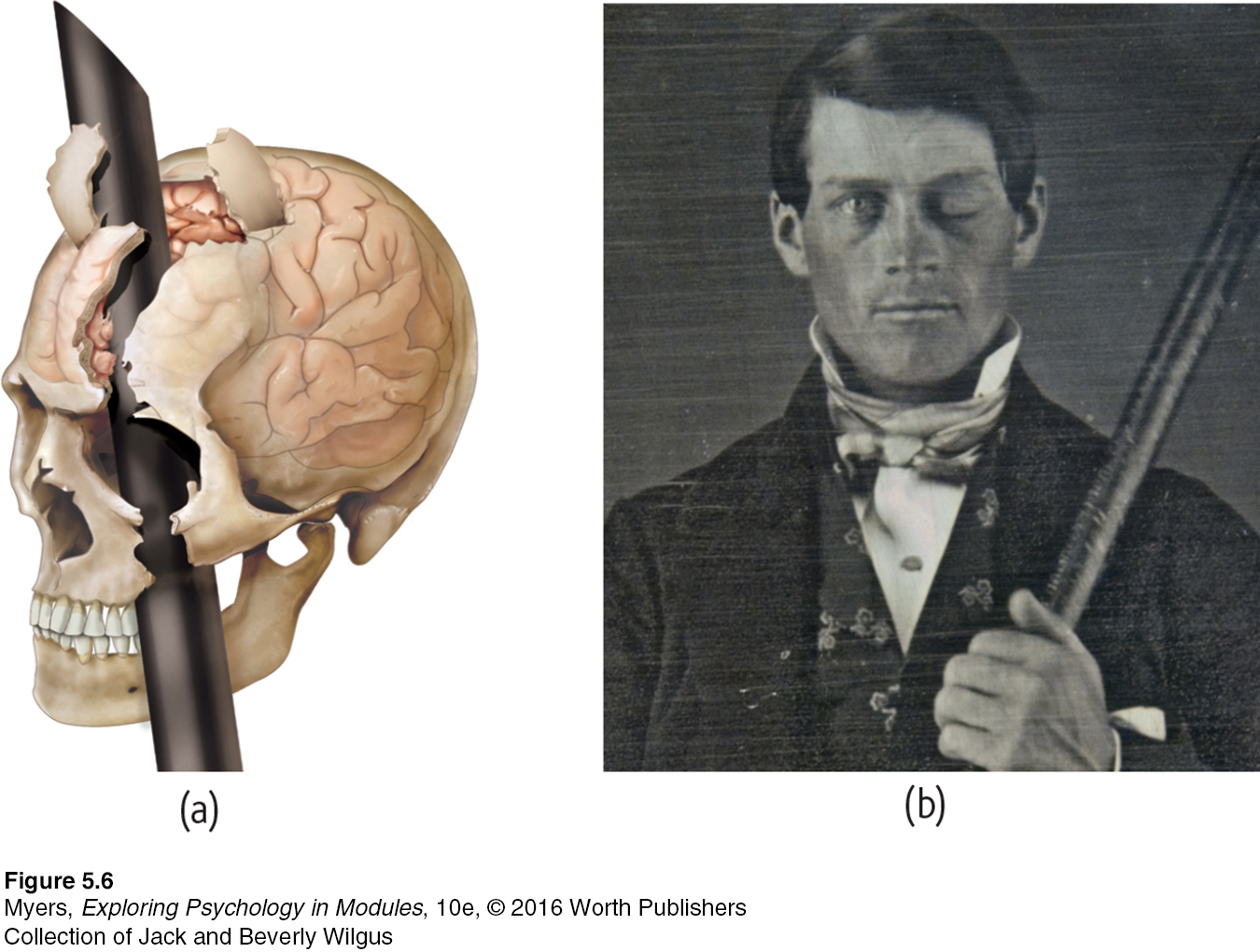
Frontal lobe damage also can alter personality and remove a person’s inhibitions. Consider the classic case study of railroad worker Phineas Gage. One afternoon in 1848, Gage, then 25 years old, was using a tamping iron to pack gunpowder into a rock. A spark ignited the gunpowder, shooting the rod up through his left cheek and out the top of his skull, leaving his frontal lobes damaged (FIGURE 5.6). To everyone’s amazement, Gage was immediately able to sit up and speak, and after the wound healed he returned to work. But having lost some of the neural tracts that enabled his frontal lobes to control his emotions (Van Horn et al., 2012), the affable, soft-spoken man was now irritable, profane, and dishonest. This person, said his friends, was “no longer Gage.” His mental abilities and memories were intact, but his personality was not. (Although Gage lost his railroad job, he did, over time, adapt to his injury and find work as a stage coach driver [Macmillan & Lena, 2010].)

image
Figure 2.23: FIGURE 5.6 A blast from the past (a) Phineas Gage’s skull was kept as a medical record. Using measurements and modern neuroimaging techniques, researchers have reconstructed the probable path of the rod through Gage’s brain (Van Horn et al., 2012). (b) This photo shows Gage after his accident. (The image has been reversed to show the features correctly. Early photos, including this one, were actually mirror images.)
Collection of Jack and Beverly Wilgus

60

image
Missing frontal lobe brakes With part of his left frontal lobe (in this downward-facing brain scan) lost to injury, Cecil Clayton became more impulsive and killed a deputy sheriff, for which, nine years later, his state executed him.
Cecil Clayton's brain scan, included with request for stay of execution filed with the Supreme Court, showing a missing portion of his frontal lobe.

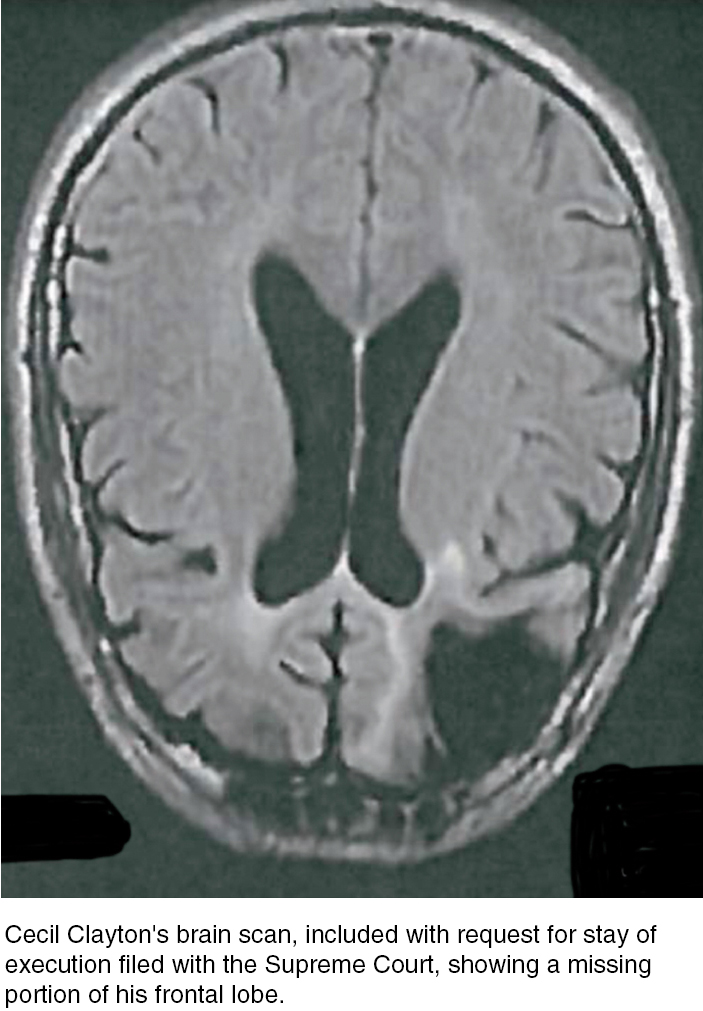
Studies of others with damaged frontal lobes have revealed similar impairments. Not only may they become less inhibited (without the frontal lobe brakes on their impulses), but their moral judgments may seem unrestrained. Cecil Clayton lost 20 percent of his left frontal lobe in a 1972 sawmill accident. Thereafter, his intelligence test score dropped to an elementary school level and he displayed increased impulsivity. In 1996, he fatally shot a deputy sheriff. In 2015, when he was 74, the State of Missouri executed him (Williams, 2015).

Would you advocate pushing one person in front of a runaway trolley to save five others? Most people would not, but those with damage to a brain area behind the eyes are often untroubled by such ethical dilemmas (Koenigs et al., 2007). The frontal lobes help steer us away from violent actions (Molenberghs et al., 2015; Yang & Raine, 2009).

Association areas also perform other mental functions. The parietal lobes, parts of which were large and unusually shaped in Einstein’s normal-weight brain, enable mathematical and spatial reasoning (Ibos & Freedman, 2014; Witelson et al., 1999). On the underside of the right temporal lobe, another association area enables us to recognize faces. If a stroke or head injury destroyed this area of your brain, you would still be able to describe facial features and to recognize someone’s gender and approximate age, yet be strangely unable to identify the person as, say, Taylor Swift, or even your grandmother.

Nevertheless, complex mental functions don’t reside in any one place. There is no one spot in a rat’s small association cortex that, when damaged, will obliterate its ability to learn or remember a maze. Your memory, language, and attention result from synchronized activity among distinct brain areas and neural networks (Knight, 2007). Ditto for religious experience. More than 40 distinct brain regions become active in different religious states, such as prayer and meditation, indicating that there is no simple “God spot” (Fingelkurts & Fingelkurts, 2009). The point to remember: Our mental experiences arise from coordinated brain activity.

RETRIEVE IT

Question

Q5DfQ3v55ARFLPefBlyESzauxV+BqTEJk3M9+COpW8wIWA2EwclIMg==
ANSWER: Association areas are involved in higher mental functions—interpreting, integrating, and acting on information processed in other areas.

The Brain’s Plasticity

5-2 To what extent can a damaged brain reorganize itself, and what is neurogenesis?

plasticity the brain’s ability to change, especially during childhood, by reorganizing after damage or by building new pathways based on experience.

Our brains are sculpted not only by our genes but also by our experiences. In other modules, we’ll focus more on how experience molds the brain. For now, let’s turn to another aspect of the brain’s plasticity: its ability to modify itself after damage.

image
Figure 2.24: FIGURE 5.7 Brain plasticity This 6-year-old had surgery to end her life-threatening seizures. Although most of an entire hemisphere was removed (see MRI of hemispherectomy above), her remaining hemisphere compensated by putting other areas to work. One Johns Hopkins medical team reflected on the child hemispherectomies they had performed. Although use of the opposite arm was compromised, the team reported being “awed” by how well the children had retained their memory, personality, and humor (Vining et al., 1997). The younger the child, the greater the chance that the remaining hemisphere can take over the functions of the one that was surgically removed (Choi, 2008; Danelli et al., 2013).
Joe McNally/Joe McNally Photography/Getty Images

Some brain-damage effects described earlier can be traced to two hard facts: (1) Severed brain and spinal cord neurons, unlike cut skin, usually do not regenerate. (If your spinal cord were severed, you would probably be permanently paralyzed.) And (2) some brain functions seem preassigned to specific areas. One newborn who suffered damage to temporal lobe facial recognition areas later remained unable to recognize faces (Farah et al., 2000). But there is good news: Some neural tissue can reorganize in response to damage. Under the surface of our awareness, the brain is constantly changing, building new pathways as it adjusts to little mishaps and new experiences.

61

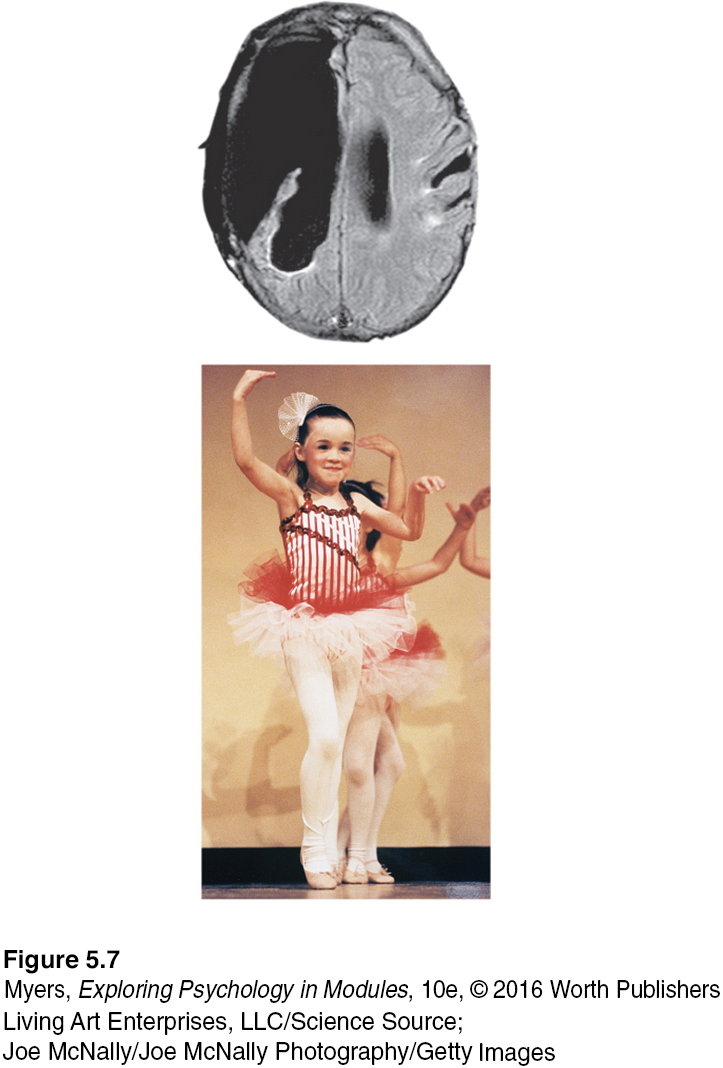
Plasticity may also occur after serious damage, especially in young children (Kolb, 1989; see also FIGURE 5.7). The brain’s plasticity is good news for those with vision or hearing loss. Blindness or deafness makes unused brain areas available for other uses (Amedi et al., 2005). If a blind person uses one finger to read Braille, the brain area dedicated to that finger expands as the sense of touch invades the visual cortex that normally helps people see (Barinaga, 1992a; Sadato et al., 1996).

Plasticity also helps explain why some studies have found that deaf people have enhanced peripheral and motion-detection vision (Bosworth & Dobkins, 1999; Shiell et al., 2014). In deaf people whose native language is sign, the temporal lobe area normally dedicated to hearing waits in vain for stimulation. Finally, it looks for other signals to process, such as those from the visual system.

Similar reassignment may occur when disease or damage frees up other brain areas normally dedicated to specific functions. If a slow-growing left hemisphere tumor disrupts language (which resides mostly in the left hemisphere), the right hemisphere may compensate (Thiel et al., 2006). If a finger is amputated, the somatosensory cortex that received its input will begin to receive input from the adjacent fingers, which then become more sensitive (Fox, 1984). So what do you suppose was the sexual intercourse experience of one patient whose lower leg had been amputated? “I actually experience my orgasm in my [phantom] foot. [Note that in FIGURE 5.2, the toes region is adjacent to the genitals.] And there it’s much bigger than it used to be because it’s no longer just confined to my genitals” (Ramachandran & Blakeslee, 1998, p. 36).

neurogenesis the formation of new neurons.

Although the brain often attempts self-repair by reorganizing existing tissue, it sometimes attempts to mend itself by producing new brain cells. This process, known as neurogenesis, has been found in adult mice, birds, monkeys, and humans (Jessberger et al., 2008). These baby neurons originate deep in the brain and may then migrate elsewhere and form connections with neighboring neurons (Aimone et al., 2010; Gould, 2007).

Master stem cells that can develop into any type of brain cell have also been discovered in the human embryo. If mass-produced in a lab and injected into a damaged brain, might neural stem cells turn themselves into replacements for lost brain cells? Might surgeons someday be able to rebuild damaged brains, much as landscapers reseed damaged lawns? Stay tuned. Today’s biotech companies are exploring such possibilities. In the meantime, we can all benefit from natural promoters of neurogenesis, such as exercise, sleep, and nonstressful but stimulating environments (Iso et al., 2007; Pereira et al., 2007; Stranahan et al., 2006).