3.2 The Nervous System
3-
nervous system the body’s speedy, electrochemical communication network, consisting of all the nerve cells of the peripheral and central nervous systems.
central nervous system (CNS) the brain and spinal cord.
peripheral nervous system (PNS) the sensory and motor neurons that connect the central nervous system (CNS) to the rest of the body.
nerves bundled axons that form neural cables connecting the central nervous system with muscles, glands, and sense organs.
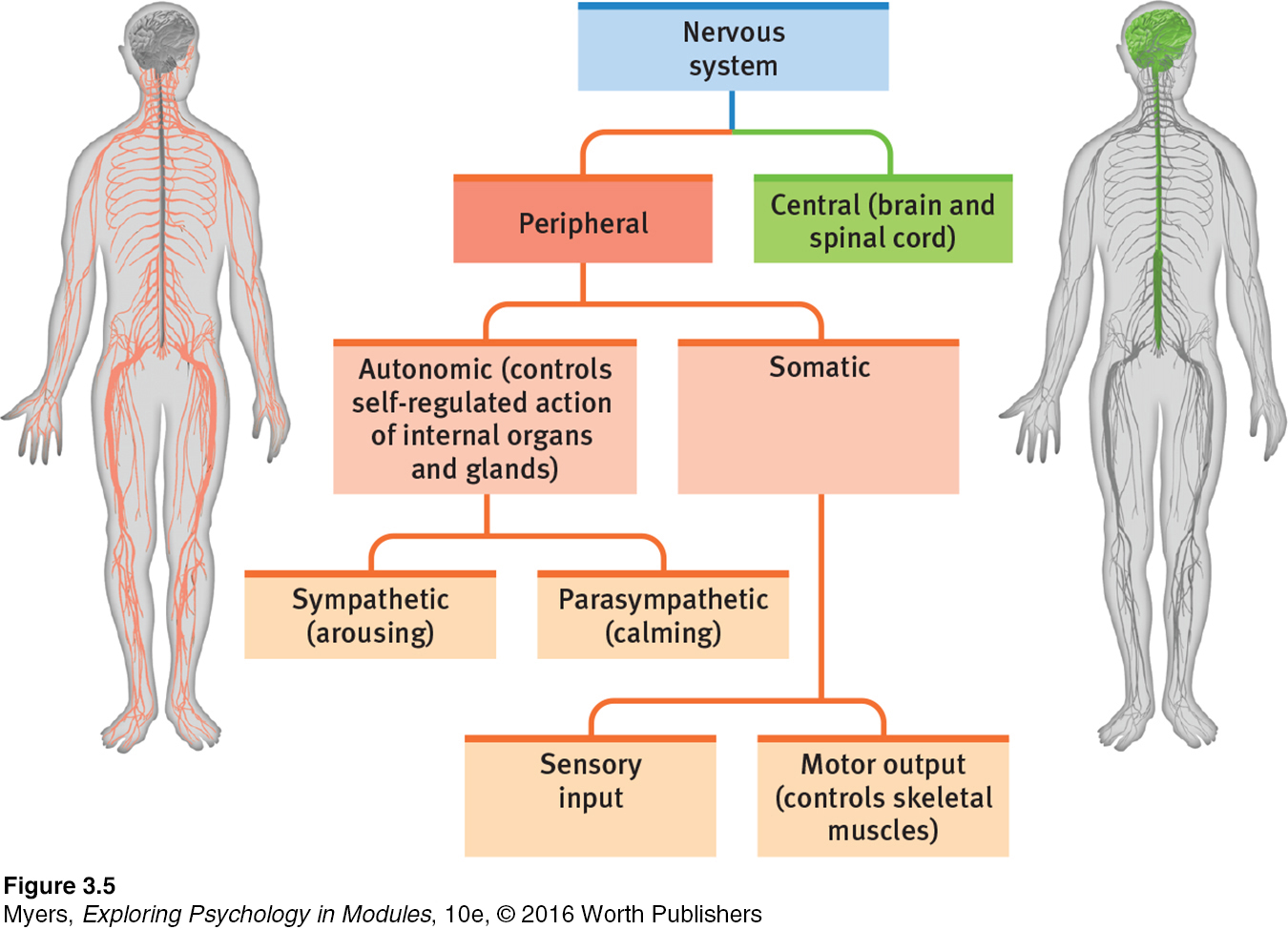
All those neurons communicating with neurotransmitters make up our body’s nervous system (FIGURE 3.5). This communication network allows us to take in information from the world and the body’s tissues, to make decisions, and to send back information and orders to the body’s tissues. A quick overview: The brain and spinal cord form the central nervous system (CNS), the body’s decision maker. The peripheral nervous system (PNS) is responsible for gathering information and for transmitting CNS decisions to other body parts. Nerves, electrical cables formed of bundles of axons, link the CNS with the body’s sensory receptors, muscles, and glands. The optic nerve, for example, bundles a million axons into a single cable carrying the messages each eye sends to the brain (Mason & Kandel, 1991).
sensory (afferent) neurons neurons that carry incoming information from the sensory receptors to the brain and spinal cord.
motor (efferent) neurons neurons that carry outgoing information from the brain and spinal cord to the muscles and glands.
interneurons neurons within the brain and spinal cord; communicate internally and process information between the sensory inputs and motor outputs.
Information travels in the nervous system through three types of neurons. Sensory neurons carry messages from the body’s tissues and sensory receptors inward (thus, they are afferent) to the brain and spinal cord for processing. Motor neurons (which are efferent) carry instructions from the central nervous system out to the body’s muscles and glands. Between the sensory input and motor output, information is processed via the interneurons. Our complexity resides mostly in these interneurons. Our nervous system has a few million sensory neurons, a few million motor neurons, and billions and billions of interneurons.
The Peripheral Nervous System
somatic nervous system the division of the peripheral nervous system that controls the body’s skeletal muscles. Also called the skeletal nervous system.
Our peripheral nervous system has two components—
Our autonomic nervous system (ANS) controls our glands and our internal organ muscles. The ANS influences functions such as glandular activity, heartbeat, and digestion. (Autonomic means “self-
autonomic [aw-
sympathetic nervous system the division of the autonomic nervous system that arouses the body, mobilizing its energy.
parasympathetic nervous system the division of the autonomic nervous system that calms the body, conserving its energy.
The autonomic nervous system serves two important functions (FIGURE 3.6). The sympathetic nervous system arouses and expends energy. If something alarms or challenges you (such as a longed-
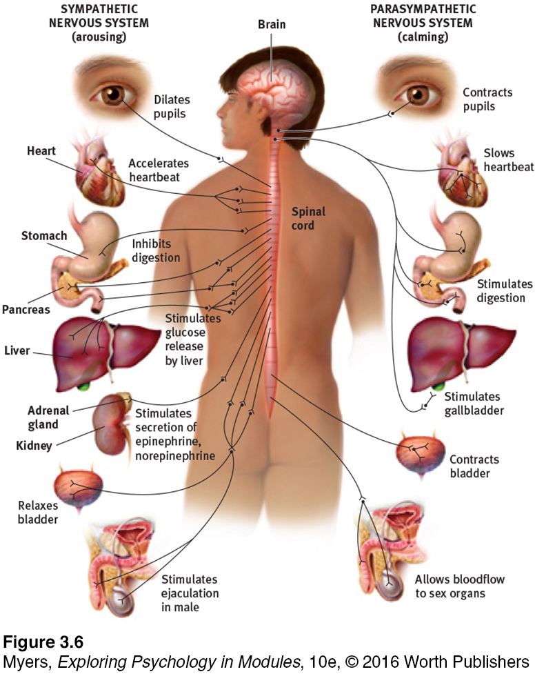
I [DM] recently experienced my ANS in action. Before sending me into an MRI machine for a shoulder scan, the technician asked if I had issues with claustrophobia. “No, I’m fine,” I assured her, with perhaps a hint of macho swagger. Moments later, as I found myself on my back, stuck deep inside a coffin-
RETRIEVE IT
Question
Match the type of neuron to its description.
Motor neurons Sensory neurons Interneurons | carry incoming messages from sensory receptors to the CNS. communicate within the CNS and process information between incoming and outgoing messages. carry outgoing messages from the CNS to muscles and glands. |
Question
What bodily changes does your ANS direct before and after you give an important speech?
The Central Nervous System
From neurons “talking” to other neurons arises the complexity of the central nervous system’s brain and spinal cord.
It is the brain that enables our humanity—
The brain’s neurons cluster into work groups called neural networks. To understand why, Stephen Kosslyn and Olivier Koenig (1992, p. 12) have invited us to “think about why cities exist; why don’t people distribute themselves more evenly across the countryside?” Like people networking with people, neurons network with nearby neurons with which they can have short, fast connections.
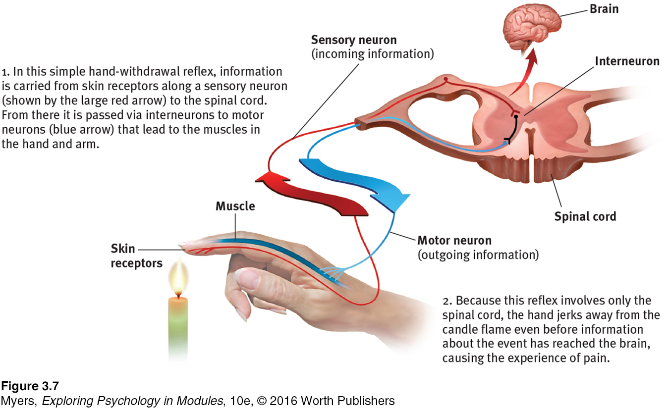
reflex a simple, automatic response to a sensory stimulus, such as the knee-
The other part of the CNS, the spinal cord, is a two-
Another neural circuit enables the pain reflex (FIGURE 3.7). When your finger touches a flame, neural activity (excited by the heat) travels via sensory neurons to interneurons in your spinal cord. These interneurons respond by activating motor neurons leading to the muscles in your arm. Because the simple pain-
“If the nervous system be cut off between the brain and other parts, the experiences of those other parts are nonexistent for the mind. The eye is blind, the ear deaf, the hand insensible and motionless.”
William James, Principles of Psychology, 1890
Information travels to and from the brain by way of the spinal cord. Were the top of your spinal cord severed, you would not feel pain from your paralyzed body below. Nor would you feel pleasure. With your brain literally out of touch with your body, you would lose all sensation and voluntary movement in body regions with sensory and motor connections to the spinal cord below its point of injury. You would exhibit the knee-