Chapter 10. Functional Anatomy of the Auditory System: From Cochlea to Cortex
10.1 Title slide
Functional Anatomy of the Auditory System: From Cochlea to Cortex
Interact with depictions of the structures and pathways from the cochlea to the auditory cortex
CLICK ANYWHERE TO BEGIN
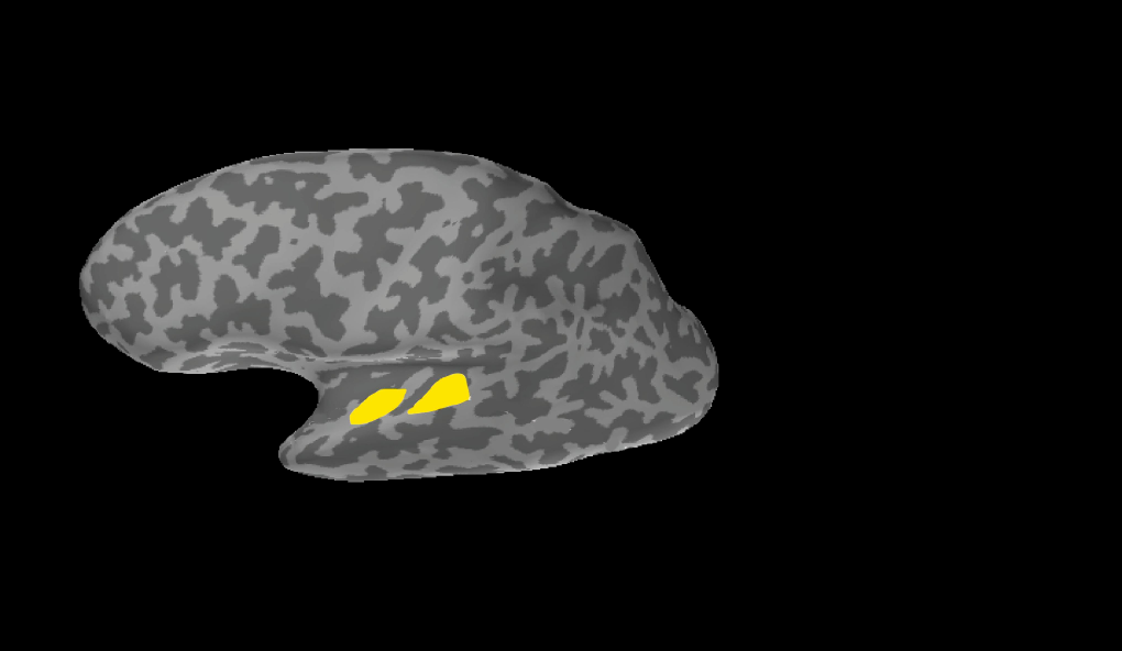
Photo: Ahveninen, J., Jääskeläinen, I.P., Raij, T., Bonmassar, G., Devore, S. Hämäläinen, M., Levänen, S., Lin, F.H., Sams, M., Shinn-Cunningham, B.G., Witzel, T., Belliveau, J.W. (2006). Task-modulated "what" and "where" pathways in human auditory cortex, Figure 3. Proceedings of the National Academy of Sciences USA, 103, 14608–14613. © 2013 National Academy of Sciences, U.S.A.
What Is the Functional Anatomy of the Auditory System?
The auditory cortex is tucked into the lateral sulcus on top of the temporal lobe. The primary auditory cortex (or A1) is one of three areas that constitute the auditory core region (the other two are the rostral core and the rostrotemporal core). Signals from the auditory core flow to two regions wrapped around it, the belt and the parabelt, each of which also contains distinct subareas.
Neurons within each of the three areas that constitute the auditory core region are arranged into a tonotopic map, as are neurons in each of the subcortical structures in the ascending auditory pathways. In the auditory core, the characteristic frequencies of neurons in the rostrotemporal core and A1 gradually shift from low at the anterior end of the region to high at the posterior end; in the rostral core, the shift is in the opposite direction.
10.2 Explain - dnd
These illustrations depict the contralateral and ipsilateral flows of neural signals from the cochlea of the right ear to the auditory cortex. To show that you understand these flows, drag the labels on the anatomical illustration into the correct blank boxes in the schematic diagram. You may use each label more than once.
10.3 Explain - dnd
Drag and drop the labels into the correct blank boxes on these illustrations of the auditory cortex.
10.4 Explain
What Is the Functional Anatomy of the Auditory System?
Type I auditory nerve fibers carry signals from inner hair cells in the cochlea to the ipsilateral cochlear nucleus in the brain stem. From there, the main pathways carry signals to the contralateral side of the brain, but signals in the secondary pathways remain mostly on the ipsilateral side. The structures on these pathways include the contralateral trapezoid body and both the ipsilateral and contralateral superior olivary complex, inferior colliculus, medial geniculate body (MGB), and auditory cortex.
The auditory cortex is tucked into the lateral sulcus on top of the temporal lobe. The primary auditory cortex (or A1) is one of three areas that constitute the auditory core region (the other two are the rostral core and the rostrotemporal core). Signals from the auditory core flow to two regions wrapped around it, the belt and the parabelt, each of which also contains distinct subareas.
Neurons within each of the three areas that constitute the auditory core region are arranged into a tonotopic map, as are neurons in each of the subcortical structures in the ascending auditory pathways. In the auditory core, the characteristic frequencies of neurons in the rostrotemporal core and A1 gradually shift from low at the anterior end of the region to high at the posterior end; in the rostral core, the shift is in the opposite direction.
10.5 Test - single choice
Select your answer to the question below. Then click SUBMIT.
Based on the flows of neural signals depicted in this demonstration, which structures do NOT receive signals carrying information the cochlea of the right ear?
10.6 Test - single choice
Select your answer to the question below. Then click SUBMIT.
What are the components of the auditory core region?
10.7 Test - single choice
Select your answer to the question below. Then click SUBMIT.
Which statement correctly describes the tonotopic organization of neurons within the auditory core region?
10.8 Activity completed
Functional Anatomy of the Auditory System: From Cochlea to Cortex.