Chapter 1. Neurons and Neural Signals
1.1 Title slide
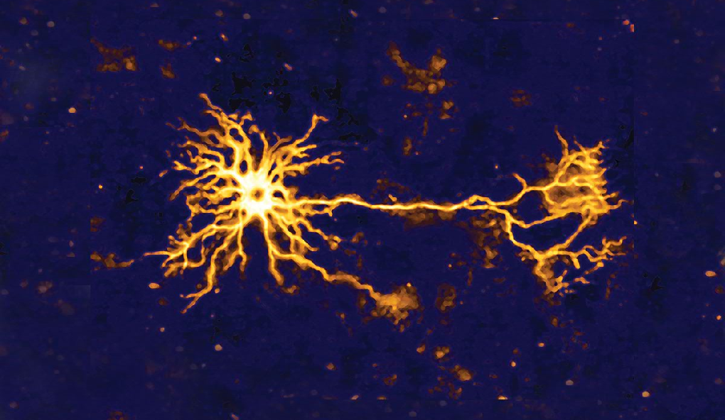
Neurons and Neural Signals
Interact with depictions of neurons, synapses, and neural signals.
CLICK ANYWHERE TO BEGIN
Photo: James Cavallini/Science Source.
Neurons and Neural Signals
An action potential is a neural signal that begins in the dendrites of a neuron that has been stimulated by a signal from another neuron. The action potential then travels down the neuron's axon to the axon terminals. The fluid within and around neurons contains positively and negatively charged ions. Differences in the concentrations of these ions give rise to a difference in electrical potential across the cell membrane, called the membrane potential (measured in millivolts, or mV). When a cell is at rest, the membrane potential is about –70 mV, a difference known as the resting potential.
Certain ions can cross the membrane through small pores called ion channels. A neuron that receives a signal from another neuron undergoes an abrupt change in membrane potential, which causes ion channels to open at the base of the axon. This allows positive ions to flow into the axon, causing the membrane potential to become markedly more positive, a change called depolarization. However, depolarization doesn't stop at 0. The inflow of positive ions continues until the membrane potential is about 130 mV.
Immediately after opening, the channels allowing the inflow of positive ions close again, and nearby channels open that allow positive ions to flow out of the axon. This pushes the membrane potential back toward its resting value of –70 mV, a process called repolarization, which continues until the membrane potential briefly exceeds the resting potential—this is termed hyperpolarization—before returning to the resting potential. This sequence of changes in membrane potential from –70 mV to 130 mV and finally back to –70 mV is known as the action potential.
The action potential at the base of the axon causes ion channels to open a little bit further down the axon, and the sequence of events repeats—in effect, the action potential travels down the axon until it arrives at the axon terminals. However, there is a tiny gap called a synapse between the axon terminal of one neuron and the dendrite of another neuron. The membrane at the axon terminal of a neuron sending a neural signal is called the presynaptic membrane; the membrane of the dendrite of the receiving neuron is called the postsynaptic membrane. Within axon terminals, tiny sacs called synaptic vesicles contain molecules of neurotransmitters.
The arrival of an action potential at an axon terminal causes synaptic vesicles to merge with the presynaptic membrane, releasing neurotransmitter molecules into the synapse. Some of these molecules float across the synapse and bind to proteins called receptors on the postsynaptic membrane. If enough neurotransmitters bind to receptors, the postsynaptic neuron can generate an action potential of its own, which then causes the neuron to release neurotransmitters into synapses with other neurons, and so on.
Neurotransmitters can be categorized on the basis of whether they have an excitatory or an inhibitory effect on the postsynaptic neuron. The binding of excitatory neurotransmitters depolarizes the membrane potential and thereby increases the probability that an action potential will be generated in the postsynaptic neuron; such a depolarization is called an excitatory postsynaptic potential (EPSP), and synapses where this happens are known as excitatory synapses. In contrast, the binding of inhibitory neurotransmitters hyperpolarizes the membrane potential and thereby decreases the probability of an action potential; such a hyperpolarization is called an inhibitory postsynaptic potential (IPSP), and synapses where this happens are known as inhibitory synapses. Depending on the balance between EPSPs and IPSPs, a postsynaptic neuron may or may not produce an action potential.
1.2 Explain - dnd
Drag the labels into the correct blank boxes to identify the principal parts of a typical neuron.
1.3 Explain - dnd
Drag the labels into the correct blank boxes to identify the phases of an action potential.
1.4 Explain - dnd
Drag the labels into the correct blank boxes to identify the principal elements and structures involved in the transmission of neural signals across a synapse.
1.5 Explain - dnd
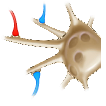
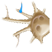
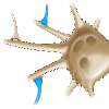
The images show a postsynaptic neuron that synapses with three presynaptic neurons. Each image shows a different pattern of signals sent to the postsynaptic neuron. A blue axon terminal indicates an excitatory signal from the presynaptic neuron, and a red axon terminal indicates an inhibitory signal. The graphs show four different possible effects on the postsynaptic membrane potential when excitatory postsynaptic potentials (EPSPs) and inhibitory postsynaptic potentials (IPSPs) result from the binding of neurotransmitter molecules at excitatory and inhibitory synapses. Drag the images into the correct blank boxes to correspond to what the graphs are showing.
1.6 Explain
What Are the Main Characteristics of Neurons; How Do Neurons Produce Action Potentials; and How Are Signals Transmitted Between Neurons?
Neurons are cells. The main characteristic that distinguishes neurons from other types of cells is their ability to receive and transmit neural signals. Any one neuron sends signals to and receives signals from as many as several thousand other neurons. A neuron’s cell body (or soma) contains the nucleus. Two prominent types of projections emanate from the cell body: dendrites, which receive signals from other neurons, and an axon, which conducts signals to axon terminals, where the signals are transmitted to other neurons.
An action potential is a neural signal that begins in the dendrites of a neuron that has been stimulated by a signal from another neuron. The action potential then travels down the neuron's axon to the axon terminals. The fluid within and around neurons contains positively and negatively charged ions. Differences in the concentrations of these ions give rise to a difference in electrical potential across the cell membrane, called the membrane potential (measured in millivolts, or mV). When a cell is at rest, the membrane potential is about –70 mV, a difference known as the resting potential.
Certain ions can cross the membrane through small pores called ion channels. A neuron that receives a signal from another neuron undergoes an abrupt change in membrane potential, which causes ion channels to open at the base of the axon. This allows positive ions to flow into the axon, causing the membrane potential to become markedly more positive, a change called depolarization. However, depolarization doesn't stop at 0. The inflow of positive ions continues until the membrane potential is about +30 mV.
Immediately after opening, the channels allowing the inflow of positive ions close again, and nearby channels open that allow positive ions to flow out of the axon. This pushes the membrane potential back toward its resting value of –70 mV, a process called repolarization, which continues until the membrane potential briefly exceeds the resting potential—this is termed hyperpolarization—before returning to the resting potential. This sequence of changes in membrane potential from –70 mV to +30 mV and finally back to –70 mV is known as the action potential.
The action potential at the base of the axon causes ion channels to open a little bit further down the axon, and the sequence of events repeats—in effect, the action potential travels down the axon until it arrives at the axon terminals. However, there is a tiny gap called a synapse between the axon terminal of one neuron and the dendrite of another neuron. The membrane at the axon terminal of a neuron sending a neural signal is called the presynaptic membrane; the membrane of the dendrite of the receiving neuron is called the postsynaptic membrane. Within axon terminals, tiny sacs called synaptic vesicles contain molecules of neurotransmitters.
The arrival of an action potential at an axon terminal causes synaptic vesicles to merge with the presynaptic membrane, releasing neurotransmitter molecules into the synapse. Some of these molecules float across the synapse and bind to proteins called receptors on the postsynaptic membrane. If enough neurotransmitters bind to receptors, the postsynaptic neuron can generate an action potential of its own, which then causes the neuron to release neurotransmitters into synapses with other neurons, and so on.
Neurotransmitters can be categorized on the basis of whether they have an excitatory or an inhibitory effect on the postsynaptic neuron. The binding of excitatory neurotransmitters depolarizes the membrane potential and thereby increases the probability that an action potential will be generated in the postsynaptic neuron; such a depolarization is called an excitatory postsynaptic potential (EPSP), and synapses where this happens are known as excitatory synapses. In contrast, the binding of inhibitory neurotransmitters hyperpolarizes the membrane potential and thereby decreases the probability of an action potential; such a hyperpolarization is called an inhibitory postsynaptic potential (IPSP), and synapses where this happens are known as inhibitory synapses. Depending on the balance between EPSPs and IPSPs, a postsynaptic neuron may or may not produce an action potential.
1.7 Test - single choice
Select your answer to the question below. Then click SUBMIT.
Which of the following is false?
1.8 Test - single choice
Select your answer to the question below. Then click SUBMIT.
Which of the following is true?
1.9 Test - single choice
Select your answer to the question below. Then click SUBMIT.
Which is the correct order of events in the transmission of signals between neurons?
1.10 Activity completed
Neurons and Neural Signals.