Chapter 2. Functional Anatomy of the Eye
2.1 Title slide
Functional Anatomy of the Eye
Interact with a depiction of the human eye.
CLICK ANYWHERE TO BEGIN
What Is the Functional Anatomy of the Eye?
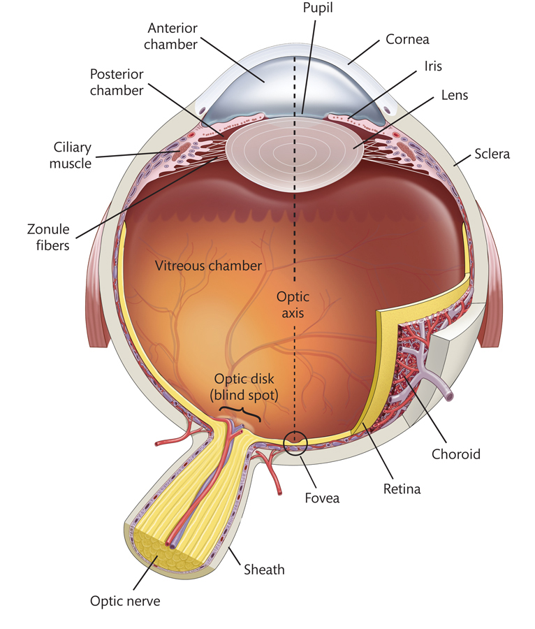
Three membranes surround the eye:
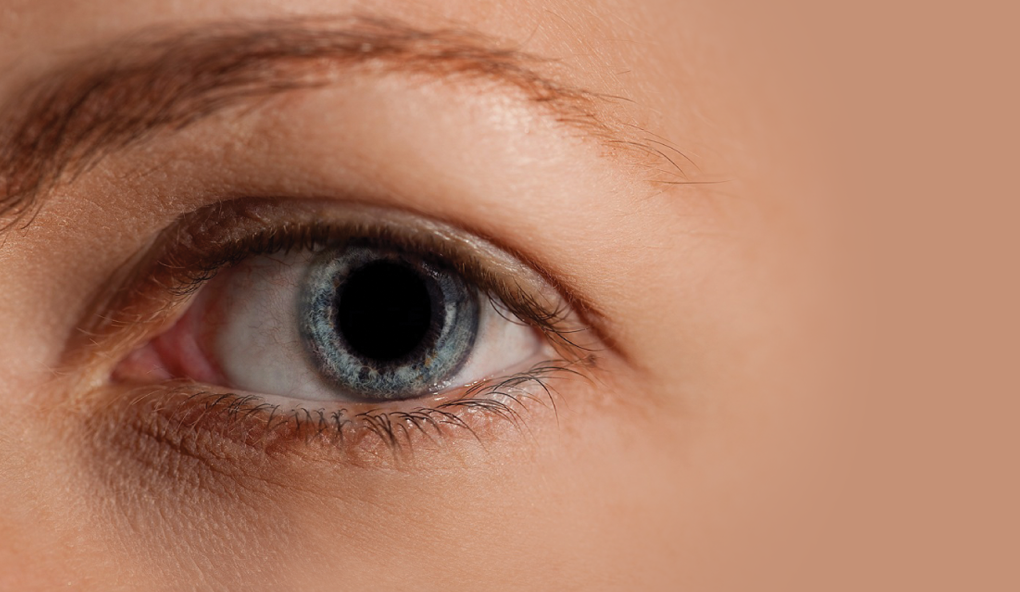
• The outer membrane is made up of the sclera (a protective covering whose visible portion is the white of your eye) and the transparent cornea at the front of the eye.
• The middle membrane is the choroid; it contains most of the blood vessels that supply the inside of the eye with oxygen and nutrients.
• The inner membrane is the retina, which is made up of neurons, including the receptors that convert the light entering the eye into neural signals.
Light enters the eye by first passing through the cornea, which sharply refracts (bends) the light. The light then passes through the pupil and into the lens, a transparent structure that further refracts the light. The shape of the lens is adjusted to ensure that light focuses properly on the retina.
The pupil is an opening in the middle of the iris, a small donut-shaped muscle. The iris controls the size of the pupil by contracting and relaxing.
The zonule fibers connect the lens to the choroid. The ciliary muscles are also attached to the choroid. When the ciliary muscles relax, the choroid pulls on the zonule fibers, stretching the lens; when the ciliary muscles contract, they oppose the pull by the choroid on the zonule fibers, and the lens isn't stretched as much.
The fovea is the area at the center of the retina, receiving light from objects at the center of gaze. The optic disk is the place where the optic nerve exits the eye and where blood vessels enter and exit the eye.
The anterior chamber is the space between the cornea and the iris; the posterior chamber is the smaller space between the iris and the lens. Both chambers are filled with a fluid called aqueous humor. The large vitreous chamber—the main interior portion of the eye—is filled with a fluid called vitreous humor. The fluids in these chambers exert outward pressure on the eye and keep it from collapsing.
2.2 Explain - dnd
Drag and drop each of the labels into the correct box on this cross-section
of the eye
viewed from above.
2.3 Explain - dnd
Drag and drop each of the labels into the correct box on this cross-section
of the eye viewed from above.
2.4 Explain - dnd
Drag each label on the illustration into the box above the matching description.
2.5 Explain
What Is the Functional Anatomy of the Eye?
Three membranes surround the eye:
• The outer membrane is made up of the sclera (a protective covering whose visible portion is the white of your eye) and the transparent cornea at the front of the eye.
• The middle membrane is the choroid; it contains most of the blood vessels that supply the inside of the eye with oxygen and nutrients.
• The inner membrane is the retina, which is made up of neurons, including the receptors that convert the light entering the eye into neural signals.
Light enters the eye by first passing through the cornea, which sharply refracts (bends) the light. The light then passes through the pupil and into the lens, a transparent structure that further refracts the light. The shape of the lens is adjusted to ensure that light focuses properly on the retina.
The pupil is an opening in the middle of the iris, a small donut-shaped muscle. The iris controls the size of the pupil by contracting and relaxing.
The zonule fibers connect the lens to the choroid. The ciliary muscles are also attached to the choroid. When the ciliary muscles relax, the choroid pulls on the zonule fibers, stretching the lens; when the ciliary muscles contract, they oppose the pull by the choroid on the zonule fibers, and the lens isn't stretched as much.
The fovea is the area at the center of the retina, receiving light from objects at the center of gaze. The optic disk is the place where the optic nerve exits the eye and where blood vessels enter and exit the eye.
The anterior chamber is the space between the cornea and the iris; the posterior chamber is the smaller space between the iris and the lens. Both chambers are filled with a fluid called aqueous humor. The large vitreous chamber—the main interior portion of the eye—is filled with a fluid called vitreous humor. The fluids in these chambers exert outward pressure on the eye and keep it from collapsing.
2.6 Test - single choice
Select your answer to the question below. Then click SUBMIT.
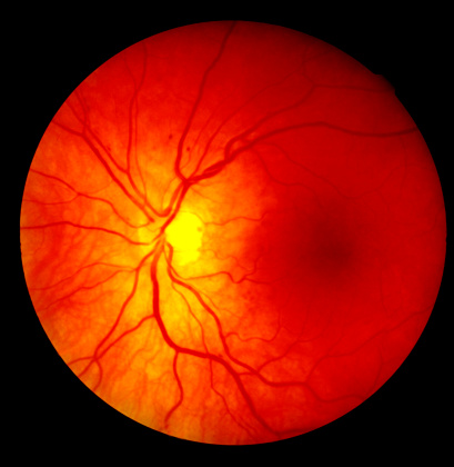
This is a normal human retina as seen through an ophthalmoscope. The fovea is the faint dark region at the right center of the photo.
When you look directly at the fovea in this image, light rays from that portion of the image strike which part of your own retina?
2.7 Test - dnd
Drag the labels into the blank boxes from left to right, in the order in which light encounters these structures on its path into the eye. Then click SUBMIT.

Retina
Vitreous chamber
Pupil
Cornea
Anterior chamber
Lens
2.8 Test - dnd
Drag each description at left to attach to the matching part of the eye. Then click SUBMIT.
Aqueous and Vitreous humor
Iris and Pupil
Eye
Sclera
Ciliary muscle and Zonule fibers
Cornea
Membrane encasing the eye
Roughly spherical, diameter about 24 mm
Sharply refacts light, more than the lens
Controls the amount of light entering the eye
Maintains the intraocular pressure
Involved in the process of accommodation
2.9 Activity completed
Functional Anatomy of the Eye.