Chapter 2. Functional Anatomy of the Retina
2.1 Title slide
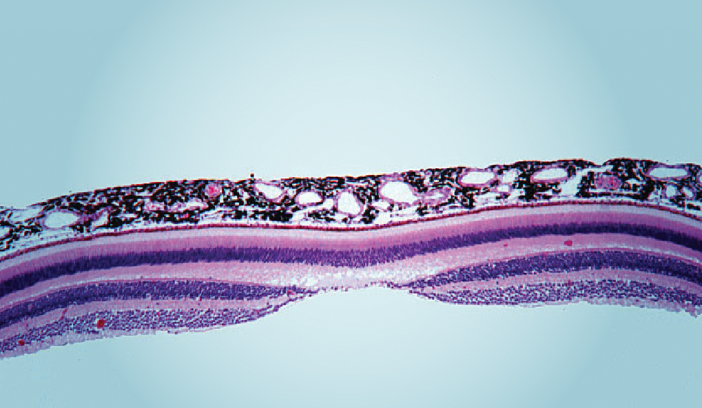
Functional Anatomy of the Retina
Interact with a depiction of the human retina.
CLICK ANYWHERE TO BEGIN
Courtesy of Dr. Deborah W. Vaughan, Histology Learning System, Boston University
What Is the Functional Anatomy of the Retina?
- The three main layers are called nuclear layers because they contain the nuclei of the various types of retinal neurons:
- The outer nuclear layer consists of the photoreceptors (but not including their inner and outer segments).
- The inner nuclear layer contains bipolar cells, horizontal cells, and amacrine cells.
- The ganglion cell layer consists of retinal ganglion cells (RGCs).
- The nuclear layers are separated by two synaptic layers, where the retinal neurons make synapses with each other:
- The synapses among the photoreceptors, bipolar cells, and horizontal cells are contained in the outer synaptic layer.
- The synapses among the bipolar cells, amacrine cells, and RGCs are contained in the inner synaptic layer.
- Closest to the back of the eye is a layer consisting of the inner and outer segments of the photoreceptors (rods and cones), the retinal neurons that transduce light into neural signals. The "business ends" of the photoreceptors, where transduction occurs, are embedded in a layer of cells called the pigment epithelium, which is itself attached to the choroid.
The axons of the RGCs exit the eye at the optic disk, where they form a bundle called the optic nerve.
The fovea, at the center of the retina, is where light from objects at the center of gaze strikes the retina. There are no rods in the fovea, and the density of cones is very high. This contributes to maximizing high-acuity vision at the center of gaze.
Within the retina, the pathways of neural signals can be broadly described as follows:
- Incoming light strikes the outer parts of the photoreceptors, where it is transduced into neural signals.
- A "through pathway" transmits signals forward from photoreceptors to bipolar cells to RGCs.
- A "lateral pathway" allows the presence of light at one location on the retina to affect the responses of photoreceptors, bipolar cells, and RGCs at adjacent locations on the retina.
- Horizontal cells receive signals from photoreceptors and other horizontal cells and send signals back to photoreceptors and laterally to other horizontal cells.
- Amacrine cells receive signals from bipolar cells and other amacrine cells and send signals back to bipolar cells, laterally to other amacrine cells, and forward to RGCs.
- Retinal ganglion cells receive signals from bipolar cells and amacrine cells and send action potentials to the brain via the optic nerve.
2.2 Explain - dnd
Drag and drop each label into the correct box on this cross-section of the retina.
2.3 Explain
What Is the Functional Anatomy of the Retina?
The retina is made up of several different classes of neurons and is structured in layers:
- The three main layers are called nuclear layers because they contain the nuclei of the various types of retinal neurons:
- The outer nuclear layer consists of the photoreceptors (but not including their inner and outer segments).
- The inner nuclear layer contains bipolar cells, horizontal cells, and amacrine cells.
- The ganglion cell layer consists of retinal ganglion cells (RGCs).
- The nuclear layers are separated by two synaptic layers, where the retinal neurons make synapses with each other:
- The synapses among the photoreceptors, bipolar cells, and horizontal cells are contained in the outer synaptic layer.
- The synapses among the bipolar cells, amacrine cells, and RGCs are contained in the inner synaptic layer.
- Closest to the back of the eye is a layer consisting of the inner and outer segments of the photoreceptors (rods and cones), the retinal neurons that transduce light
The axons of the RGCs exit the eye at the optic disk, where they form a bundle called the optic nerve.
The fovea, at the center of the retina, is where light from objects at the center of gaze strikes the retina. There are no rods in the fovea, and the density of cones is very high. This contributes to maximizing high-acuity vision at the center of gaze.
Within the retina, the pathways of neural signals can be broadly described as follows:
- Incoming light strikes the outer parts of the photoreceptors, where it is transduced into neural signals.
- A "through pathway" transmits signals forward from photoreceptors to bipolar cells to RGCs.
- A "lateral pathway" allows the presence of light at one location on the retina to affect the responses of photoreceptors, bipolar cells, and RGCs at adjacent locations on the retina.
- Horizontal cells receive signals from photoreceptors and other horizontal cells and send signals back to photoreceptors and laterally to other horizontal cells.
- Amacrine cells receive signals from bipolar cells and other amacrine cells and send signals back to bipolar cells, laterally to other amacrine cells, and forward to RGCs.
- Retinal ganglion cells receive signals from bipolar cells and amacrine cells and send action potentials to the brain via the optic nerve.
2.4 Test - dnd
Drag each label on the left to connect to the correct description on the right. When you're finished,
click SUBMIT.
Contains the synapses among photo receptors, horizontal cells, and bipolar cells
Contains the synapses among bipolar cells, amacrine cells, and retinal ganglion cells
Embedded in the pigment epithelium
Attached to the choroid
Contains bipolar cells, horizontal cells, and amacrine cells
Contains photoreceptors (but not their inner and outer segments)
Contains the cells whose axons form the optic nerve
2.5 Test - dnd
Drag the correct cell types into the blank boxes next to the matching descriptions. When you're finished, click SUBMIT.
Receive signals from rods and cones
Send signals to rods and cones
Receive signals from horizontal cells
Send signals to horizontal cells
Receive signals from bipolar cells
Send signals to bipolar cells
Receive signals from amacrine cells
Send signals to amacrine cells
Send signals to retinal ganglion cells
2.6 Explain
Click on the arrows to rotate the cross-sections of the retina so each is oriented in the appropriate direction. Assume that the eye containing the retina is looking toward the outside light. When you have finished positioning both cross-sections click 'Submit' to submit your answer and get feedback.
2.7 Activity completed
Functional Anatomy of the Retina.