Chapter 2. Blind Spot
2.1 Title slide
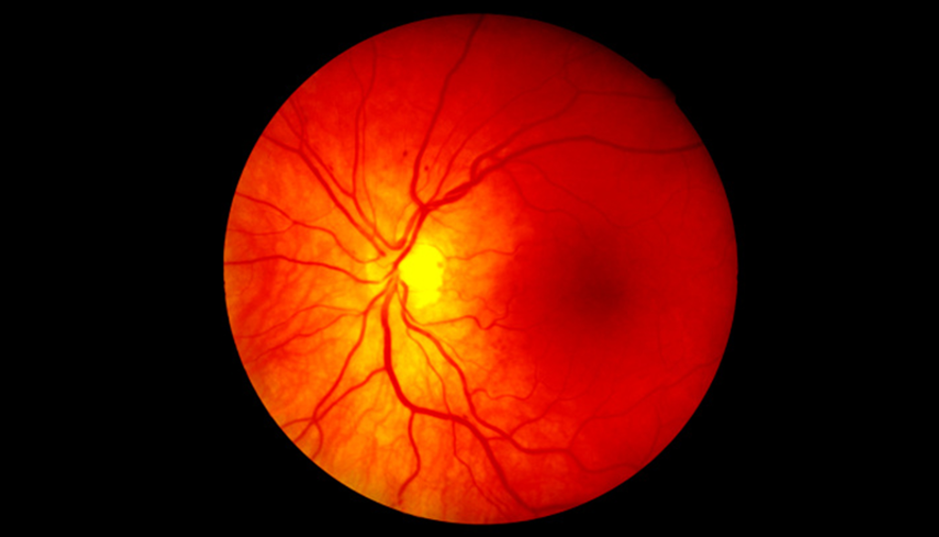
Blind Spot
Explore the blind spot interactively, and map the blind spots in your own eyes.
CLICK ANYWHERE TO BEGIN
Photo: Steve Allen/Getty Images
What Is the Blind Spot, and How Does It Affect Perception?
The animation below shows the location of the blind spot to the left of the fovea in the right eye (in the left eye, the blind spot is to the right of the fovea). Play the animation to see what happens when the eye remains fixated on the black cross while moving progressively closer to the cross. Note how light reflected from the cross always falls directly on the fovea, whereas light from the red disk strikes the retina progressively closer to the blind spot, until the light actually falls on the blind spot.
Then play the animation again, stopping and restarting it to note how the person's perception matches up with the retinal image:
• When the light reflected from the red disk isn't falling on the blind spot, the person perceives the red disk. During this time, perception matches the retinal image.
• When the light reflected from the red disk falls on the blind spot, there is a "hole" in the retinal image at that location; that is, the red disk isn't in the retinal image. But the person doesn't perceive a hole in that location, so at that time, perception doesn't match the retinal image. Instead, the visual system automatically fills in the hole with its "best guess" about what's there—in this case, the visual system guesses that the two vertical lines are really the two ends of one continuous line, and that's what the person perceives.
This filling-in of the blind spot shows that the visual system doesn't just report what's in the retinal image but constructs the likeliest "explanation" of the optic array. In specially constructed situations like the ones in this demonstration, the filling-in can result in an illusory perception—for example, in the animation, the two vertical lines seem to be a single line. But in almost all real-life scenes, the visual system's guesses are essentially correct, and we perceive what's actually there.
2.2 Explain
Calibrate your monitor before proceeding
Make sure that you'll be able to see the phenomena in this demonstration.
Drag the slider so the dollar bill below is the same size as an actual dollar bill.

2.3 Explain
"See" your blind spot.
1. At a comfortable reading distance from your monitor, cover your left eye.
2. Use your right eye to focus on the black cross.
3. Slowly move your face toward the monitor while staying focused on the cross.
The red disk should seem to disappear when your right eye is about a foot away from the monitor, indicating that the red disk is in your blind spot—that is, light emitted from the red disk is striking the retina on the blind spot in your right eye. When that occurs, note what happens to your perception of the elements around the red disk.

2.4 Explain
Mapping your blind spot.
Important! Read these instructions to the end before starting the activity on the screen.
- Cover your left eye, focus your right eye on the black cross, and move your face toward the monitor until the red circle in the array at the right is in your blind spot.
- Press the spacebar. A timer will overlay the black cross.
- Keeping your left eye covered, your right eye focused on the black cross, and the red circle in your blind spot, press the spacebar whenever you detect a flashing light in the array at the right.
- To start over, press RESTART and then follow these instructions again from the beginning.
Flashes that you saw.
Flashes that you missed. These map your blind spot.
CAN'T GET THE RED CIRCLE TO DISAPPEAR? TRY THIS:
- If you haven't calibrated your monitor, do so now.
- Make sure you keep your left eye covered and your night eye focused on the black cross. The red disk won't disappear if you're looking at it with your eye or if your left eye is uncovered.
- Start with your right eye about 2 feet (60 cm) from the monitor and move closer slownly.
- The smaller your monitor, the closer you may have to get to it before the red disk will disappear.
2.5 Explain
What Is the Blind Spot, and How Does It Affect Perception?
"Blind spot" is an alternative name for the optic disk, the portion of the retina
where the axons of the retinal ganglion cells exit the eye in a bundle, forming the optic nerve, and where blood vessels enter and exit the eye.
The term "blind spot" refers to the fact that there are no rods or cones in the retina there, which means that we cannot sense that part of the
retinal image.
The animation at right shows the location of the blind spot to the left of the fovea in the right eye (in the left eye, the blind spot is
to the right of the fovea). Play the animation to see what happens when the eye remains fixated on the black cross while moving progressively
closer to the cross. Note how light reflected from the cross always falls directly on the fovea, whereas light from the red disk strikes the
retina progressively closer to the blind spot, until the light actually falls on the blind spot.
Then play the animation again, stopping and restarting it to note how the person's perception matches up with the retinal image:
• When the light reflected from the red disk isn't falling on the blind spot, the person perceives the
red disk. During this time, perception matches the retinal image.
• When the light reflected from the red disk falls on the blind spot, there is a "hole" in the retinal image at that location; that is, the
red disk isn't in the retinal image. But the person doesn't perceive a hole in that location, so at that time, perception doesn't match the
retinal image. Instead, the visual system automatically fills in the hole with its "best guess" about what's there—in this case, the visual
system guesses that the two vertical lines are really the two ends of one continuous line, and that's what the person perceives.
This filling-in of the blind spot shows that the visual system doesn't just report what's in the retinal image but constructs the likeliest
"explanation" of the optic array. In specially constructed situations like the ones in this demonstration, the filling-in can result in an
illusory perception—for example, in the animation, the two vertical lines seem to be a single line. But in almost all real-life scenes, the
visual system's guesses are essentially correct, and we perceive what's actually there.
2.6 Test - single choice
Select your answer to the question below. Then click SUBMIT.
When we look at a scene with one eye closed, why don't we perceive a hole in the retinal image at the location of the blind spot in the open eye?
The correct answer is A.
Click EXPLAIN if you want to review this topic.
2.7 Test - single choice
Select your answer to the question below. Then click SUBMIT.
Why is the optic disk also referred to as the "blind spot"?
The correct answer is B.
Click EXPLAIN if you want to review this topic.
2.8 Test - single choice
Select your answer to the question below. Then click SUBMIT.
When might you have an illusory perception related to the visual system's automatic filling-in of the blind spot?
The correct answer is B.
Click EXPLAIN if you want to review this topic.
2.9 Test - single choice
Suppose you had one eye closed and the central green circle in the top array were in the
blind spot of your open eye.
Click on the array in the bottom row that you would be most likely to perceive. Then click SUBMIT.

Suppose you had one eye closed and the central green circle in the top array were in the
blind spot of your open eye. Click on the array in the bottom row that you would be most likely to perceive.



The correct answer is B.
Click EXPLAIN if you want to review this topic.
2.10 Activity completed
Blind Spot.