Chapter 9. Functional Anatomy of the Ear
9.1 Title slide

Functional Anatomy of the Ear
Interact with depictions of the human ear.
CLICK ANYWHERE TO BEGIN
Photo: © Blend Images / Alamy.
What Is the Functional Anatomy of the Ear?
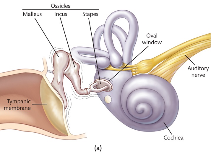
The ossicles transmit sound energy from the tympanic membrane to the inner ear, which contains the cochlea and the semicircular canals (involved in the sense of balance and acceleration, not audition). The malleus is connected to the tympanic membrane, so when the tympanic membrane vibrates, the malleus pushes on the incus, which in turn displaces the stapes, which then pushes in on the oval window, a membrane-covered opening at the base of the cochlea. This creates vibrations within the cochlea, and neurons in the cochlea transduce the vibrations into neural signals that are sent to the brain via the auditory nerve.
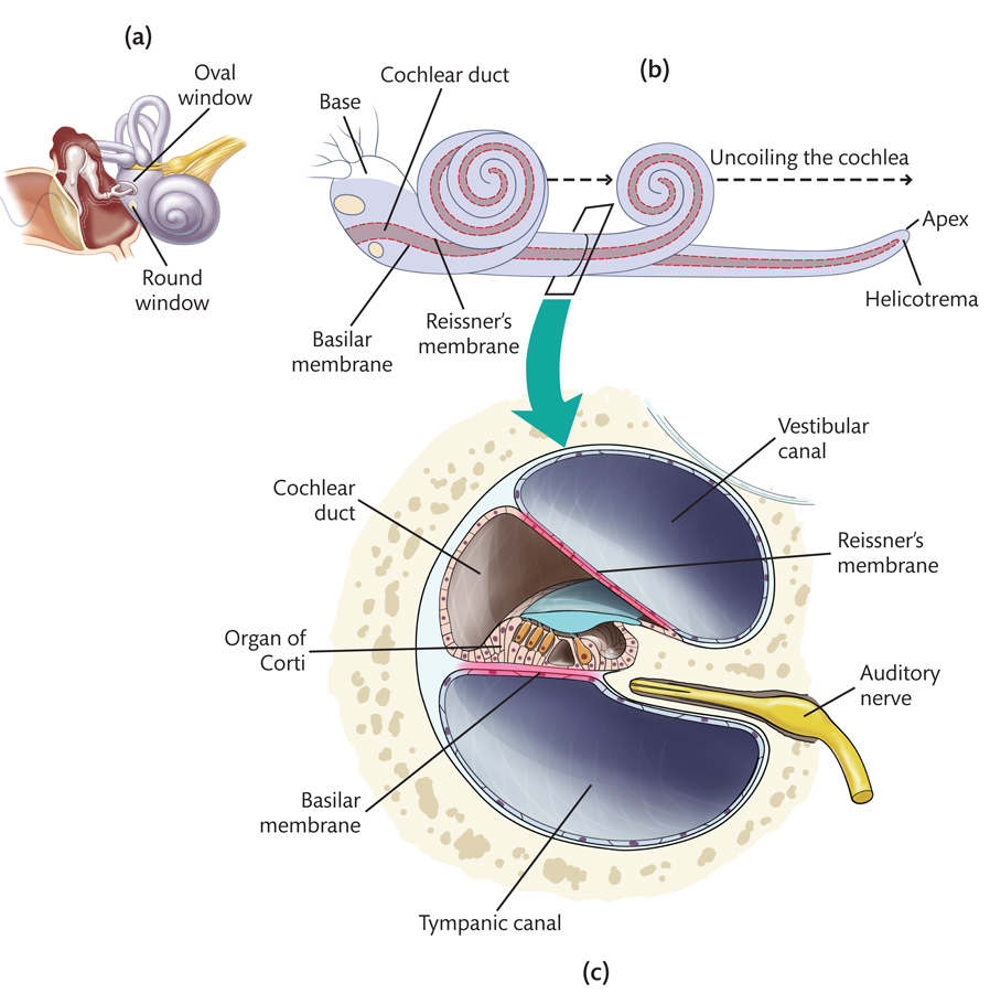
The cochlea is a snail-shaped compartment partitioned along its length into three fluid-filled chambers—the vestibular canal, the cochlear duct, and the tympanic canal. Reissner's membrane separates the vestibular canal from the cochlear duct, and the basilar membrane separates the cochlear duct from the tympanic canal. Within the cochlear duct, resting on the basilar membrane, is the structure responsible for auditory transduction, the organ of Corti.
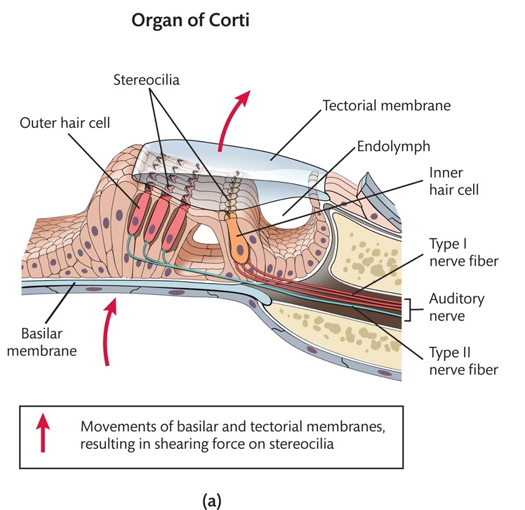
The organ of Corti contains two sets of neurons—inner hair cells and outer hair cells—and the tectorial membrane, which lies above the hair cells. Each hair cell has stereocilia protruding from the top. Open spaces within the organ of Corti are filled with endolymph, the fluid inside the cochlear duct. The stereocilia atop the hair cells are bent back and forth as a result of the movements of the basilar and tectorial membranes, and this bending of the stereocilia causes the hair cells to produce neural signals that are conveyed to the brain. Inner hair cells are connected to Type I auditory nerve fibers; outer hair cells are connected to Type II auditory nerve fibers.
9.2 Explain - dnd
Drag and drop each label into the correct box on this illustration of the ear.
9.3 Explain - dnd
Drag and drop each label into the box with the correct definition of these parts of the ear.
9.4 Explain - dnd
Drag and drop each label into the correct box on these views of the cochlea.
9.5 Explain - dnd
Drag and drop each label into the box with the correct definition of these parts of the cochlea.
9.6 Explain - dnd
Drag and drop each label into the correct box on this illustration of the organ of Corti.
9.7 Explain
What Is the Functional Anatomy of the Ear?
The ear is conventionally divided into three parts—the
outer ear, middle ear, and inner ear. The outer ear consists of the pinna, the auditory canal, and the tympanic
membrane. The pinna gathers sound waves from the environment, and the auditory canal funnels the sound waves onto
the tympanic membrane, which vibrates in response. Those vibrations are transmitted into the middle ear, a tiny
air-filled chamber containing the ossicles—three small bones called the malleus, the incus, and the stapes. The
air pressure on the outer side of the tympanic membrane must be approximately equal to the air pressure on the
inner side. This is accomplished via the Eustachian tube, which connects the middle ear to the top part of the
throat.
The ossicles transmit sound energy from the tympanic membrane to the inner ear, which contains the cochlea
and the semicircular canals (involved in the sense of balance and acceleration, not audition). The malleus is
connected to the tympanic membrane, so when the tympanic membrane vibrates, the malleus pushes on the incus, which
in turn displaces the stapes, which then pushes in on the oval window, a membrane-covered opening at the base of
the cochlea. This creates vibrations within the cochlea, and neurons in the cochlea transduce the vibrations into
neural signals that are sent to the brain via the auditory nerve.
The cochlea is a snail-shaped compartment partitioned along its length into three fluid-filled chambers—the
vestibular canal, the cochlear duct, and the tympanic canal. Reissner's membrane separates the vestibular canal
from the cochlear duct, and the basilar membrane separates the cochlear duct from the tympanic canal. Within the
cochlear duct, resting on the basilar membrane, is the structure responsible for auditory transduction, the organ
of Corti.
The organ of Corti contains two sets of neurons—inner hair cells and outer hair cells—and the tectorial
membrane, which lies above the hair cells. Each hair cell has stereocilia protruding from the top. Open spaces
within the organ of Corti are filled with endolymph, the fluid inside the cochlear duct. The stereocilia atop the
hair cells are bent back and forth as a result of the movements of the basilar and tectorial membranes, and this
bending of the stereocilia causes the hair cells to produce neural signals that are conveyed to the brain. Inner
hair cells are connected to Type I auditory nerve fibers; outer hair cells are connected to Type II auditory nerve
fibers.
9.8 Test - dnd
The ear transduces sound waves into neural signals. Drag the labels into the empty boxes to show the order of events in this process. Then click SUBMIT.
Stereocilia on hair cells bend
Tympanic membrane vibrates
Hair cells produce neural signals
Ossicles vibrate
Basilar membrane is displaced
Waves travel in cochlear fluid
9.9 Test - single choice
Select your answer to the question below. Then click SUBMIT.
Which type of damage do you think would be most likely to result in total deafness?
9.10 Test - single choice
Select your answer to the question below. Then click SUBMIT.
Hearing aids and cochlear implants are two types of devices that help people with hearing impairments.
Hearing aids amplify the sounds coming into the ear. Cochlear implants convert sounds into electrical impulses
that directly stimulate auditory nerve fibers. Based on this information, which of the following statements do
you think is true?
9.11 Activity completed
Functional Anatomy of the Ear.