5.3 Functional Organization of the Nervous System
So far in this chapter we have examined the basic workings of individual neurons and have overviewed the main methods for learning about the functions of large groups of neurons in the brain. Now it is time to tour the whole nervous system. We will look into each of its major divisions with an eye toward understanding its role in the broad task of governing the person’s behavior. The section will conclude with an examination of how hormones interact with the nervous system.
165
As we proceed, it will be helpful to keep in mind that the nervous system is hierarchically organized. Indeed, it contains two distinct but interacting hierarchies. One, the sensory-perceptual hierarchy, is involved in data processing. It receives sensory data about a person’s internal and external environment, and it analyzes those data to make decisions about the person’s bodily needs and about threats and opportunities in the outside world. The flow of information in this hierarchy is primarily from bottom (sensory receptors) to top (perceptual centers in the brain). The other hierarchy, the motor-control hierarchy, is involved in control of movement. The flow of information here is primarily from top to bottom. At the top of this hierarchy are executive centers that make decisions about the activities that the person as a whole should engage in, and at lower levels are centers that translate those decisions into specific patterns of muscle movement.
Most of the large anatomical divisions of the nervous system are involved in both hierarchies, so we will examine both at once. We’ll pay most attention here, though, to the motor hierarchy; the sensory-perceptual system is discussed in detail in Chapters 7 and 8. Let’s start at the bottom and work our way upward. The bottom parts of the hierarchy are evolutionarily the most primitive and are most directly tied to the muscles and the sensory organs.
Peripheral Nerves: The Nervous System’s Interface with the World
The peripheral nervous system, as you may recall, consists of the entire set of nerves, which connect the central nervous system to the body’s sensory organs, muscles, and glands (refer back to Figure 5.2). Nerves are divided into two classes that correspond to the portion of the central nervous system from which they protrude. Cranial nerves project directly from the brain. Spinal nerves project from the spinal cord. Like most other structures in the body, nerves exist in pairs; there is a right and a left member in each pair. Humans have 12 pairs of cranial nerves and 31 pairs of spinal nerves. With their many branches, these nerves extend to all portions of the body.
Three pairs of cranial nerves contain only sensory neurons and five pairs contain only motor neurons; the remaining four pairs of cranial nerves and all 31 pairs of spinal nerves contain both sensory and motor neurons. The total number of sensory and motor neurons combined is “only” about 5 or 6 million, a tiny fraction of the roughly 100 billion neurons of the nervous system as a whole (the rest are all interneurons). Yet these neurons are crucially important. Without them, the rest of the nervous system would be useless because it would have no way of receiving sensory input or controlling the person’s actions.
Sensory Neurons Provide Data Needed for Governing Behavior
As noted earlier, sensory neurons are activated at their dendritic ends by the effects of sensory stimuli (such as light in the eye, chemicals on the tongue or in the nose, sound waves in the ear, or pressure on the skin). They send their action potentials all the way into the central nervous system by way of their very long axons. The rates and patterns of action potentials in sensory neurons are the data that perceptual areas of the central nervous system use to figure out the state of the external and internal environment. Without such input, the central nervous system would have no information on which to base its behavior-controlling decisions.
Sensory input from the specialized sensory organs of the head—the eyes, ears, nose, and tongue—enters the brain by way of cranial nerves. Sensory input that comes from the rest of the body—from the skin, muscles, tendons, and various internal organs—enters the central nervous system by way of all of the spinal nerves and some of the cranial nerves. The sensations conveyed by these inputs, which include touch and pain, are referred to collectively as somatosensation.Soma means “body,” and somatosensation is the set of sensations that derive from the whole body as opposed to those that come just from the special sensory organs of the head. (A full discussion of the processes of sensation and perception is found in Chapters 7 and 8.)
166
Motor Neurons Are the “Final Common Path” for Control of Behavior
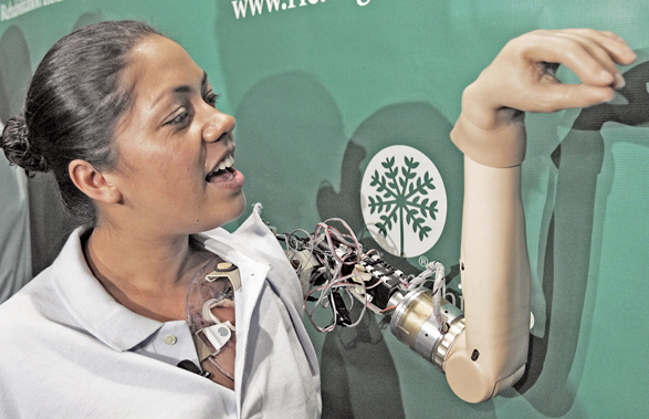
Motor neurons, as noted earlier, have their cell bodies in the central nervous system and send their long axons out, by way of cranial or spinal nerves, to terminate on muscles or glands. Ultimately, all of the behavioral decisions of the nervous system are translated into patterns of action potentials in the axons of motor neurons, and those patterns determine our behavior. In the words of the pioneering neurophysiologist Charles Sherrington (1906), motor neurons are the “final common path” of the nervous system. Through them, and only through them, can the nervous system control behavior. All of the brain’s calculations—including those that we experience consciously as perceptions, thoughts, emotions, desires, and intentions—would be useless if they could not act on muscles and glands. The 100 billion neurons of the central nervous system are all involved, ultimately, in controlling the 2 or 3 million motor neurons, which in turn control behavior. For a wonderful example of how knowledge of motor neurons has been put to medical use, see Figure 5.16 and its caption.
The Motor System Includes Skeletal and Autonomic Divisions
12
How do the autonomic and skeletal motor systems differ from one another in function? How do the sympathetic and parasympathetic divisions of the autonomic system differ from one another in function?
Motor neurons act on two broad classes of structures. One class consists of the skeletal muscles, the muscles that are attached to bones and produce externally observable movements of the body when contracted. The other class consists of the visceral muscles and glands. Visceral muscles are muscles that are not attached to bones and do not move the skeleton when they contract. They form the walls of such structures as the heart, arteries, stomach, and intestines. Glands are structures that produce secretions, such as the salivary glands and sweat glands. Neurons that act on skeletal muscles make up the skeletal portion of the peripheral motor system. Those that act on visceral muscles and glands make up the autonomic portion.
Whereas skeletal motor neurons initiate activity in the skeletal muscles, autonomic motor neurons typically modulate (modify) rather than initiate activity in the visceral muscles. Skeletal muscles are completely inactive in the absence of neural input, but visceral muscles have built-in, nonneural mechanisms for generating activity. The heart continues to beat and the muscular walls of such structures as the intestines and arteries continue to contract in response to local influences, even if all the nerves to these organs are destroyed. Most visceral muscles and glands receive two sets of neurons, which produce opposite effects and come from two anatomically distinct divisions of the autonomic system: sympathetic and parasympathetic (see Figure 5.17).
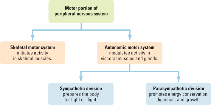
The sympathetic division responds especially to stressful stimulation and helps prepare the body for possible “fight or flight.” Among its effects are (a) increased heart rate and blood pressure, (b) the release of energy molecules (sugars and fats) from storage deposits to permit high energy expenditure, (c) increased blood flow to the skeletal muscles (to help prepare them for action), and (d) inhibition of digestive processes (which helps explain why a heated argument at the dinner table can lead to a stomachache). Conversely, the parasympathetic division serves regenerative, growth-promoting, and energy-conserving functions through effects that include the opposites of those just listed for the sympathetic division. If you are relaxed while reading this book, your parasympathetic activity probably predominates over your sympathetic, so your heart is beating at a slow, normal rate and your digestion is working fine. If you are cramming for a test that is coming up in an hour or so, the opposite may be true.
167
The Spinal Cord: A Conduit and an Organizer of Simple Behaviors
13
What are three categories of functions of the spinal cord?
The spinal cord (depicted in Figure 5.2 on p. 149) connects the spinal nerves to the brain. It also organizes some simple reflexes and rhythmic movements.
The Spinal Cord Contains Pathways to and from the Brain
The spinal cord contains ascending tracts, which carry somatosensory information brought in by the spinal nerves up to the brain, and descending tracts, which carry motor control commands down from the brain to be transmitted out by spinal nerves to muscles. A person whose spinal cord is completely severed will be completely paralyzed and lacking in sensation in those parts of the body that are innervated by nerves that come from below the place of injury. The closer the place of injury is to the head, the greater the number of spinal nerves that are cut off from the brain and the greater the extent of paralysis and insensitivity. Thus, an injury that severs the spinal cord just below the brain will result in paralysis and insensitivity including the arms, trunk, and legs; but if the injury is farther down, only the legs may be paralyzed.
168
The Spinal Cord Organizes Simple Reflexes
Some reflexive behaviors do not require the brain; they are organized by the spinal cord alone. Such reflexes are most clearly demonstrated in animals whose spinal cords have been surgically separated from the brain. (Experiments of this sort may seem cruel, but the knowledge gained from them has been extremely valuable in helping people who have spinal cord injuries.) Animals that have had this operation—referred to as spinal animals—still have both a brain and a spinal cord, but these structures no longer communicate with each other.
If the paw of a spinal cat is pricked with a pin, the animal does not hiss or show facial signs of pain, as a normal cat would, because the stimulus input cannot reach the pain and vocalization centers of the brain. The cat cannot feel sensations from below the neck because feeling is mediated by the brain. Nevertheless, the animal’s paw quickly withdraws from the pin. This reflex is called the flexion reflex because it involves contraction of the flexor muscles—the muscles that bend the limb at each joint, causing it to be pulled inward (flexed) toward the body. The adaptive advantage of the flexion reflex is obvious: It quickly and automatically moves the limb away from potentially damaging stimuli. In the intact, normal cat, this reflex occurs quickly, even before the cat shows evidence of feeling pain. It occurs quickly precisely because it occurs at the level of the spinal cord and does not require that messages be sent up to the brain for further processing and then be sent back down again.
The Spinal Cord Contains Pattern Generators for Locomotion
The old saying “running around like a chicken with its head cut off” refers to the fact that a freshly decapitated chicken will flap its wings and run around the barnyard if not restrained. This occurrence demonstrates the fact that the spinal cord is capable of generating sustained, organized movements without the involvement of the brain. The spinal cord contains networks of neurons that stimulate one another in a cyclic manner and thereby produce bursts of action potentials that wax and wane in a regular, repeating rhythm (Kiehn, 2006). These networks, called pattern generators, activate motor neurons in the spinal cord in such a way as to produce the rhythmic sequence of muscle movements that results in walking, running, flying (in birds), or swimming (in fish). In some animals (but not in humans), the pattern generators become active when released from the brain’s inhibitory control over them, which accounts for the wing flapping and running motions of the headless chicken. Normally, in intact animals, pattern generators are controlled by neurons descending from the brain. They can be either inhibited, producing a motionless animal, or activated to varying degrees, producing varying rates of locomotion.
Subcortical Structures of the Brain
We now leave the spinal cord and enter the brain itself. The lower, more primitive parts of the brain are referred to as subcortical structures because of their position beneath the cerebral cortex, the topmost part of the brain. Working our way upward from the bottom, we begin our tour of the brain with the brainstem.
The Brainstem Organizes Species-Typical Behavior Patterns
14
How is the brainstem similar to and different from the spinal cord? What role does the brainstem play in the control of behavior?
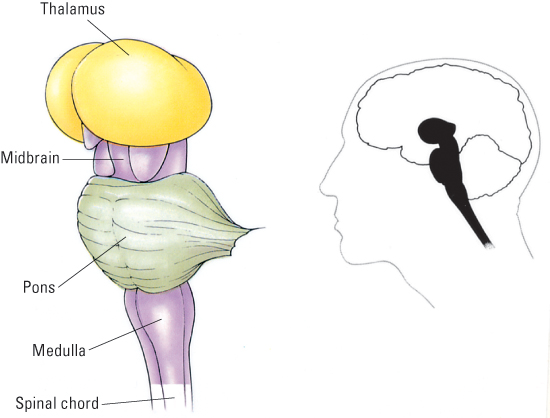
As it enters the head, the spinal cord enlarges and becomes the brainstem. The parts of the brainstem, beginning closest to the spinal cord and going upward toward the top of the head, are the medulla [mo-du´-la], pons, and midbrain (see Figure 5.18). The brainstem is functionally and anatomically quite similar to the spinal cord, but is more elaborate. The spinal cord is the site of entry of spinal nerves, and the brainstem is the site of entry of most (10 of the 12 pairs) of the cranial nerves. Both the spinal cord and the brainstem contain ascending (sensory) and descending (motor) tracts that communicate between nerves and higher parts of the brain. Also like the spinal cord, the brainstem has some neural centers that organize reflexes and certain species-typical behavior patterns.
169
The medulla and pons organize reflexes that are more complex and sustained than spinal reflexes. They include postural reflexes, which help an animal maintain balance while standing or moving, and certain so-called vital reflexes, such as those that regulate breathing rate and heart rate in response to input signaling the body’s metabolic needs. The midbrain contains neural centers that help govern most of an animal’s species-typical movement patterns, such as those involved in eating, drinking, attacking, or copulating (Klemm, 1990). Also in the midbrain are neurons that act on pattern generators in the spinal cord to increase or decrease the speed of locomotion (Pearson & Gordon, 2000).
An animal (such as a cat) whose central nervous system is cut completely through just above the brainstem, referred to as a brainstem animal, is a fascinating creature to watch. It can produce most of the species-typical behaviors that a normal animal can produce (Klemm, 1990; Schmidt, 1986). It can walk, run, jump, climb, groom itself, attack, produce copulatory movements, chew, swallow, and so on. Unlike a normal animal, however, it makes these responses only when provoked by immediate stimuli; it does not behave in either a spontaneous or a goal-directed manner. If placed on a pole, for example, the animal automatically climbs, but it does not itself choose to climb a pole that has food at the top or avoid one that does not. The animal behaves like a machine that responds to certain triggers rather than like an intelligent, decision-making mammal. Such behavior indicates that the midbrain and the structures below it contain neural systems that organize species-typical patterns of movement but do not contain neural systems that permit deliberate decisions to move or refrain from moving in accordance with the animal’s long-term interests or needs.
The Thalamus Is a Relay Station for Sensory, Motor, and Arousal Pathways
15
What are the main functions of the thalamus?
Directly atop the brainstem is the thalamus [thă´-lǝ-mǝs] (Figure 5.18). This structure, seated squarely in the middle of the brain, is most conveniently thought of as a relay station that connects various parts of the brain with one another. Most of the sensory tracts that ascend through the brainstem terminate in special nuclei in the thalamus; those nuclei, in turn, send their output to specific areas in the cerebral cortex. The thalamus also has nuclei that relay messages from higher parts of the brain to movement-control centers in the brainstem.
In addition to relaying specific sensory and motor signals, the thalamus also plays a role in the arousal of the brain as a whole. Arousal pathways in the midbrain converge in the center of the thalamus and then project diffusely to all areas of the cerebral cortex. The arousal function of the thalamus was nicely exemplified when medical researchers were able to awaken a patient who, because of a brain injury, had spent the previous 6 years in a minimally conscious state (Schiff et al., 2007). The researchers implanted electrodes deep into the central nuclei of the patient’s thalamus. In response to prolonged weak electrical stimulation through those electrodes, the patient would open his eyes, respond to simple requests, recognize and respond to family members, chew and swallow food placed in his mouth, and could begin a course of physical therapy that had previously been impossible.
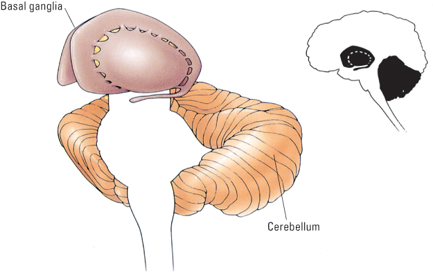
The Cerebellum and the Basal Ganglia Help to Coordinate Skilled Movements
16
What are the functional similarities and differences between the cerebellum and the basal ganglia?
We move now to two portions of the brain that are anatomically distinct but closely related in function—the cerebellum and the basal ganglia (see Figure 5.19). Cerebellum [sěr´-ǝ-běl´-um] means “little brain” in Latin, and indeed this structure looks something like a smaller version of the rest of the brain, riding piggyback on the rear of the brainstem. The basal ganglia [bă´-sǝl găng´-lē-a] are a set of interconnected structures lying on each side of the thalamus. Damage to either the cerebellum or the basal ganglia can greatly interfere with a person’s ability to produce learned, skilled, well-coordinated movements.
170
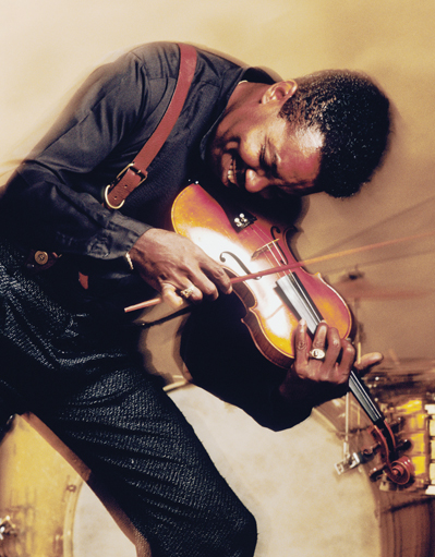
Damage to the cerebellum is especially associated with loss in ability to behave in ways that require rapid, well-timed sequences of muscle movements, such as pitching a baseball, leaping over a hurdle, playing a musical instrument, or typing a series of words at a computer (Houk & Mugnaini, 2003). In contrast, damage to the basal ganglia is especially associated with loss of ability to coordinate slower, deliberate movements, such as reaching out to pick up an object (Packard & Knowlton, 2002).
Both structures are specialized to use sensory information to guide movements, but they apparently use that information in different ways. The basal ganglia appear to use sensory information primarily in a feedback manner. In other words, sensory input pertaining to an ongoing movement (such as the sight of how the hand is moving) feeds back into the basal ganglia and is used to adjust the movement as it progresses. The cerebellum, in contrast, uses sensory information primarily in a feed-forward manner (Ohyama et al., 2003). That is, it uses sensory information to program the appropriate force and timing of a movement before the movement is initiated. That is why the cerebellum is especially crucial for movements that occur too rapidly to be modified once they are in progress.
These characterizations are useful, but they do not describe the full range of functions of these large brain structures. People with damage in either the cerebellum or the basal ganglia can show a wide range of motor deficits, depending on the precise location of damage. Moreover, neuroimaging studies (using PET and fMRI) show that portions of the cerebellum, basal ganglia, and certain motor-planning areas of the cerebral cortex become active not just when people are producing movements but also when they are imagining themselves producing movements (Houk & Mugnaini, 2003). Perhaps when divers, gymnasts, or pianists “visualize” their performance before they act, what they are doing, partly, is warming up specific neurons in these motor areas, thereby setting up the neural programs that will eventuate in their best performance.
171
The Limbic System and the Hypothalamus Play Essential Roles in Motivation and Emotion
17
Why is the limbic system so named, and what functions does it perform?
The term limbic comes from the Latin word limbus, meaning “border.” The limbic system can be thought of as the border dividing the evolutionarily older parts of the brain below it from the newest part (the cerebral cortex), above it. The limbic system consists of several distinct structures that interconnect with one another in a circuit wrapped around the thalamus and basal ganglia (see Figure 5.20). Some of these structures—including especially the amygdala [ǝ-mĭg´-dǝ-lǝ]—are involved in the regulation of basic drives and emotions. But the limbic system also plays other roles. One of its most prominent structures, the hippocampus, is crucial for keeping track of spatial location (the direction-sensitive place cells, noted earlier in the chapter, are located there) and for encoding certain kinds of memories.

The limbic system is believed to have evolved originally as a system for the sophisticated analysis of olfactory input (Thompson, 1985), and its connections with the nose remain strong. This may help explain the special influence that smells—such as the aroma of good food or perfume, or the stench of vomit, or the scent of freshly mown grass—can have on drives, emotions, and memories. But the limbic system also receives input from all the other sensory organs. In addition, it is intimately connected to the basal ganglia, and that connection is believed to help translate emotions and drives into actions.
18
What are three ways by which the hypothalamus controls the body’s internal environment?
The hypothalamus [hī-pō-thăl´-ǝ-mǝs] is a small but vitally important structure. Its name derives from its position directly underneath the thalamus (hypo in this case means “beneath”). The hypothalamus is not technically part of the limbic system, but is intimately connected to all the structures of that system (see Figure 5.20). Its primary task is to help regulate the internal environment of the body. This it accomplishes by (a) influencing the activity of the autonomic nervous system, (b) controlling the release of certain hormones (to be described later), and (c) affecting certain drive states, such as hunger and thirst. In addition, through its connections with the limbic system, the hypothalamus helps regulate emotional states, such as fear and anger. You will read in Chapter 6 about the role of the hypothalamus in drives and emotions.
If you had to give up a cubic millimeter (a tiny speck) of tissue from some part your brain, the last place you would want it taken from is the hypothalamus. Depending on just which part was taken, you could be left without one or more of your basic drives, without a normal cycle of sleep and wakefulness, or without the ability to regulate your rate of metabolism.
The Cerebral Cortex
We now move up to the anatomically outermost and evolutionarily newest part of the brain, the cerebral cortex.Cerebrum is the Latin word for “brain,” and the term now generally refers to all parts of the brain other than the brainstem and cerebellum. Cortex is the Latin word for “bark,” and in anatomical usage it refers to the outside layer of any structure. So, the cerebral cortex is the outside layer of the major portion of the brain. It is by far the largest part of the human brain, accounting for approximately 80 percent of its total volume (Kolb & Whishaw, 2009). Its surface area is much greater than it appears because it folds inward in many places. If the cortex were unfolded and spread out as a single sheet, it would be only 2 to 3 millimeters thick and would occupy a surface area equivalent to a square that is half a meter long on each side (Kolb & Whishaw, 2009).
172
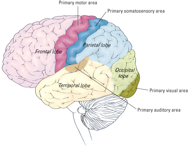
The entire folded cerebral cortex is divided into left and right hemispheres, and each hemisphere is further divided into four lobes, or segments, demarcated at least partly by rather prominent inwardly folding creases, referred to as fissures. The lobes, whose positions you can see in Figure 5.21, are, from back to front, the occipital [ȯk-sĭ´-pǝ-tǝl], temporal, parietal [pa-rī´- ǝ-tǝl], and frontal lobes.
The Cortex Includes Sensory, Motor, and Association Areas
19
What are the four lobes of the cortex, and what are three functional categories of areas that exist within these lobes?
Researchers who study the functions of the cortex divide it into three categories of functional regions, or areas. One category consists of the primary sensory areas, which receive signals from sensory nerves and tracts by way of relay nuclei in the thalamus. As shown in Figure 5.21, primary sensory areas include the visual area in the occipital lobe, the auditory area in the temporal lobe, and the somatosensory area in the parietal lobe. A second category is the primary motor area, which sends axons down to motor neurons in the brainstem and spinal cord. As shown in Figure 5.21, this area occupies the rear portion of the frontal lobe, directly in front of the somatosensory area. The third category consists of all the remaining parts of the cortex, which are called association areas. These areas receive input from the sensory areas and lower parts of the brain and are involved in the complex processes that we call perception, thought, and decision making.
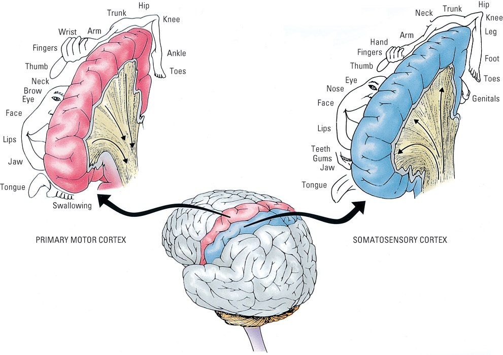
The Primary Sensory and Motor Areas Are Topographically Organized
20
What does it mean to say that cortical sensory and motor areas in the cortex are topographically organized?
The primary sensory and motor areas of the cortex are organized in such a way that adjacent neurons receive signals from or send signals to adjacent portions of the sensory or muscular tissue to which they are ultimately connected. This fact is referred to as the principle of topographic organization. For example, neurons that are near one another in the visual cortex receive signals from receptor cells that are near one another in the retina of the eye. Similarly, neurons that are near one another in the somatosensory cortex receive signals from adjacent areas of the skin, and neurons that are near one another in the primary motor cortex send signals to adjacent sets of muscle fibers. It is possible to map onto the somatosensory cortex the parts of the body from which each portion of somatosensory cortex receives its signals, and onto the motor cortex the part of the body to which each portion of motor cortex sends its signals.
173
The resulting maps, depicted in Figure 5.22, show a distorted view of the human body. This is because the amount of cortex devoted to each part of the body corresponds not to the size of the body part but to the degree of sensitivity of that part (in the case of the sensory map) or the fineness of its movements (in the case of the motor map). As you can see, huge areas of the human primary motor cortex are devoted to control of the fingers and vocal apparatus, where fine control is needed (Grillner, 2012). In other animals, other body parts have greater representation, depending on the range and delicacy of their movements. In a cat, for example, large portions of the somatosensory and primary motor areas of the cortex are devoted to the whiskers, and in a spider monkey—a creature that uses its tail as a fifth arm and hand—large areas are devoted to the tail (Walker, 1973).
21
What is some evidence that the primary motor cortex comes relatively late in the chain of command preceding an action and that its function is to refine the more delicate parts of the action?
The primary motor cortex is part of the chain of command in controlling movements, but it is not at the top of that chain and is not involved in all types of movements. It receives input from the basal ganglia and cerebellum and is specialized to fine-tune the signals going to small muscles, such as those of the fingers and tongue, which must operate in a finely graded way (Lemon, 2008).
Experiments conducted as long ago as the 1970s, in which monkeys made well-controlled hand movements to obtain food rewards, revealed that each movement was preceded first by a burst of activity in the basal ganglia and then by a burst in the primary motor cortex (Evarts, 1979; Kornhuber, 1974). This is part of the evidence that the primary motor cortex generally comes later than the basal ganglia and the cerebellum in the chain of command. In other experiments, monkeys whose primary motor cortex had been entirely destroyed behaved normally in most respects but were unable to make delicate hand movements, such as those needed to lift a small piece of food out of a narrow hole (Passingham et al., 1983). More recently, researchers have found that electrical stimulation of the motor cortex produces not just muscle twitches, but also well-organized movements. For example, stimulating the hand area of the motor cortex in a monkey can result in a well-coordinated grasping response, or pushing response, depending on just where the stimulus is applied (Graziano, 2006).
174
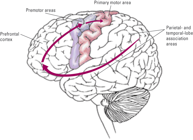
Premotor Areas Help Organize Specific Patterns of Movement
22
What is the role of the premotor areas in the control of behavior? How has activity there been used to assess consciousness in individuals with brain damage?
Directly in front of the primary motor area lie a set of other cortical areas devoted to motor control, referred to collectively as premotor areas (see Figure 5.23). These areas set up neural programs for producing organized movements or patterns of movements. To choose what program to set up, they use information sent to them from anterior (forward) portions of the frontal lobe that are involved in overall behavioral planning. To execute an action program they send information out to the cerebellum, basal ganglia, and motor cortex, which refine the program further before sending messages down toward the muscles.
Like the cerebellum and basal ganglia, premotor areas become active during the mental rehearsal of coordinated movements as well as during the actual production of such movements (Cisek & Kalaska, 2004). Medical researchers have begun to exploit this fact in order to learn about the mental capacities and potential for recovery of individuals with severe brain injuries who are in a so-called “vegetative state.” Patients in such a state show evidence of sleep–wake cycles but are unable to speak or move in a deliberate fashion. Until recently, there was no way to know whether or not they were conscious and able to hear and understand what people said to them. Recently, researchers in the United Kingdom began testing the consciousness of patients in a vegetative state by assessing premotor neural activity, with fMRI neuroimaging, while asking them to imagine specific movements—such as swinging a tennis racket or kicking a soccer ball (Owen & Coleman, 2008). In early tests they identified 2 patients (out of 17 tested) who repeatedly, in response to such instructions, exhibited patterns of neural activity in premotor regions that were indistinguishable from those produced by people with intact brains who were given the same instructions. The results showed clearly that those 2 patients could understand the instructions and were able to imagine the specified movement. More recent work showed similar results, with 5 of 54 patients in a vegetative state displaying evidence of consciousness as reflected by changes in fMRI images (Monti et al., 2010).
175
Prefrontal Association Areas Create General Plans for Action
23
What is the role of the prefrontal cortex in the control of behavior?
The portion of the cerebral cortex that has expanded the most in human beings compared to other animals is the prefrontal cortex, consisting of the entire frontal lobe anterior to (in front of) the premotor areas (see Figure 5.23). This part of the brain is involved in what has been termed executive function: the processes involved in regulating attention and in determining what to do with information just gathered or retrieved from long-term memory. It plays a central role in planning and behaving flexibly, particularly when dealing with novel information (Miyake et al., 2000). We’ll examine executive function in later chapters (especially Chapters 9 and 11) and see how individual and developmental differences in the prefrontal cortex are related to differences in executive functioning and performance on a host of cognitive tasks.
To review the general flow of information in the cortex involved in the control of movement, notice the arrows in Figure 5.23. Association areas in the rear parts of the cortex, especially in the parietal and temporal lobes, analyze information that comes to them from sensory areas. These areas, in turn, send output to prefrontal association areas, which also receive information about the internal environment through strong connections with the limbic system. Combining all this information, the prefrontal areas set up general plans for action that can be put into effect through connections to the premotor cortex and through downward links to the basal ganglia and cerebellum.
Consistent with this model of cortical control, damage in the prefrontal lobe does not, as a rule, reduce one’s ability to extract information from the environment, but does reduce one’s ability to use that information effectively to control behavior. Depending on the location and extent of the injury, such damage can destroy either short-range planning, such as working out the series of movements needed to solve a particular puzzle, or long-range planning, such as organizing one’s day, week, or life (Kolb & Whishaw, 2009).
Hierarchical Organization in the Control of Movement: A Summary
24
How are the movement-control functions of the nervous system summarized as a hierarchical, top-down flow of information? How is the hierarchy illustrated by an imaginative tour through the nervous system of a person who decides to eat some fresh cherries?
We have ascended the nervous system, from bottom to top, and glimpsed the functions of each of its divisions in the overall task of controlling behavior. The parts, as we said at the outset, work together in a hierarchical manner. To review the movement-control functions of the various parts and to visualize their hierarchical organization, look at Figure 5.24. Structures are organized there according to their general roles in controlling movement, not according to their anatomical positions. The highest structures are involved in motivation and planning, and the lower structures are involved in refining and executing the plans, turning them into action. Notice that both subcortical and cortical structures (shown, respectively, on the left and right sides of the diagram) are involved at each of the top three levels in the hierarchy.
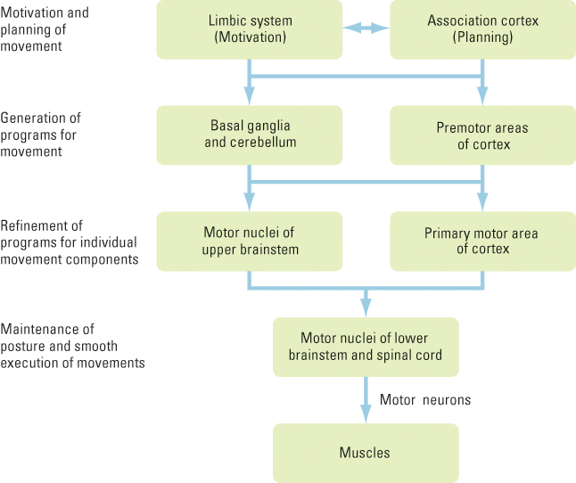
To illustrate the hierarchy further, imagine what might occur in the nervous system of a person who has not eaten in a while and sees some fresh cherries. At the top of the hierarchy, the limbic system (which most directly monitors the internal state of the body) senses that food is needed and sends a message of “hunger” to cortical association areas with which it is connected. These areas, which share the top of the hierarchy with the limbic system, analyze information coming to them from the visual cortex and determine that fresh cherries are available in a bowl across the room. Other information is also considered by the association areas, including memories about the taste of fresh cherries, the propriety of eating them in this room at this time, and how to eat them, including seeking a receptacle for disposing of the stems and pits. Such information, integrated by prefrontal association areas, leads to a decision to cross the room, pick up some cherries, and eat them.
176
At the second level, the basal ganglia, cerebellum, and premotor cortex receive the program of planned action from the limbic system and prefrontal cortex. They also receive somatosensory input about the exact position of parts of the body and visual input about the exact location of the cherries. They use this information to refine the motor program—that is, to work out the specific timing and patterning of the movements to be made.
At the third level, the motor program is conveyed through two pathways for further refinement. The program for larger movements, such as walking toward the cherries, is sent directly down to a set of motor nuclei in the upper part of the brainstem. The program for delicate movements, such as those needed for removing the stems and eating the fruit while avoiding swallowing the pits, is conveyed to the motor cortex, which, in turn, sends its output down to the brainstem and spinal cord.
www.cornered.com
177
Finally, at the fourth level of the hierarchy, in the lower brainstem and spinal cord, are motor neurons—the final common pathway in control of behavior. Skeletal motor neurons send their messages out to the skeletal muscles of the body to trigger all the movements needed to reach the cherries, pick up a handful, and put them in the mouth. Autonomic motor neurons (of the parasympathetic division) send their messages out to the salivary glands and muscular walls of the gastrointestinal system to prepare the digestive tract for the receipt of cherries.
A Word of Caution
The hierarchical model just described is useful as a first approach to understanding the nervous system, and it is consistent with the kinds of behavioral deficits that occur when different parts of the nervous system are damaged. However, there is a possible danger in this portrayal: It can seduce us into believing that we know more than we actually do know.
25
What is the difference between knowing where a brain function occurs and knowing how it occurs?
Specifically, the knowledge that certain parts of the brain are essential for certain aspects of behavioral control can be mistaken for knowledge about how those processes are accomplished. But the discovery of “where” does not explain “how.” In illustrating the hierarchy, we spoke of “decisions” made in prefrontal areas of the cortex and of programs for action developed and refined by other brain areas. What do such statements mean? They mean only that individuals who suffer damage in one part of the brain lose the ability to make reasonable choices for action, and that those who suffer damage in another part retain the ability to make reasonable choices but lose the ability to carry them out in a coordinated manner. Such statements don’t address the far more difficult question of how the association cortex makes decisions or how various other structures develop and refine programs for action.
How Hormones Interact with the Nervous System
When the ancient Greeks argued that the heart is the seat of thought, feeling, and behavioral control, they were not without reasons. Like the brain, the heart has long protrusions (blood vessels, in this case) that connect it with other parts of the body. Blood vessels are easier to see than nerves, and because they can be found in all the sense organs and muscles, as well as in other tissues, early theorists believed that blood vessels were the conduits of sensory and motor messages. Today we know that the circulatory system does indeed play a vital communicative role in the body, though not the one envisioned by the ancients. A slower messenger system than the nervous system, it carries chemicals that affect both physical growth and behavior. Among these chemicals are hormones, which are secreted naturally into the bloodstream, and drugs, which may enter the bloodstream artificially through various routes.
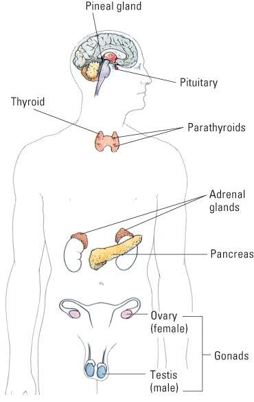
Hormones are chemical messengers that are secreted into the blood. They are carried by the blood to all parts of the body, where they act on specific target tissues. Dozens of hormones have been identified. The classic hormones—the first to be identified and the best understood—are secreted by special hormone-producing glands called endocrine glands (see Figure 5.25). But many other hormones are secreted by organs that are not usually classified as endocrine glands, such as the stomach, intestines, kidneys, and brain.
How Hormones Affect Behavior
Hormones influence behavior in many ways. They affect the growth of peripheral bodily structures, including muscles and bones, and in that way influence behavioral capacity. Hormones also affect metabolic processes throughout the body and thereby influence the amount of energy that is available for action. Of greatest interest to psychologists is the fact that hormones also act in the brain in ways that influence drives and moods.
178
26
What are some examples of long-term and short-term effects of hormones?
Some effects of hormones are long term or irreversible, and some of these occur before birth. For example, almost all the anatomical differences between newborn boys and girls are caused by the hormone testosterone, which is produced by the male fetus but not by the female fetus. These anatomical differences are evident in the brain as well as in the genitals, and the brain differences provide one basis for sex differences in behavior throughout life. At puberty, the increased production of sex hormones—especially testosterone in the male and estrogen in the female—stimulates a new set of growth processes that further differentiate males and females anatomically and thereby influence their behavior.
The short-term effects of hormones range in duration from a few minutes to many days. In response to stressful stimulation, for example, the adrenal cortex (the external layer of the adrenal gland) secretes various hormones, including cortisol, which are sometimes referred to as “stress hormones.” These hormones produce a variety of effects throughout the body that help the animal in a stressful situation. For example, they release sugar and fat molecules into the blood to supply extra energy for possible “fight or flight,” and they suppress inflammation caused by wounds. These hormones are also taken up by neurons in certain parts of the brain and apparently act there to help the animal adapt behaviorally to the stressful situation (McEwen, 1989).
How Hormones Are Controlled by the Brain
27
How does the brain control the release of hormones from the two lobes of the pituitary and thereby control the release of other hormones as well?
The pituitary, which sits at the base of the brain, is sometimes called the master endocrine gland because it produces hormones that, in turn, stimulate the production of other hormones in other glands, including the adrenal cortex and the gonads (ovaries in the female and testes in the male).
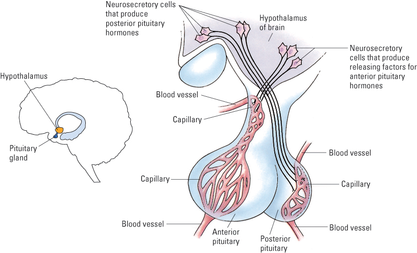
To visualize the intimate relationship between the brain and the pituitary, look at Figure 5.26. The rear part of the pituitary, the posterior lobe, is in fact a part of the brain. The posterior lobe consists mainly of modified neurons, referred to as neurosecretory cells, which extend down from the hypothalamus. When these neurosecretory cells are activated, by brain neurons that lie above them, they release their hormones into a bed of capillaries. Once these hormones enter the capillaries, they are transported into the rest of the circulatory system to affect various parts of the body.
179
The remainder of the pituitary, the anterior lobe, is not part of the brain (no neurons descend into it) but is intimately connected to the brain by a specialized set of capillaries, as shown in Figure 5.26 on page 178. Neurosecretory cells in the brain’s hypothalamus produce releasing factors, hormones that are secreted into the special capillary system and are carried to the anterior pituitary, where they stimulate the anterior pituitary cells to synthesize and release hormones into capillaries that carry the hormones into the general bloodstream. Different releasing factors, produced by different sets of neurosecretory cells in the hypothalamus, act selectively to stimulate the production of different anterior pituitary hormones.
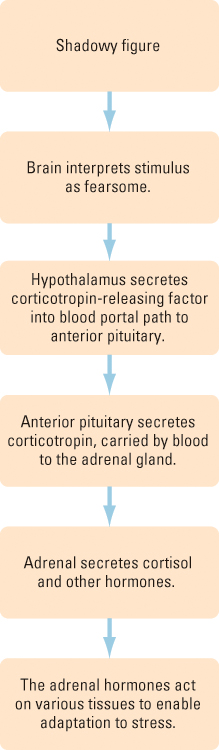
Consider the sequence of hormonal events, triggered by the brain, that is diagrammed in Figure 5.27: (1) A shadowy figure is seen at night, and the brain interprets it as fearsome. (2) The association cortex sends a neural message to the viewer’s hypothalamus that causes it to secrete corticotropin-releasing factor. (3) The specialized capillary system transports this releasing factor to the anterior pituitary, where it stimulates the release of another hormone, corticotropin, into the bloodstream. (4) From the bloodstream, corticotropin enters the adrenal cortex, where it causes the release of adrenal cortical hormones, including cortisol. (5) These adrenal hormones are carried throughout the body to help prepare it for a possible emergency. At the same time, many other brain-controlled effects are also occurring to deal with the possible emergency suggested by the sight of the shadowy figure. They range from the activation of the sympathetic portion of the autonomic nervous system to the development of a plan to escape.
SECTION REVIEW
Divisions of the nervous system are organized hierarchically to control behavior.
Peripheral Nervous System
- This division consists of spinal and cranial nerves and their various branches, which contain sensory and motor neurons.
- Motor neurons include skeletal motor neurons, which contract skeletal muscles, and sympathetic and parasympathetic autonomic motor neurons, which act in opposing ways on internal organs.
Spinal Cord
- The spinal cord acts as a conduit, carrying somatosensory information to the brain and motor commands from the brain.
- It also organizes spinal reflexes and contains pattern generators that produce rhythmic movement sequences, such as walking.
Subcortical Structures
- The brainstem is similar to the spinal cord but more elaborate.
- The thalamus is a major sensory and motor relay station.
- The cerebellum and basal ganglia are crucial for the production of coordinated actions.
- The hypothalamus and limbic system are critically involved in motivation and emotion as well as other functions.
Cerebral Cortex
- The cortex is divided into two hemispheres, each with four lobes.
- Cortical areas include primary sensory and primary motor areas, which are organized topographically, and association areas, which are crucial for thought.
- Prefrontal and premotor association areas plan and establish neural programs for actions.
Hormones
- Hormones are chemical messengers secreted into the bloodstream.
- Hormones can influence behavior by affecting bodily growth, metabolism, and brain activity, including brain activity responsible for drives and moods.
- Hormonal effects can be long term or even permanent, as in the case of prenatal testosterone’s effects on brain development. They can also be short term, as in the case of stress hormones’ effects in preparing for an emergency.
- The pituitary gland controls hormone production by other glands, but is itself controlled by the brain.
180