10.1 TISSUES AND ORGANS
At the beginning of Chapter 9, we considered the features that enable a group of individuals to function as a community. One of these is the presence of members able to perform specialized tasks. In addition, community members stick together and respond to their surrounding environment. Now, think of our own tissues and organs—a heart, a stomach, a kidney, a brain—all are communities of cells that work together to perform highly specific and important tasks in response to cues from the environment.
10.1.1 Tissues and organs are communities of cells.
A biological tissue is a collection of cells that work together to perform a specific function. Two or more tissues often combine and function together as an organ, such as a heart or lung. Animals and plants have tissues and organs that allow them to carry out the various processes necessary to sustain them. In animals, for example, four types of tissue—epithelial, connective, nervous, and muscle—combine to make up all the organs of the body. Plants are made up of just three types of tissue: dermal (the outer protective layer), ground (the bulk of the plant body), and vascular (the channels that transport nutrients and water throughout the plant).
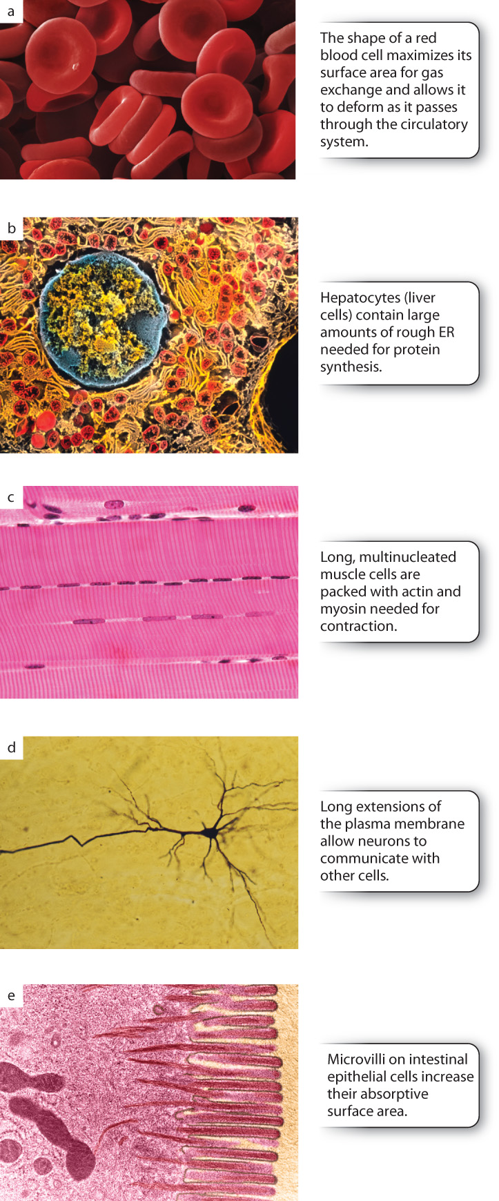
Tissues and organs have distinctive shapes that reflect how they work and what they do. In the same way, the different cell types that make up these organs have distinctive shapes based on what they do in the organ. In animals, the shape of cells is determined and maintained by structural protein networks in the cytoplasm called the cytoskeleton. The shape and structural integrity of tissues and organs depend on the ability of cells to adhere to one another. In turn, the adhesion of cells to one another depends on cell adhesion molecules and other cytosolic proteins that assemble into structures called cellular junctions. Equally important to a strong, properly shaped tissue or organ is the ability of cells to adhere to a meshwork of proteins and polysaccharides outside the cell called the extracellular matrix. In plants, the extracellular matrix provides both a mechanism for cells to adhere to one another and structural support that maintains their shape.
10.1.2 The structure of skin relates to its function.
To start our investigation of the cytoskeleton, cellular junctions, and the extracellular matrix, let’s consider a community of cells very familiar to all of us—our own skin (Fig. 10.2). The structure of mammalian skin is clearly tied to its function. Skin has two main layers. The outer layer, the epidermis, serves as a water-resistant, protective barrier. The layer beneath the epidermis is the dermis. This layer of the skin supports the epidermis, both physically and by supplying it with nutrients. It also provides a cushion surrounding the body.
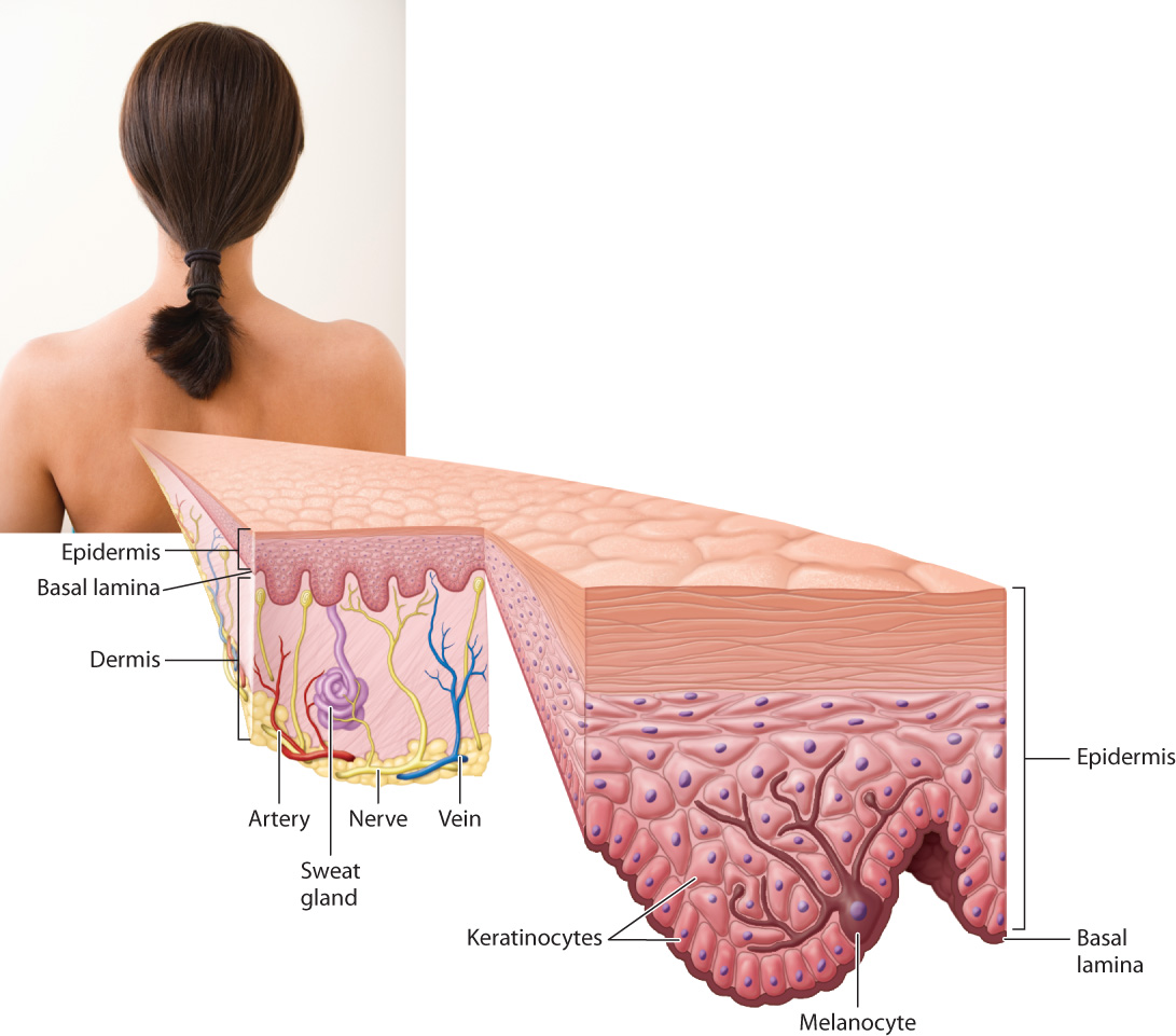
As you can see in Fig. 10.2, the epidermis is several cell layers thick. Cells arranged in one or more layers are called epithelial cells and together make up a type of animal tissue called epithelial tissue. Epithelial tissue covers the outside of the body and lines many internal structures, such as the digestive tract and vertebrate blood vessels. The epidermal layer of skin is primarily composed of epithelial cells called keratinocytes. The epidermis also contains melanocytes that produce the pigment that gives skin its coloration. Different kinds of epithelial cells are specialized to carry out different functions, such as protection, secretion, and absorption.
Keratinocytes in the epidermis are specialized to protect underlying tissues and organs. They are able to perform this function in part because of their elaborate system of cytoskeletal filaments. These filaments are often connected to the cellular junctions that hold adjacent keratinocytes together. Cellular junctions also connect the bottom layer of keratinocytes to a specialized form of extracellular matrix called the basal lamina (also called the basement membrane, although it is not in fact a membrane), which underlies and supports all epithelial tissues.
The dermis is made up mostly of connective tissue, a type of tissue characterized by few cells and substantial amounts of extracellular matrix. The dermis is strong and flexible because it is composed of tough protein fibers of the extracellular matrix. The dermis also has many blood vessels and nerve endings. The main type of cell in the dermis is the fibroblast. Fibroblasts synthesize the extracellular matrix and repair wounds.
We will come back to the skin several times in this chapter to show more precisely how these multiple connections among cytoskeleton, junctional complexes, and the extracellular matrix keep the skin a watertight and strong protective barrier.