41.2 EXCRETION OF WASTES IN RELATION TO ELECTROLYTE BALANCE
In most organisms, the regulation of water and solute levels is closely tied to waste excretion. Excretion is the elimination of waste products and toxic compounds from the body. Some of these wastes are generated as a by-product of metabolism, particularly protein and nucleic acid breakdown. Others were originally ingested, like the undigested remains of food, and need to be eliminated from the organism.
Most multicellular animals have evolved specialized excretory organs, such as the vertebrate kidney, to isolate, store, and eliminate toxic compounds and metabolic waste products. Because these compounds are eliminated as solutes that are dissolved and transported within the blood or other body fluids, their elimination from the body is intimately tied to water and electrolyte balance. The excretory organs of most animals therefore function to maintain water and electrolyte balance in addition to eliminating waste products.
Both the gastrointestinal tract (Chapter 40) and excretory organs like the vertebrate kidneys eliminate wastes from the body. However, the gastrointestinal tract is continuous with the outside world, and therefore its role in the elimination of wastes is quite different from that of excretory organs that separate toxic compounds from essential nutrients, electrolytes, and cells circulating in the blood.
41.2.1 The excretion of nitrogenous wastes is linked to an animal’s habitat and evolutionary history.

When proteins and nucleic acids are broken down by metabolism, one of the by-products is ammonia (NH3). Ammonia contains nitrogen and is toxic to organisms, so it is a form of nitrogenous waste (Fig. 41.7). By far, most ammonia is produced from the breakdown of proteins. Ammonia can disturb the pH balance of cells and cause neuronal cell injury. At high levels, it is lethal to organisms. It is either eliminated from the body or converted into a less toxic form. In either case, ammonia is kept at very low levels in the body.
Some organisms, such as fish, are able to excrete ammonia directly into the surrounding water, primarily through their gills. As a result, ammonia does not accumulate to toxic levels. This process is not energetically expensive, but it requires high volumes of water in the surrounding environment to dilute the highly toxic ammonia.
The movement of animals from water to land posed many challenges, among them the problem of waste elimination. No longer could animals rely on simple diffusion of ammonia into the surrounding water. Instead, organisms evolved biochemical pathways to convert ammonia to less toxic forms. One of these forms is urea (Fig. 41.7). Because urea is less toxic than ammonia, it can be stored in a fairly concentrated form before being excreted. Mammals (including humans), many amphibians, sharks, and some bony fish excrete nitrogenous waste in the form of urea. In mammals, urea is produced in the liver and is then carried by the blood to the kidneys, where it is eliminated. Urea is less toxic than ammonia but requires energy to produce it and water to eliminate it.
Ammonia can also be converted into uric acid (Fig. 41.7). Uric acid is the least toxic of the three forms of nitrogenous waste, so animals can store it at a higher concentration than urea. Birds and many reptiles, arthropods (including insects), and land snails excrete nitrogenous waste in the form of uric acid. These groups evolved the ability to convert ammonia to uric acid independently of one another as an adaptation to living on land, providing a good example of convergent evolution (Chapter 23). Uric acid precipitates from solution and forms a semisolid paste (familiar as white bird droppings). Because uric acid is not dissolved in water, it does not exert osmotic pressure and is eliminated with minimal water loss for the animal. This allows many reptiles and insects to live in extremely hot and dry environments. However, uric acid is energetically expensive to produce.
Many amphibians, like frogs, excrete ammonia early in their development, when they live in water, but then excrete urea when they move onto land after metamorphosis. Alligators and turtles also adjust what they excrete: ammonia when in the water and uric acid when on land.
Ammonia, urea, and uric acid are the three major forms of nitrogenous wastes excreted by animals. However, they are not the only forms. Animals excrete nitrogen in the forms of trimethylamine oxide, creatine, creatinine, and even amino acids. The form of nitrogenous waste that an animal excretes is ultimately linked to its environment and evolutionary history.
Quick Check 3
Why do mammals convert ammonia to urea rather than simply excreting it, as fish do?
41.2.2 Excretory organs work by filtration, reabsorption and secretion.
Excreting wastes does not occur by simply pumping the wastes out of the body. Instead, animals filter or secrete wastes out of the blood along with water, electrolytes, and even essential nutrients, but then reabsorb the nutrients they need, excreting the waste and enough water and electrolytes to maintain appropriate levels of water and electrolytes for hydration and osmotic balance. While this process may seem inefficient, it has several advantages compared to simply pumping wastes out. First, as we will see, it allows organisms to excrete wastes and at the same time balance their water and electrolyte levels. Second, it allows any toxic compound that ends up in the blood to be excreted. If organisms relied on molecule-specific active transport of wastes, novel toxic compounds ingested by organisms could not be excreted.
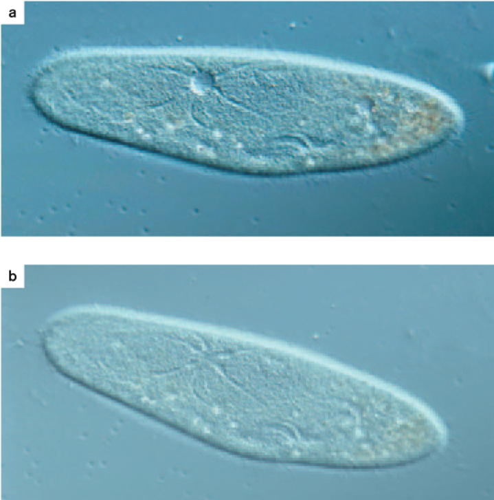
The first step is to isolate the waste into an extracellular space. In the case of unicellular organisms such as Paramecium and simple multicellular animals such as sponges and cnidarians (jellyfish and sea anemones), waste compounds are isolated in a cellular compartment called a contractile vacuole (Fig. 41.8). Fusion of the contractile vacuole with the cell membrane eliminates the waste contents by exocytosis from the cell (Chapter 5).
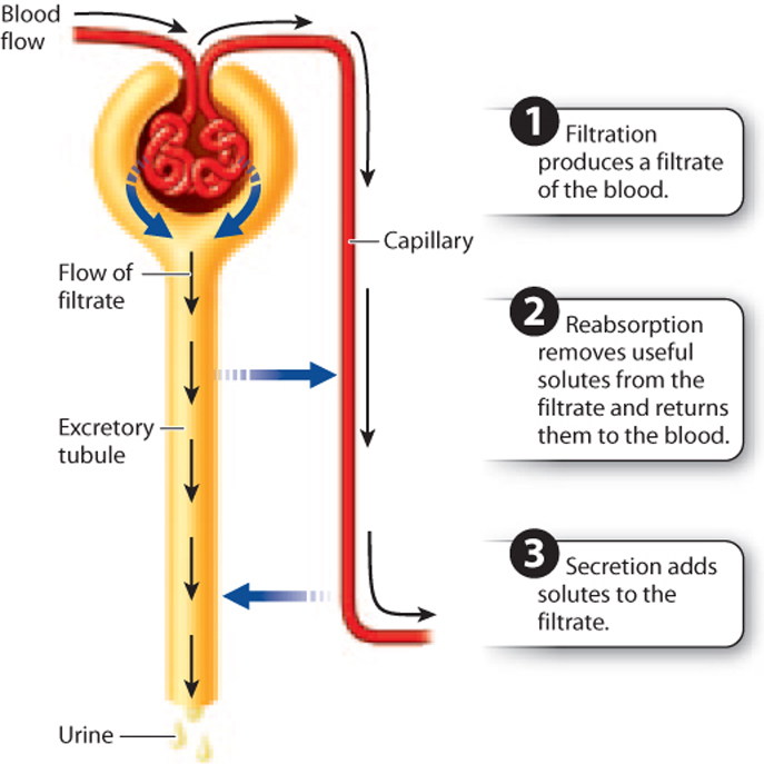
Multicellular animals with pressurized circulatory systems (annelid worms, mollusks, and vertebrates) also isolate wastes from the blood, but they do so by filtration (Fig. 41.9). Circulatory pressure pushes fluid containing the wastes through specialized filters into an extracellular space. The filtered fluid, called the filtrate, contains waste products along with water, electrolytes, and other solutes, which drain into excretory tubules that connect to the outside. The excretory filter, like a cell’s plasma membrane, is selectively permeable. It allows smaller ions and solutes to pass into the tubule lumen along with water but retains larger solutes such as proteins and cells.
Filtration is followed by reabsorption of key ions and solutes (Fig. 41.9). Reabsorption is an active process for some molecules and a passive one for others. In both cases, substances that are important for the animal to retain, such as electrolytes, amino acids, vitamins, and simple sugars, are taken up by cells of the excretory tubule and returned to the bloodstream. Water is reabsorbed into the cells by osmosis, driven by the osmotic pressure established by the active transport of solutes into the cell.
The final step in waste removal is the secretion of additional toxic compounds and excess ions (Fig. 41.9). Secretion is also an active process and involves eliminating substances that were not filtered from the blood earlier. Filtration, reabsorption, and secretion are the three steps that all animal excretory systems perform to eliminate toxic wastes, retain key electrolytes and solutes, and regulate water volume.
41.2.3 Animals have diverse excretory organs.
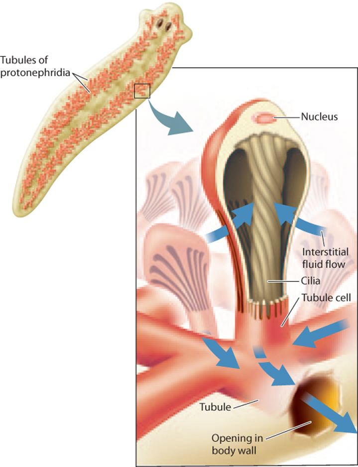
The processes of filtration, reabsorption, and secretion serve two important and complementary functions: They allow wastes to be isolated and subsequently removed from the body, and they allow the organism to adjust the amounts of water and electrolytes required to meet its osmoregulatory needs. These two functions are carried out by different excretory organs in different animals. For example, in flatworms, excretion involves isolating the fluid from the body cavity in excretory organs called protonephridia without filtering it first (Fig. 41.10). Cells with beating cilia move the fluid down the lumen of the tubule to an excretory pore. Freshwater flatworms, such as Planaria, have an extensive network of tubules that eliminate excess fluid along with waste products. As the fluid passes down the tubule, cells lining the tubule modify the fluid’s contents through reabsorption and secretion. The urine leaving a freshwater flatworm contains nitrogenous waste and is less concentrated (that is, more dilute) than its body to balance the water it ingests as it feeds.
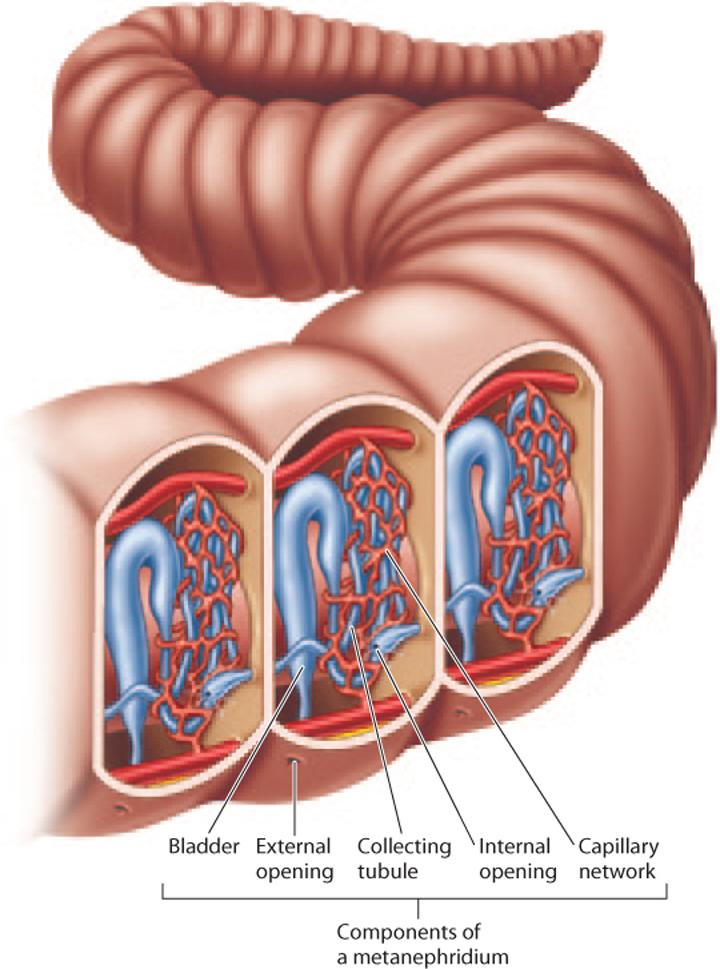
In segmented annelid worms such as earthworms, the body fluid is filtered through small capillaries into a pair of excretory organs called metanephridia within each body segment (Fig. 41.11). The fluid enters a metanephridium at a funnel-shaped opening surrounded by cilia that drains body fluid into a series of tubes in the next body segment and then out of the animal through an excretory pore. Blood vessels and cells surrounding the tubules reabsorb and secrete additional compounds as urine is formed. Like freshwater flatworms, earthworms reabsorb electrolytes and eliminate dilute urine containing dissolved nitrogenous wastes.
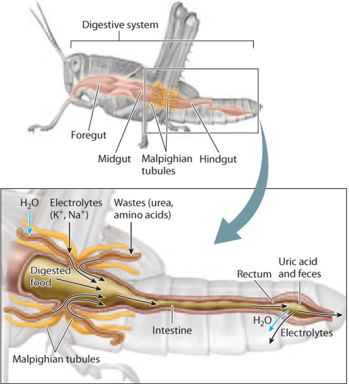
In insects and other terrestrial arthropods, fluid passes from the main body cavity into a series of tubes called Malpighian tubules that empty into the hindgut (Fig. 41.12). (These structures are named for Marcello Malpighi, the seventeenth-century Italian physician who first described them.) An insect can have two Malpighian tubules or more than 100. The tubules actively secrete uric acid and excess electrolytes from the animal’s tissues while retaining key solutes. Muscle fibers in the walls of the tubules help push the fluid to the hindgut. In the hindgut, the fluid becomes more acidic because of the buildup of uric acid, causing uric acid to precipitate from solution when it enters the rectum. Because uric acid is removed from the fluid, it does not influence the movement of water. Water is therefore free to diffuse into the tissues with the reabsorbed electrolytes that are transported from the rectum before the uric acid and remaining dried digestive wastes are eliminated. Malpighian tubules represent one of the most successful adaptations for excreting nitrogenous wastes with minimal water loss, enabling insects to live in diverse and arid terrestrial environments.
41.2.4 Vertebrates filter blood under pressure through paired kidneys.
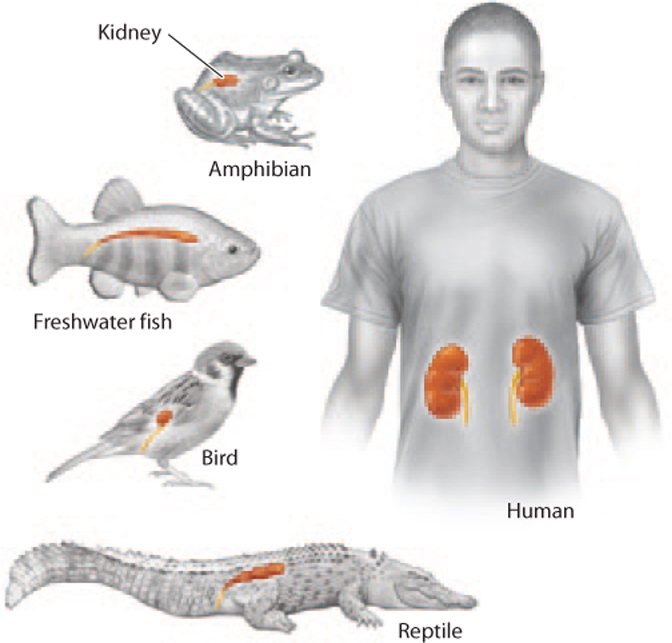
The excretory organs of vertebrates are the kidneys. Like all excretory organs, they not only serve to eliminate wastes, but also play an important role in water and electrolyte balance. Vertebrate kidneys also reflect the evolutionary transition of vertebrates from an aquatic to a terrestrial environment, where there is a need to minimize water loss. Mammals and birds, for example, can excrete urine that is more concentrated than their blood and tissues.
The kidneys lie posterior to the abdominal cavity. Whereas the kidneys of humans and other mammals are bean-shaped, the kidneys of birds and other vertebrate animals are elongated and segmented (Fig. 41.13). Paired vertebrate kidneys receive blood supply from the heart by renal arteries that branch from the abdominal aorta.
Vertebrates filter their blood through specialized capillaries that have openings in their walls. These porous capillaries form a tufted loop called a glomerulus (Fig. 41.14). There are thousands to millions of glomeruli in each kidney. Driven by circulatory pressure, nitrogenous wastes, electrolytes, other small solutes, and water move through small holes in the capillary walls into an extracellular space surrounded by a capsule. The filtrate then moves through a series of renal tubules, which further process the filtrate by reabsorption and secretion before it enters the collecting ducts as urine. The collecting ducts converge on a larger tube called the ureter, which brings urine from the kidneys to a hollow organ called the bladder in mammals and fishes, or the cloaca in the case of amphibians, reptiles, and birds, for storage and elimination from the body.
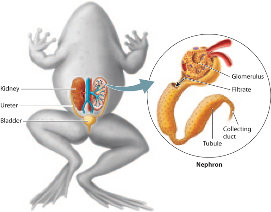
The glomerulus, capsule, renal tubules, and collecting ducts make up a nephron, the functional unit of the kidney. Nephrons perform the three basic steps of excretion and osmoregulation: filtration of blood passing through the glomerulus, reabsorption from the renal tubule back to the bloodstream of key electrolytes and solutes, and secretion of additional wastes by the renal tubules.
In freshwater and saltwater fish, the kidneys run along the length of the body, with nephrons arranged segmentally. Because fish excrete nitrogenous waste in the form of ammonia through their gills, the kidney is primarily an organ of water and electrolyte balance rather than one of waste elimination. In saltwater fish, the renal tubules are readily permeable to water, allowing water to diffuse into and out of the tubule passively as the cells of the tubules actively reabsorb electrolytes and other valuable solutes. However, no net water uptake occurs. Consequently, the urine produced has the same concentration as the fish’s body fluids. By contrast, the renal tubules in freshwater fish are relatively impermeable to water while allowing reabsorption of valuable electrolytes, sugars, and amino acids. This enables freshwater fish to excrete very dilute urine, eliminating the excess water that continually enters through their gills.
The kidneys of amphibians are similar to those of freshwater fish, reflecting their shared evolutionary history. Like freshwater fish, amphibians take in excess water across their gills and skin as they breathe. Amphibian kidneys therefore produce extremely dilute urine, enabling them to maintain an electrolyte concentration in their body that exceeds that of their freshwater environment.
Reptiles are found in a variety of habitats, from oceans to deserts, and are well adapted to their environments. Marine iguanas, for example, ingest salt water with their food, and so rely on specialized salt glands to eliminate excess electrolytes from their body. Terrestrial reptiles living in desert regions face the challenge of water loss and limited water availability in their environment. Like saltwater fish, these animals produce urine with the same salt content as the rest of their bodies after reabsorbing needed compounds in their bodies and secreting toxic wastes not filtered by their kidneys. However, because reptiles excrete nitrogenous waste in the form of uric acid, they are able to reabsorb most of the water from the urine after it drains into their cloaca. This process allows many reptiles to inhabit extremely dry environments.
Mammals also live on land, but excrete most of their nitrogenous waste in the form of urea dissolved in water. Their kidneys are well adapted for excreting waste and balancing their water and electrolyte levels, as we discuss now.