The experiment described below explores the same concepts as the one described in Fig. 47.8. Read the description of the experiment and answer the questions below the description to practice interpreting data and understanding experimental design.
Mirror Experiment activities practice skills described in the brief Experiment and Data Analysis Primers, which can be found by clicking on the “Resources” button on the upper right of your LaunchPad homepage. Certain questions in this activity draw on concepts described in the Experimental Design primer. Click on the “Key Terms” buttons to see definitions of terms used in the question, and click on the “Primer Section” button to pull up a relevant section from the primer.
Background
You may be familiar with leaf-cutter ants, which cut pieces from leaves and use these fragments as bases on which they “farm” certain fungal species. This is an example of a mutualism: the ants are provided with food in the form of the fungus, and the fungus receives nutrients (derived from leaf pieces) in the ant nest. Leaf-cutter ants are actually one species of a much larger group of ants that grow fungus - the Attine ants. Fungus-ant interactions are well-studied examples of coevolution, but do Attine ants share mutualisms with other (non-fungal) species? Is it possible for an organism to coevolve with multiple species depending on selective pressures?
Hypothesis
The fungus grown by Attine ants can be infected by parasitic Escovopsis fungus. Much like a farmer’s wheat crop can be ruined by the presence of rust fungi (Chapter 34), so too can infection by Escovopsis ruin the fungal “crops” of Attine ants. Matías Cafaro and his colleagues (along with other research groups) noticed that many species of Attine ants have visible bacterial colonies growing on their exoskeletons. Since certain types of gram-positive bacteria (such as Streptomycetes) can synthesize antifungal compounds, Cafaro et al. hypothesized that Attine ants are in a mutualistic relationship with bacteria that can produce compounds toxic to Escovopsis. Researchers also predicted that the phylogenetic trees of Attine ants and their bacteria would be similar, indicating that these organisms coevolved.
Experiment
Cafaro and colleagues collected different species of Attine ants and isolated bacteria from their exoskeletons. Escovopsis fungus was introduced onto petri dishes containing colonies of ant-derived bacteria, and researchers evaluated whether Escovopsis could grow on these dishes. Scientists also sequenced two genes in Attine ant bacteria: an rRNA-encoding gene and the tuf gene (involved in translation). They used these sequences to identify the species of bacteria carried by Attine ants, and create a bacterial phylogenetic tree (Figure 1). The phylogenetic trees of Attine ants (determined by previous studies of these ants) and their associated bacteria were then compared.
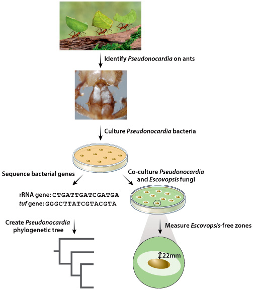
Photo credits: Gail Shumway/Getty Images, Carafo, M.J., et al. 2011. Specificity in the symbiotic association between fungus-growing ants and protective Pseudonocardia bacteria. Proc Biol Sci. 278: 1814-22.
Results
Cafaro and colleagues determined that most Attine ants carry Pseudonocardia bacteria, which belong to the same bacterial order as Streptomycetes (such as, Streptomyces). When researchers compared their Pseudonocardia phylogenetic tree to that of Attine ants, they noted that these two trees appeared very similar; certain groups of Pseudonocardia bacteria always appear to associate with certain groups of Attine ants. Furthermore, when Escovopsis fungus was plated on petri dishes containing Attine ant-derived Pseudonocardia, “Escovopsis-free” regions were observed surrounding bacterial colonies. This observation suggested that Pseudonocardia bacteria could produce antifungal compounds. Collectively, these results indicated that a mutualism exists between Attine ants and Pseudonocardia bacteria, and that these organisms coevolved.
Source
Currie, C. R., et al., 2006. Coevolved crypts and exocrine glands support mutualistic bacteria in fungus-growing ants. Science. 311, 81-3.
Cafaro, M. J., et al., 2011. Specificity in the symbiotic association between fungus-growing ants and protective Pseudonocardia bacteria. Proc Biol Sci. 278, 1814-22.
Experimental Design
Testing Hypotheses: Variables
When performing experiments, researchers manipulate the test group differently from the control groups. This difference is known as a variable. There are two types of variables. An independent variable is the manipulation performed on the test group by the researchers. It is considered “independent” because the researchers could choose any variable they wish. The dependent variable is the effect that is being measured. It is considered “dependent” because the expectation is that it depends on the variable that was changed. In our example of the headache medicine, the independent variable is the type of medicine (new medicine, no medicine, placebo, or medicine known to be effective). The dependent variable is the presence or absence of headache following treatment.
In designing experiments, there is an additional issue to consider: the size of each of our groups. In order to draw conclusions from our data, we need to make sure that our results are valid and reproducible, and not merely the result of chance. One way to minimize the effect of chance is to include a large number of patients in each group. How many? The sample size is the number of independent data points and is determined based on probability and statistics, the subject of the next primer.
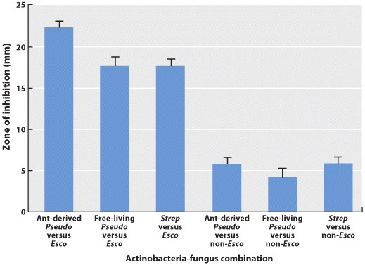
During the course of their experiments, Cafaro and colleagues cultured different combinations of bacteria and fungi on the same petri dishes (that is, they performed co-cultures). Researchers actually used three different types of bacteria for this work: free-living Pseudonocardia bacteria that can be found in soil and which do not require a host to survive; Pseudonocardia bacteria derived from Attine ants; andStreptomyces bacteria, which are also found in soil and are known to produce antifungal compounds. Each type of bacteria was cultured with either Escovopsisfungus or other fungal species, which researchers classified as non-Escovopsis. Cafaro et al. then determined if bacterial colonies were surrounded by fungus-free zones, called zones of inhibition. The larger the size of the inhibition zone, the more effective that bacteria are at killing a particular fungus. Cafaro and colleagues collected the following inhibition zone data. Which of the following statements is true of these data?
Positive Control | A group in which a variable is introduced that has a known effect to be sure that the experiment is working properly. |
Experimental Design
Testing Hypotheses: Controls
Hypotheses can be tested in various ways. One way is through additional observations. There are a large number of endemic species on the Galápagos Islands. We might ask why and hypothesize that it has something to do with the location of the islands relative to the mainland. To test our hypothesis, we might make additional observations. We could count the number of endemic species on many different islands, calculate the size of each of these islands, and measure the distance from the nearest mainland. From these observations, we can understand the conditions that lead to endemic species on islands.
Hypotheses can also be tested through controlled experiments. In a controlled experiment, several different groups are tested simultaneously, keeping as many variables the same among them. In one group, a single variable is changed, allowing the researcher to see if that variable has an effect on the results of the experiment. This is called the test group. In another group, the variable is not changed and no effect is expected. This group is called the negative control. Finally, in a third group, a variable is introduced that has a known effect to be sure that the experiment is working properly. This group is called the positive control.
Controls such as negative and positive control groups are operations or observations that are set up in such a way that the researcher knows in advance what result should be expected if everything in the study is working properly. Controls are performed at the same time and under the same conditions as an experiment to verify the reliability of the components of the experiment, the methods, and analysis.
For example, going back to our example of a new medicine that might be effective against headaches, you could design an experiment in which there are three groups of patients—one group receives the medicine (the test group), one group receives no medicine (the negative control group), and one group receives a medicine that is already known to be effective against headaches (the positive control group). All of the other variables, such as age, gender, and socioeconomic background, would be similar among the three groups.
These three groups help the researchers to make sense of the data. Imagine for a moment that there was just the test group with no control groups, and the headaches went away after treatment. You might conclude that the medicine alleviates headaches. But perhaps the headaches just went away on their own. The negative control group helps you to see what would happen without the medicine so you can determine which effects in the test group are due solely to the medicine.
In some cases, researchers control not just for the medicine (one group receives medicine and one does not), but also for the act of giving a medicine. In this case, one negative control involves giving no medicine, and another involves giving a placebo, which is a sugar pill with no physiological effect. In this way, the researchers control for the potential variable of taking medication. In general, for a controlled experiment, it is important to be sure that there is only one difference between the test and control groups.
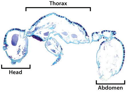
The ant depicted in Figure 1 is actually a leaf-cutter ant flipped on its back; the white dots are Pseudonocardia bacterial colonies localized to the underbelly of this insect. One researcher, Cameron Currie (in whose laboratory Matías Cafaro later carried out his work), and his colleagues decided to rigorously explore the localization of Pseudonocardia in Attine ants. You may recall from Chapter 26 that gram-positive bacteria like Pseudonocardia have thick peptidoglycan cell walls, which can be stained by certain dyes. Currie and colleagues took thin tissue sections from Cyphomyrmex Attine ants (which are not leaf-cutter ants but members of a related genus) and stained them with methylene blue, a dye that is used to visualize gram-positive bacteria and stains these bacteria very dark blue. Figure 2 below shows the staining pattern they observed (note that here, the ant exoskeleton appears light blue).
Photo credit: Cruuie, C.R., et al., SCIENCE 311:81 (2006). Coevolved crypts and exocrine glands support mutualistic bacteria in fungus-growing ants. Reprinted with permission.
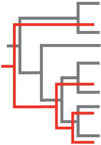
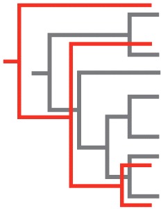
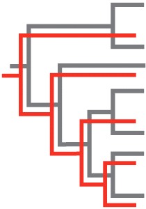
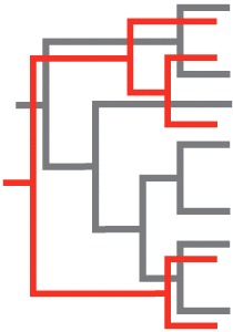
EACc/TnptU3OLetxtD+6ibu2IMEMMb34tuskN2csC5u/t5qiHt3iOm+0w3nFhzfj4savbtfiakM5VJ9Zxe+oJAj/zHAW84TlmvShOiLZHbk5i9kVwCQel0NebwcetrXQ98vWPJeTJA4I8az577fxTJYHNnxEk+/J+Gw+mpDRaJocnnKGmfHEkI+eXa36QpyPcu48NzTuRBZHi+4ynSuOx+YuR1X9JfOfHNeuAi5a9yI2eeIicRARdkktYfKStaguuScoSRklpWPdP65DJ+8xdrwuESKo8/vMV8VZrQovyh6f3tRCPn7FILmCC37HTvyhvTnNABUeEgAHntSh6PrRFoTOa64l91dx+uPaTF4Ea962Wx9kULomo3LXAZPjaPR8CltMSEHW+VUGqfADtHuid9DcCPFdWybADj5kfszCufoz1ai9y+PS/8WrC+khPGW8ss+ZAzq8LlBq2wpfldST9vselMvOSZ3ovn1fN9y8guWJZl4i/19ZiHDbYwvh3E4igIvsr+12qMqMTkdP4AZC0SBzVYd1xY4V6kALQyHEUDZLusTcV+JXzQ3dXrgsCu+gkLfB1cl7gRmFNLThue7lBUNwQjqj9Cy6txDvJwX0NFIEz8/W5bS+I9zf+4gpvUHJtCVpisMg3OLmN3noH8vxzLhZHuG+mwuBUu0hwsZkYbxe43bePRQ+LNCZhWgKecVtZt3F9MrTbigK95ZBLkvUyeHXfKDVk6Kd+G4nvXJjcn34ZBmL2SDlI6KR1zvPnNGxc/F55JC4GNPRq3A+PN8xln+NMzQrZ6xMCNJsN5j9eQrzkxZ9gDbDbvxWvcSXkhfCLO0m50Iq5Q5UL9KFqDHuCLrdoy3A3TDumtzezPwzuFj8Vz9sih7i+/zBF8mvfq8n7k7E64MMqUoXARgrAO4WoZXbm+R1Ge8zrqyX2hWlI8wI8+He+NjEtzErNoTjB2PTAOvp98XgVPSvmxxQPBbS0Lww1L6mKF23HrezoQj6Ith/jMKzHmd5nHHIUbG+3TPcgAQvR1vS5Mc5nYiTRh1ZVUjtlHmkSJT/VpfdC64eWTFQ2wPM61GnEFn3ldfHpS7dmF9TXLgJzWZDU/en4/K6ldCXTcA2DS1eMr2iYIRhhYkHGtrEC2X6G3jeP+h7u50UEX/H7FL48aj0GJ21T0FfFRWMXph9HCE6UZ0tJtIh7vkAqz6qA8Zp9ys0x25kiW5uFa8MHGExA/I+XUNGjlW+B9EthsHB8xHbCQGE6RDQHZsEDxPuZp9y1hL13ZENLO4cIvrDQuaXk+HOwvdn4qA/YV2T0EoWtT083CtZkxY2waVZtDngv8huI1AFQbv4tOgUpSZ8AKslxb9Zi6gEGp/wURtH8ykQVbYNQ5YDA8oqRe10z7rBUJXfst6zGt7eYbXGFlNpc3/TgQnSiGcqVpgm9KLxZOA4xSqMAWq/gf2yIG6OaGZ0STFvXCb6WNC5QayE8UPueWOxJ3ZJsXzGRm2Rw7WW8QjnhU2cgRIaghLI0OE/Ql0zr5CpcQaFX2lC/UBGAVvzQXSbACDO3qQPboC4rnoOpHIFnNugR/nzkTV/CKu1CPQd06H4mUpUHtvuaVSOHnqDmXS2YJm1LgC9X348z5MzJXHrvP6Wi4iKLe6fbA1E6Psiy0THC/x78h8MVRB4nxFwVOCPho86+RmeXumJOUq5/Tw6hodlbu343lSyn4YuXob7p7FGduD569ztek9HUYmix6xtxFEK+lEogDC/Y0xVvVWKC7Gcar7o9ITxD+d/zMRUmnPIw4PlI9gOK5mZr2Ora7Tq+28XKwVQfq5O1kIrD5T2nxs8eAFWzqt/e8Yzf3p2rqMtsDbNBK0PheITvmEG1yjA9dSJ3vVbz80IuZbJUShWfvVbFbZqhneEfbY521PVTCOif+l5sbk=During the course of their experiments, Cafaro et al. and Currie et al. speculated that Attine ants and Pseudonocardia bacteria coevolved. If these organisms evolved alongside one another, which of the following pairs of phylogenetic trees best represent those of Attine ants and Pseudonocardia bacteria? Assume that the grey tree represents Attine ants, and the red tree represents Pseudonocardia bacteria (and that these trees have been overlain).