Chapter 2. Molecular Biology II
General Purpose
This pre-lab will present some of the general concepts related to some of the techniques used in the fields of microbiology and molecular biology and how these techniques relate to understanding the relationship between DNA, genes, and traits expressed by an organism.
Learning Objectives
General Purpose
- Develop a basic understanding of some of the techniques used in the field of molecular biology.
Procedural
- Gain proficiency interpreting fragment patterns in gels.
Background Information
There are two related parts of interpreting the fragment patterns produced by gel electrophoresis. The first part is determining the sizes of the fragments in base pairs (bp). This is done by comparing the relative positions of the fragment with fragments of known size.
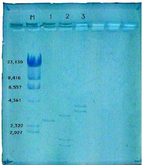
The three different plasmids in your experiment were all digested with the restriction enzyme EcoRI and the fragments were separated by electrophoresis using an agarose gel. In Figure 12-1 is a picture of a gel from a similar type of experiment.

In lane M are the molecular weight markers which are generated by digesting bacteriophage lambda DNA with the restriction enzyme HindIII. The numbers listed beside the fragments are the sizes in base pairs (bp). From the gel you determine the number of fragments and estimate the sizes of the fragments.
The second part of the analysis is determining the number of cuts in the plasmid. The assumption is that all of the fragments are shown by the gel. If that is true, then the number of cut sites in the plasmid is equal to the number of fragments in that sample. This is often a place of some misconception; however, this can be easily visualized by imaging a circular piece of string. If you cut it once there is one length of string, whereas if you cut it twice then there will be two lengths of string.
Pre-Lab Quiz
Proceed to the Pre-Lab Quiz