Chapter 1.
Nervous Tissue
Neuroglial Cells
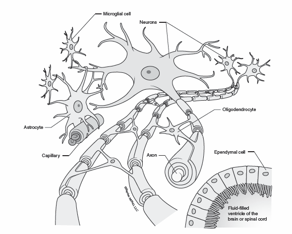
Neuroglial cells are small cells of the nervous system that function to support, nourish, and protect neurons. Astrocytes are star shaped cells found in the central nervous system. They form an important part of the blood-brain barrier and help to pass materials back and forth between capillaries and neurons. Ependymal cells are found lining the ventricles of the brain and central canal of the spinal cord. They help to circulate cerebrospinal fluid and regulate the chemical composition of tissue fluid in the central nervous system. Oligodendrocytes and Schwann cells form myelin sheaths around the axons of neurons. Oligodendrocytes do this for cells in the central nervous system (CNS), while Schwann cells are found in the peripheral nervous system (PNS). Myelin sheaths help to electrically insulate the axons and allow axons to conduct electrical impulses faster.
Neuron
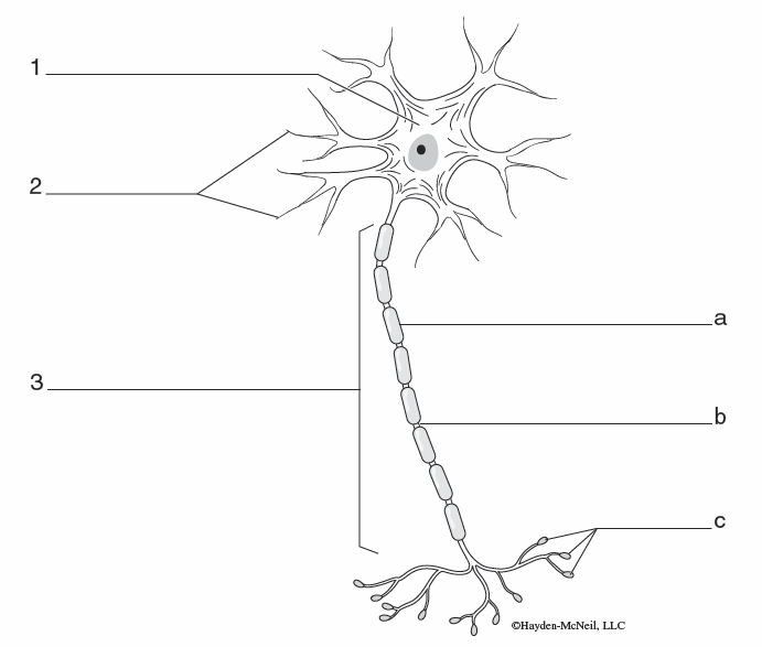
Neurons are responsible for sensing changes inside and outside of the body, processing information, and stimulating responses. Their structure is specialized for collecting and sending impulses. The cell body of a neuron contains the nucleus and most of the cytoplasm of the cell. It also contains many mitochondria for carrying out aerobic metabolism to provide the neuron with energy. Dendrites respond to stimuli in their environment and carry signals toward the cell body. The axon carries electrical impulses away from the cell body toward the next cell. Axons in the peripheral nervous system may be surrounded by myelin sheaths produced by Schwann cells. There are small gaps between adjacent Schwann cells along the length of the axon called nodes of Ranvier. Ions move in and out of the axon at the nodes of Ranvier when the axon is conducting an electrical impulse in a process called saltatory conduction. The axon ends at the axon terminal where neurotransmitters are released to influence the next cell. Each axon has multiple axon terminals to impact many cells simultaneously.
Neuron Types
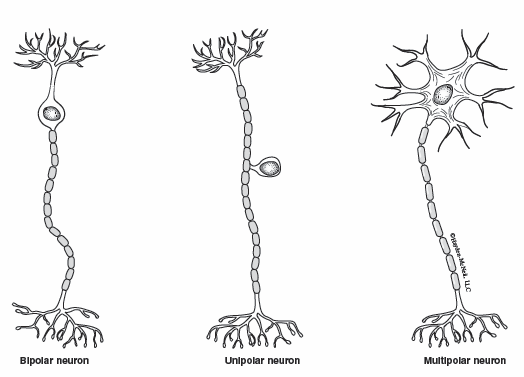
Neurons can be classified by their general structure and how many processes extend from the cell body. Bipolar neurons are neurons with one dendrite and one axon extending from the cell body. Neurons found in the eye and olfactory areas are bipolar neurons. Unipolar neurons have only one process, an axon, extending from the cell body. Many sensory neurons responsible for monitoring skin sensations and muscle length are unipolar. Multipolar neurons have one axon and more than one dendrite extending from the cell body. Motor neurons controlling activity of other tissues are typically multipolar. There are two types of motor neurons. Those that influence skeletal muscle tissue are under voluntary control and called somatic motor neurons. Autonomic motor neurons are controlled involuntarily, and stimulate smooth muscle, cardiac muscle, and glands. Most of the neurons in the CNS are multipolar. Neurons in the CNS are called interneurons and are responsible for processing information and creating perceptions.
Neural Activity
Resting Membrane Potential
When a neuron is at rest, there is an unequal distribution of ions on the outside of its membrane compared to the inside. The inside of the neuron has a higher concentration of potassium ions (K+), and a lower concentration of sodium ions (Na+) than the extracellular fluid. There are sodium/potassium pumps on the membrane of the neuron that actively shuttle sodium ions outside of the neuron and potassium inside in order to maintain this difference in ion distribution. The difference in ion concentrations across the membrane of a neuron allows the neuron to generate a negative membrane potential when at rest as K+ constantly leaves.
Electrical Impulse
The electrical impulse that can be measured when a neuron “fires” is a switch in the membrane potential of the neuron. When at rest, the neuron has a negative membrane potential. When it sends a signal, the charge along the membrane of the axon switches to a positive charge and then back to negative. This change in charge is a result of the movement of sodium and potassium ions across the membrane of the neuron. The first part of the electrical impulse is the switch from negative to positive charge. This is called depolarization, and is a result of the rapid movement of sodium ions into the neuron. The second step of the electrical impulse returns the neuron to a negative charge. This stage, called repolarization, is caused by the rapid diffusion of potassium ions out of the neuron.
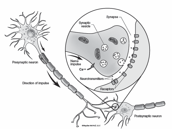
Chemical Synapse
When the electrical impulse reaches the axon terminal, the neuron releases chemical messengers known as neurotransmitters. The neurotransmitter diffuses across a small space between the neuron that released it (presynaptic neuron), and the next neuron (postsynaptic neuron). This space is called the synaptic cleft. Once it crosses the synaptic cleft, the neurotransmitter will bind to receptors on the postsynaptic neuron. The binding of neurotransmitter to receptors on the postsynaptic neuron influences the membrane potential of the postsynaptic neuron. Changes in the membrane potential of the postsynaptic neuron that make it more likely to send its own electrical impulse are excitatory; changes in the membrane potential that make it less likely to send its own signal are inhibitory. Several important neurotransmitters and their characteristics are listed in the table below.


Nervous System Organization
Peripheral Nervous System (PNS)
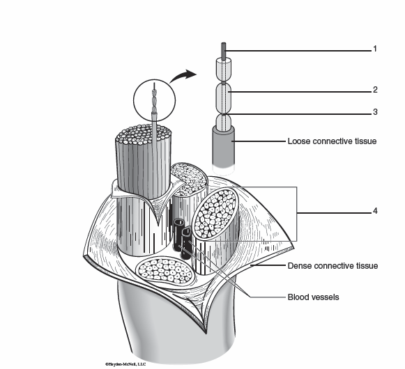
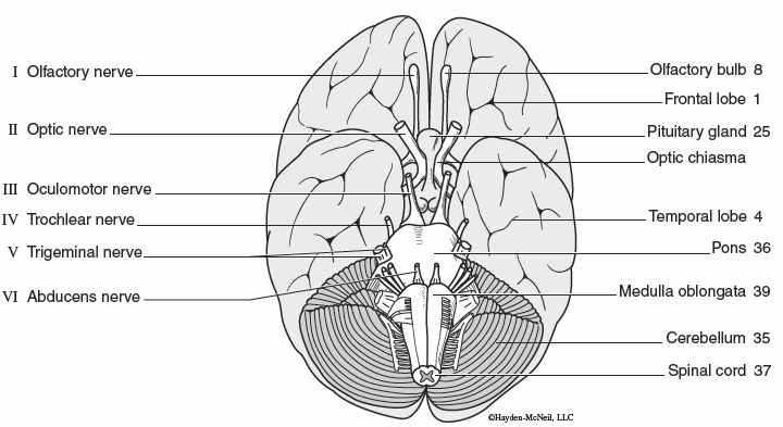
The peripheral nervous system consists of all of the sensory and motor neurons that carry information to and from the brain and spinal cord. A nerve is a collection of axons from many neurons that are bundled together by layers of connective tissue that carry information between the same body tissues and the brain or spinal cord. There are twelve pairs of cranial nerves that connect body tissues directly with the brain. The characteristics of the twelve cranial nerves are listed in the table below. There are 31 pairs of spinal nerves that connect body tissues with the spinal cord. Ganglia are collections of the cell body of neurons whose axons travel through the peripheral nerves.
Central Nervous System (CNS)
The central nervous system consists of the brain and spinal cord. The main function of the CNS is the integration of sensory information, and initiation of motor impulses. The cerebrum is the largest part of the brain and is responsible for memories, thinking, problem solving, sensations, and initiating skeletal muscle movements. The cerebellum aids in coordinating muscle movements. The thalamus plays an important role in sorting sensory information to the correct part of the brain. The hypothalamus is an important regulator of homeostasis. The pineal gland produces melatonin which influences the body’s biological clock. The brain stem connects the brain with the spinal cord and plays important roles in regulating breathing, heart rate, and blood pressure. The spinal cord travels through the vertebral canal, controls spinal reflexes, and connects the brain with much of the peripheral nervous system.
Cranial Nerves (12 pairs)
The cranial nerves are nerves that connect directly to the brain. The table below summarizes the major functions of the cranial nerves.

Spinal Nerves and Nerve Plexus
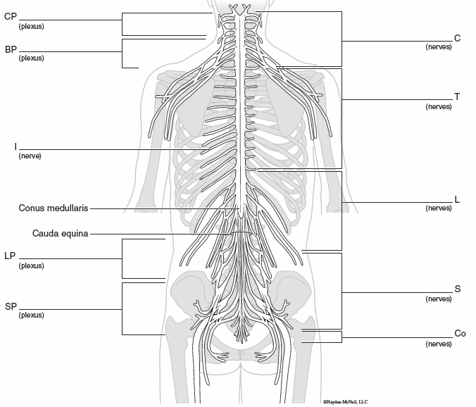
There are eight pairs of cervical spinal nerves that connect with the spinal cord between the cervical vertebrae. The cervical plexus is formed from parts of the first four cervical nerves. Nerves in this area transmit sensory impulses from the skin of the neck and back of the head. The phrenic nerve originates here and stimulates the diaphragm for breathing. The brachial plexus is formed from parts of the fifth through eighth cervical nerves, and the first thoracic nerve. The brachial plexus carries sensory information from skin of the arm, and motor impulses to the muscles of the arm. There are twelve pairs of thoracic spinal nerves that connect with the spinal column between the thoracic vertebrae. Generally, thoracic spinal nerves do not form a nerve plexus. Instead they travel along the ribs as intercostal nerves to the muscles and skin of the chest and abdominal wall. Lumbar spinal nerves connect with the spinal cord through openings between the lumbar vertebrae. The lumbar plexus is formed from parts of the first four lumbar nerves. Nerves from the lumbar plexus innervate muscles of the abdominal wall and thigh. The five sacral spinal nerves travel through the sacral foramen to connect with the spinal cord. The sacral plexus is formed from branches of the fourth lumbar nerve through the fourth sacral nerve. Nerves from this plexus travel all the way down the leg and also innervate the pelvic structures and perineum. There is one pair of coccygeal nerves that connect with the spinal cord near the coccyx.
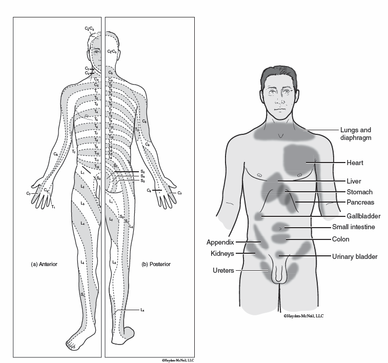
Dermatomes are areas of the skin that are innervated by a single spinal nerve. Physicians can use dermatomes to determine where damage to the spinal cord occurred based upon which regions of the skin are impacted. This is particularly useful when nerves that go out to the upper or lower limbs are involved as there is very little overlap in the innervation of dermatomes in the limbs compared to the torso region.
Phantom pain is the feeling of pain coming from a limb or portion of the limb that is absent. Referred pain occurs when pain is felt in a body part other than the one that is actually being stimulated. It is caused because pain signals from your organs or viscera often travel along the same pathways as somatic pain. Our perception of the visceral pain signals can cause us to feel like the pain was coming from the associated body region. A good example of this is the pain that is often associated with a heart attack. Impulses from the heart travel along the same spinal segments as the left chest and arm. The brain may interpret pain signals from the heart as coming from the left chest or medial side of the left arm.
Peripheral Nerves
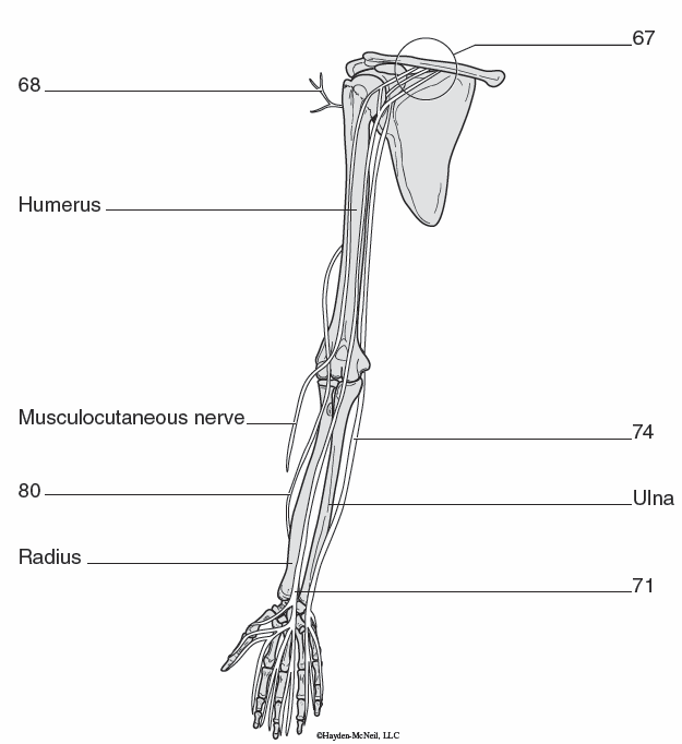
The brachial plexus is formed from parts of the fifth through eighth cervical nerves, and the first thoracic nerve. The brachial plexus carries sensory information from skin of the arm, and motor impulses to the muscles of the arm. The axillary nerve stimulates the deltoid muscle. The musculocutaneous nerve stimulates the biceps brachii. The ulnar nerve stimulates pronator teres. The median nerve stimulates muscles of the hand and the triceps brachii. The radial nerve stimulates the supinator and brachioradialis.
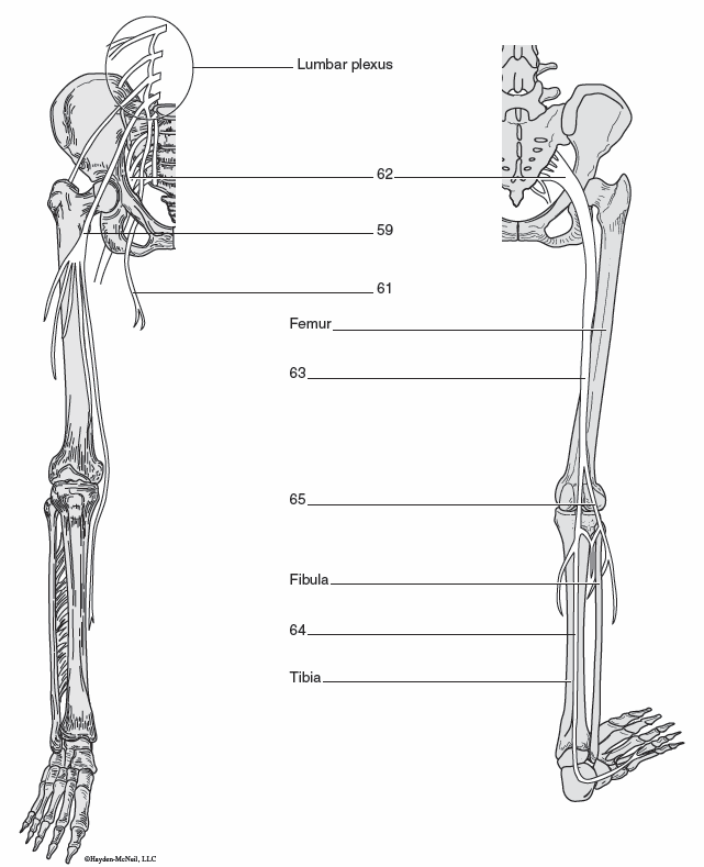
The lumbar plexus is formed from parts of the first four lumbar nerves, and gives rise to two important nerves. The femoral nerve stimulates the quadriceps femoris muscles, and the obturator nerve stimulates the medial muscles of the thigh such as gracilis.
The sacral plexus is formed from branches of the fourth lumbar nerve through the fourth sacral nerves. The sciatic nerve originates from the sacral plexus. It is the thickest and longest nerve in the body, and stimulates the hamstrings among other muscles. The common fibular nerve extends from the sciatic nerve anteriorly to stimulate the tibialis anterior muscle. The tibial nerve continues from the sciatic nerve to stimulate calf muscles such as gastrocnemius.
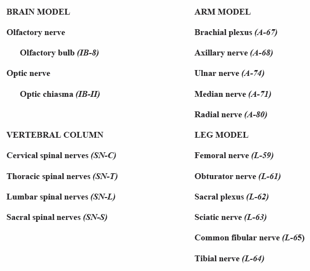
Exercise: Peripheral Nervous System Identification
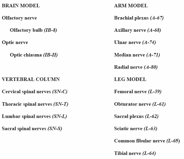
Examine the vertebral column, arm, and leg models for the nerves and nerve plexus listed in the Peripheral Nervous System Review (page 113).
Spinal Cord
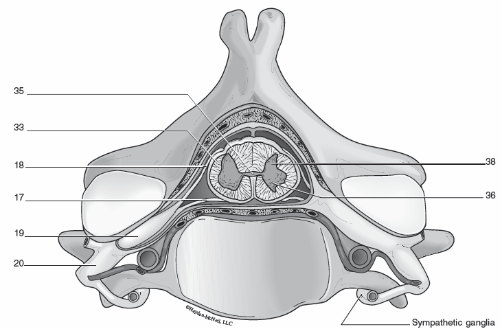
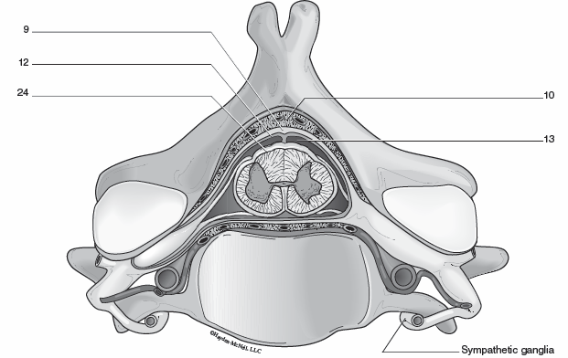
The spinal cord is a cylindrical mass of nervous tissue that extends from the brain stem through the vertebral canal to the second lumbar vertebrae. It is organized into regions of white matter surrounding central portions of grey matter. The white matter is made up of axons that allow for communication between different regions of the spinal cord and the brain. Ascending tracts carry sensory information toward the brain, while descending tracts carry motor impulses from the brain.
The gray matter is a butterfly shaped region of neural cell bodies located in the center of the spinal cord. Dorsal horns are the projections of the gray matter that extend towards the back. The dorsal horns contain mainly interneurons. The dorsal root ganglion is a cluster of the cell bodies of sensory neurons from the spinal nerves of the peripheral nervous system. Axons from these neurons form the dorsal root to meet the spinal cord. Axons in the dorsal root carry sensory impulses into the spinal cord. The ventral horn contains the cell bodies of motor neurons. The axons from these neurons exit the spinal cord forming the ventral roots of the spinal nerves. The central canal is a small cavity filled with cerebrospinal fluid located in the center of the spinal cord. The spinal cord extends from the brain stem through the vertebral canal. It ends in a cone shaped structure around the second lumbar vertebra. Nerves continue inferiorly through the lower lumbar vertebrae and sacrum forming the cauda equina.
Exercise: Spinal Cord Idenitification
Examine the spinal cord model for the structures listed in the Central Nervous System Review (page 114).
Reflex Activity
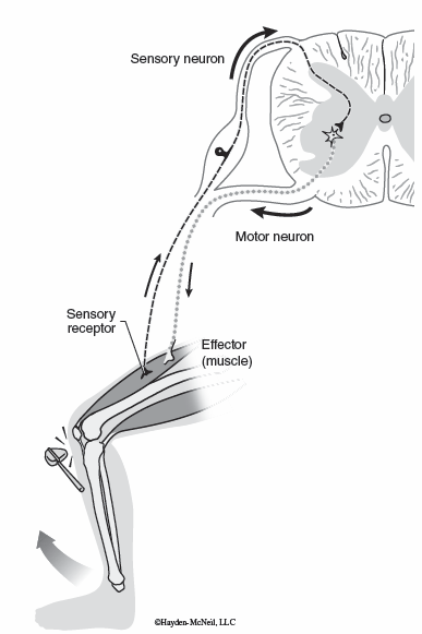
A reflex is an automatic, involuntary response to a stimulus. These rapid and predictable responses help to protect us from harm, as in the case of the withdrawal reflex. The withdrawal reflex causes a rapid and forceful movement of a body part away from a painful stimulus, such as moving your hand quickly away after touching a hot stove. The quick response of the reflex removes your finger before any further damage can occur even before you become consciously aware that you have touched the stove. Other reflexes help to regulate organ activity without conscious thought, such as the gastrocolic reflex. The gastrocolic reflex simulates the movement of waste or feces through the colon in response to stretching of the stomach when you consume more food.
A reflex arc refers to the pathway that the signal travels to stimulate a reflex. The sensory receptor detects the stimulus. Then the signal travels along a sensory neuron to the central nervous system. In a reflex arc the integration center can be the synapse between a sensory neuron and a motor neuron, or can involve additional synapses between neurons within the CNS. From the central nervous system, the signal travels along a motor neuron toward an effector. An effector is the organ or structure that is responsible for carrying out the action that is the response. Reflexes that activate skeletal muscle are somatic reflexes. Reflexes that activate smooth muscle, cardiac muscle, or glands are autonomic reflexes.
Exercise: Reflex
Examine the knee jerk reflex, a simple stretch reflex, on your lab partner.
1. Have your lab partner sit on the table with their legs hanging off.
2. Locate the patellar ligament between the patella and tibial tuberosity.
3. Strike the patellar ligament with the reflex hammer and observe the reflex.
4. Test the knee jerk reflex of the other leg following the same procedure.
Now repeat the test of the knee jerk reflex of each leg while your lab partner performs the Jendrassik maneuver. This maneuver involves clasping the hands together and pulling against each arm. Did the Jendrassik maneuver change the response strength of the knee jerk reflex?
Central Nervous System
Protection
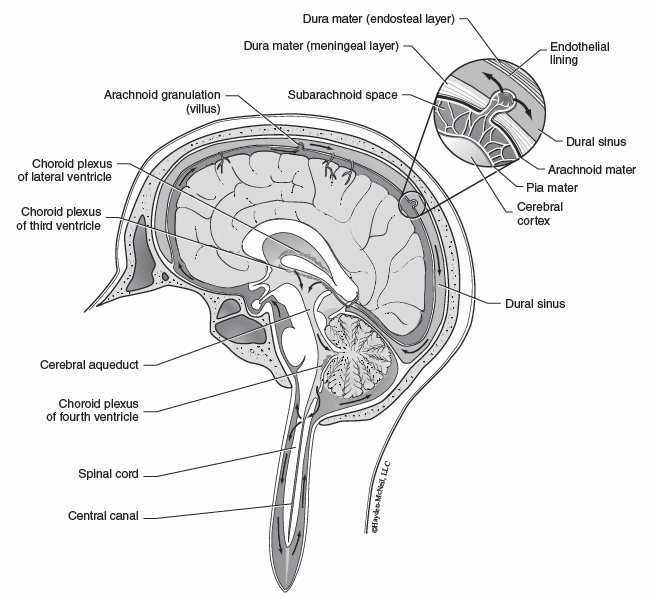
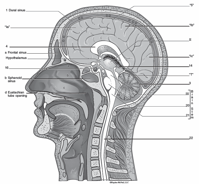
The structures of the central nervous system are protected by the bones of the skeleton and by three layers of dense connective tissue called the meninges. The skull protects the brain and the vertebral column protects the spinal cord. The outer most layer of the meninges is called the dura mater. Superficial to the dura mater in the vertebral cavity is the epidural space. The epidural space contains adipose tissue and larger blood vessels. It is the site where epidural anesthetics are injected. The dural sinuses are found in the dura mater of the brain. They collect blood from the brain and drain into the internal jugular veins of the neck. The middle layer of the meninges is the arachnoid mater. Internal to the arachnoid mater is the subarachnoid space that contains cerebrospinal fluid. The subarachnoid space is the site of injections of spinal anesthetics. The innermost layer of the meninges laying directly on the brain and spinal cord is the pia mater.
Cerebrospinal Fluid (CSF) Cerebrospinal fluid (CSF) is found in the subarachnoid space of the meninges surrounding the brain and the spinal cord. It can also be found in the fluid filled ventricles of the brain and central canal of the spinal cord. The main functions of CSF are to support the CNS, cushion it from shock, and transport nutrients and metabolic wastes.
Cerebrum
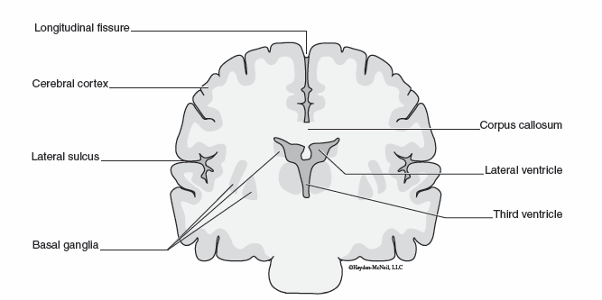
The cerebrum is the largest, most superior portion of the brain. It is divided into left and right halves or cerebral hemispheres at the top by the longitudinal fissure. Each hemisphere of the cerebrum has the ability to specialize to carry out tasks that are not done by the opposite hemisphere. As a result, the left side of the brain has greater control over language, math and logic. The right hemisphere is dominant in tasks relating to artistic expression. This is known as lateralization, and the corpus callosum connects the two halves of the cerebrum allowing them to communicate with each other. The transverse fissure separates the cerebrum from the cerebellum. The central sulcus is a major groove found in the cerebrum that separates the frontal and parietal lobes. The lateral sulcus is the groove that separates the temporal lobe of the cerebrum.

Cerebral Gray Matter
The cerebral cortex is the outer most layer of the cerebrum. It consists of gray matter and is the site of most of the neural activity in the cerebrum. The cerebral cortex is important in conscious thought, memory, reasoning, judging consequences, planning, and initiating voluntary movements. Other areas of gray matter embedded deep within the cerebrum are called basal ganglia. Parkinson’s disease is a degenerative brain disorder caused by the death of dopamine producing neurons in the basal ganglia.
Cerebral Lobes
The different regions or lobes of the cerebrum carry out specialized functions. The frontal lobe is the most anterior region of the cerebrum. The primary motor cortex is responsible for initiating skeletal muscle movements. The premotor cortex controls learned, complex movements of skeletal muscle such as how to play the piano. Broca’s area is the part of the brain that is responsible for producing speech. The prefrontal cortex is the area of the brain that functions in planning complex behaviors, personality expression, and moderating correct social behavior. The parietal lobe of the cerebrum is located under the parietal bones of the skull. It contains the primary somatosensory cortex which interprets sensory information from the skin such as touch and temperature. The temporal lobe contains the auditory cortex which receives and interprets impulses from the ear. Wernicke’s area is important in interpreting the meaning of speech by recognizing words and translating thoughts into words. The occipital lobe is located in the back of the cerebrum. It contains the visual cortex which receives impulses from the eye.
Cerebellum
The cerebellum is located inferior to the cerebrum. The cerebellum helps to coordinate voluntary muscle movements, maintain balance and muscle tone. It receives information about muscle tension, balance, joint position, and movement from sensory receptors and determines the best way to coordinate muscle activity to carry out desired movements. It compares the intended movements to what is actually occurring and makes necessary adjustments to correct and smooth muscle movements. More recent studies of the cerebellum indicate that it may also function in other types of sensory perception such as comparing the textures of objects, spatial relationships, and the amount of time that has passed. Finally, the cerebellum may play a role in language and emotions.
Exercise: Cerebellar Function
The cerebellum assists with coordination of muscle movement. You will examine this cerebellar function with the following series of steps.
1. Sit in your lab chair or favorite comfortable chair at home.
2. Close your eyes and keep them closed while sitting.
3. Raise your right arm straight into the air.
4. Extend your right index finger.
5. Touch your right index finger to the tip of your nose.
Discuss the significance of this muscle movement accomplished without looking.
Thalamus
The thalamus is located near the center of the brain. One of its primary functions is to route sensory information to the correct part of the cerebrum. The thalamus is part of the limbic system which determines our emotions. As sensory impulses travel through the thalamus, we begin to perceive them as pleasant or unpleasant, even before we are aware of what the sensation is. The thalamus also aides muscle coordination by relaying messages between the cerebrum and the cerebellum.
Hypothalamus
The hypothalamus is located inferior to the thalamus. It is the main control center for the autonomic nervous system. The hypothalamus connects directly to the pituitary gland, and influences the activity of the pituitary gland and the endocrine system. As a result, the hypothalamus plays a central role in regulating homeostasis of body systems. The hypothalamus plays an important role in regulating body temperature, hunger, and thirst. The hypothalamus is also an important part of the limbic system which influences emotions such as pleasure, pain, fear, and rage.
Pineal Gland
The pineal gland is located posterior to the thalamus. It produces melatonin which influences the body’s internal clock and sleep/wake cycles. Seasonal affective disorder is a mood disorder where people experience the winter blues. Because the pineal gland secretes more melatonin in dim light or dark conditions, it may contribute to seasonal affective disorder.
Brain Stem
The brain stem contains three major parts: the midbrain, pons and medulla oblongata. The midbrain is the most superior portion of the brain stem and controls reflexes of the eyes and ears such as tracking objects as they move or turning toward a sudden sound. The pons is located inferior to the midbrain. It contains a respiratory center which helps regulate the rate and depth of breathing. The most inferior portion of the brain stem is the medulla oblongata. The medulla oblongata contains the main respiratory center that influences breathing, a cardiac center that influences heart rate, and a vasomotor center that influences blood pressure and flow. The medulla oblongata is also involved in reflexes such as coughing, sneezing, vomiting and swallowing.
Exercise: Brain Identification
Examine the brain and median head models for the regions and structures listed in the Central Nervous System Review (page 114).
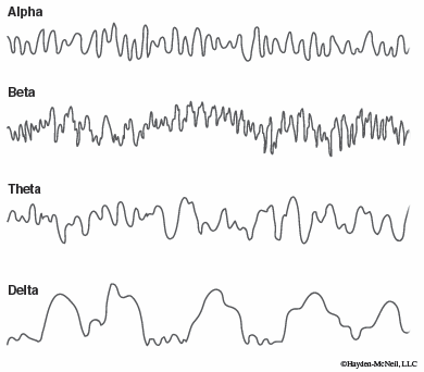
Electroencephalogram (EEG)
An electroencephalogram (EEG) records the brain’s electrical activity. Alpha waves indicate relaxation and closing of the eyes. Beta waves are the busiest, and indicate activities such as concentration, anxiety, or analyzing the importance of biology in everyday life. Theta waves indicate drowsiness. They can also be seen when waking from sleep. Delta waves are produced during deep sleep.
Functional Brain Regions
The reticular formation is found in the brain stem. It works with the cerebrum and cerebellum to coordinate muscle movements and maintain balance. The reticular activating system is a part of the reticular formation and filters sensory input from all the sensory receptors. This allows us to focus on important and meaningful information and disregard background chatter and noise. It is also important in sleep and arousal from sleep. There are two general categories of sleep. Non-REM sleep is a deep, restful sleep where the body is relaxed and there is little cerebral activity. REM sleep occurs when a person is dreaming, and is characterized by increased brain activity and rapid eye movements. During REM sleep the body is less responsive to outside stimuli.
The limbic system consists of parts of the hypothalamus, thalamus, brain stem, and cerebrum. It controls emotions and motivations. Because the hypothalamus is a part of the limbic system and it is also the main regulator of the autonomic nervous system, there is a connection between our emotions and our body systems. Psychosomatic illnesses are illnesses caused by emotions such as stress. They can result in a variety of conditions or symptoms such as high blood pressure. The amygdyla is part of the limbic system, and is responsible for our ability to recognize facial expressions such as anger or fear.
The gnostic area of the brain is located in the parietal lobe. Its major function is to correlate sensory impulses with memories to interpret sensations. Memory is the ability to recall ideas, thoughts, and past experiences. There are two types of memory. Short-term memory lasts for only a few seconds to minutes. It is our working memory. Information that is stored in long-term memory can be recalled at a later time. Repetition and alertness aid in transferring information from short-term to long-term memory. Short-term memories are stored in the limbic system as they are being used, long-term memories are stored in the cerebral cortex.
Exercise: Sheep Brain Dissection
Many anatomical features of the brain are similar in different mammals and dissection of the sheep brain will demonstrate several characteristics you have already examined.
1. You will need one entire sheep brain, a dissecting tray, and a dissecting kit for your group of laboratory partners. Each laboratory partner will need gloves and goggles.
2. Several layers of connective tissue or meninges normally surround and protect the mammalian brain. The tough, outermost layer is made up of dense connective tissue and is known as duramater. The arachnoid mater lies between the dura mater and innermost pia mater. The pia mater is a thin layer lying directly on the brain. Carefully remove the meninges that still may be associated with the brain. The pituitary gland on the ventral surface of the brain will probably be removed along with the meninges.
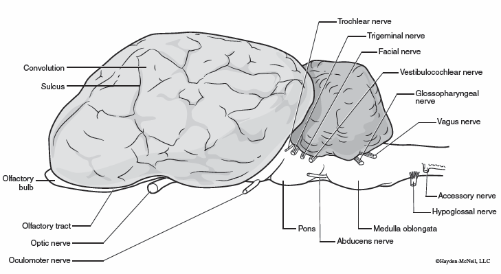
3. Examine the external anatomy of the entire sheep brain. Identify the cerebrum, cerebellum and brain stem. Locate the frontal, parietal, temporal and occipital lobes of the cerebrum. Note the convolutions in the cerebrum; the folds are called gyri and are separated by grooves or depressions called sulci.
4. Gently bend the cerebellum away from the cerebrum along the transverse fissure, being careful not to break or cut the brain stem. The worm-like structure along the midline of the cerebellum is the vermis. Looking deep within the transverse fissure, notice the single projection, the pineal gland, along the upper midline just beneath the occipital lobe. This is the pineal gland and it lies superior to midbrain.
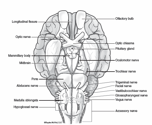
5. Rotate the sheep brain such that the ventral surface faces upward. Identify the olfactory bulb, olfactory tract and the optic chiasma on the ventral surface. The optic chiasma is a partial crossing over of the two optic nerves.
6. Position the sheep brain with the dorsal surface upward. Gently separate the parietal lobes along the longitudinal fissure until you can see the band of white fibers connecting the cerebral hemispheres. This is the corpus callosum. Make a midsagittal incision through the cerebrum, brain stem, cerebellum and spinal cord along the longitudinal fissure to separate the brain into two equal halves.
7. Examining the midsagittal sections of the brain, notice the two cerebral hemispheres are connected by several bundles of white fibers that can be seen in the midsagittal view. The largest and most superior of the commissures is the corpus callosum. The wall of the hemisphere just inferior to this is the thin septum pellucidum. Behind this membrane is part of a large chamber or cavity, the lateral ventricle, within each hemisphere. There are a total of four ventricles in the brain, each connected to the next. The third ventricle is surrounded by the thalamus and the fourth ventricle separates the cerebellum from the pons and medulla oblongata. T he ventricles contain cerebrospinal fluid secreted by choroid plexuses. The fourth ventricle is continuous with the central canal of the spinal cord.
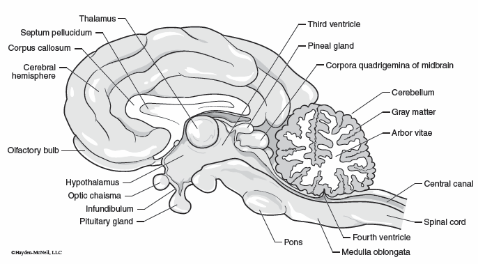
8. On the posterior of the brain, identify the large cerebellum in midsagittal section. The cerebellum resembles the cerebrum in being highly convoluted and in having a cortex of gray matter. The white matter of the cerebellum is in the shape of a branching shrub and is called the arbor vitae.
9. The thalamus appears as a round mass in the midsagittal section. All sensory information is relayed through it before sending them to various regions of the cerebrum to be interpreted. The hypothalamus is the area inferior to the thalamus. The hypothalamus integrates many autonomic functions: sleep, body temperature, water balance, appetite, and others. The pineal gland is located posterior to the thalamus. The pineal gland is associated with the influence of day length on the sleep wake cycle.
10. The brain stem is the upward continuation of the spinal cord. The midbrain is the most superior portion of the brain stem. It coordinates movements of the head related to vision and hearing (like turning your head toward a sudden noise). The pons appears as a large oval shaped bulge on the brain stem. Posterior to the pons and merging with the spinal cord is the medulla oblongata. Many visceral activities are regulated by centers in the medulla including heart rate, blood pressure, breathing movements and swallowing.
11. Make a frontal section across the cerebral hemisphere with your scalpel. Notice that the surface is gray matter surrounding deeper white matter. The gray matter is referred to as the cerebral cortex and is composed largely of the cell bodies of neurons. The white matter consists of the myelinated processes (axons) of neurons. The convolutions of the cerebral surface greatly increase the area available for neurons.
12. Dispose of the dissected brain into the appropriate container. Wash and dry the dissecting tray. Wash, dry and return the dissecting instruments to the dissecting kit. Wipe down your table with a disinfecting wipe. Dispose of your gloves and return your goggles to the sterilization storage unit. Wash your hands before continuing any other activity.
Peripheral Nervous System Review
Central Nervous System Review