2.2 The Neuron
THE BASIC UNIT OF COMMUNICATION
KEY THEME
Information in the nervous system is transmitted by specialized cells, called neurons.
KEY QUESTIONS
What are the basic components of the neuron, and what are their functions?
What are glial cells, and what is their role in the nervous system?
What is an action potential, and how is it produced?
Communication throughout the nervous system takes place via neurons—cells that are highly specialized to receive and transmit information from one part of the body to another. Most neurons, especially those in your brain, are extremely small. A bit of brain tissue no larger than a grain of rice contains about 10,000 neurons! Your entire brain contains an estimated 100 billion neurons.
neuron
A highly specialized cell that communicates information in electrical and chemical form; a nerve cell.
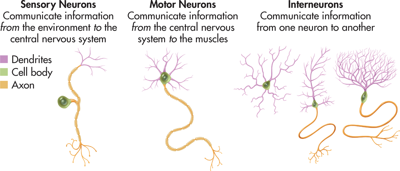
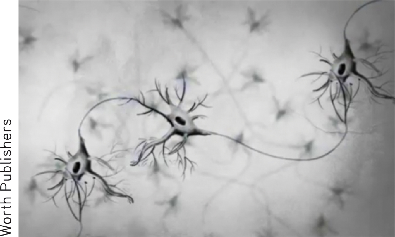
Neurons vary greatly in size and shape, reflecting their specialized functions (see FIGURE 2.1). There are three basic types of neurons, each communicating different kinds of information. Sensory neurons convey information about the environment, such as light or sound, from specialized receptor cells in the sense organs to the brain. Sensory neurons also carry information from the skin and internal organs to the brain. Motor neurons communicate information to the muscles and glands of the body. Simply blinking your eyes activates thousands of motor neurons. Finally, interneurons communicate information between neurons. By far, most of the neurons in the human nervous system are interneurons. Many interneurons connect to other interneurons.
sensory neuron
The type of neuron that conveys information to the brain from specialized receptor cells in sense organs and internal organs.
motor neuron
The type of neuron that signals muscles to relax or contract.
interneuron
The type of neuron that communicates information from one neuron to the next.
One type of neuron deserves special mention. Mirror neurons are not structurally different from other motor neurons. They are a distinct type of motor neuron that becomes activated both when individuals perform a motor act and when they observe the same motor act done by another individual (Cook & others, 2014; Rizzolatti & others, 2008). We discuss mirror neurons in greater detail in Chapter 5, Learning (see p. 217).
Characteristics of the Neuron
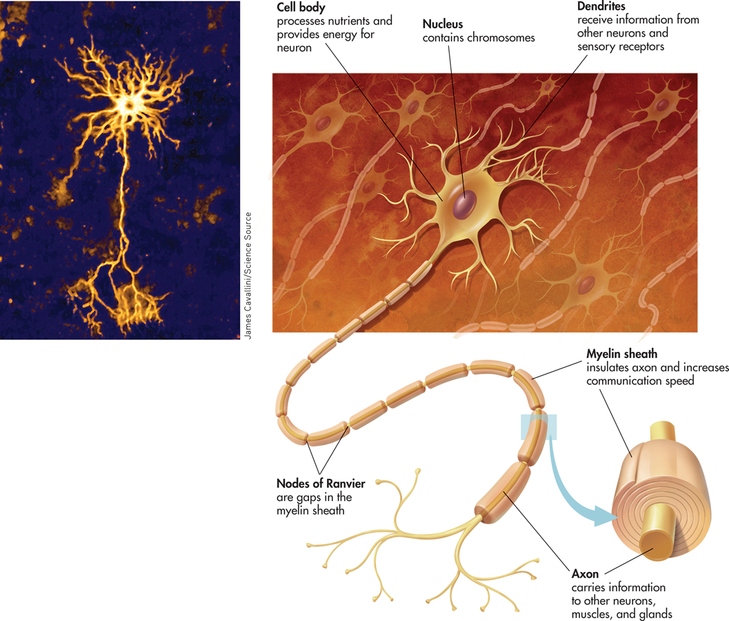
Most neurons have three basic components: a cell body, dendrites, and an axon (see FIGURE 2.2 below). The cell body, also called the soma, contains structures that manufacture proteins and process nutrients, providing the energy the neuron needs to function. The cell body also contains the nucleus, which in turn contains the cell’s genetic material—twisted strands of DNA called chromosomes.
cell body
The part of a cell that processes nutrients and provides energy for the neuron to function; contains the cell’s nucleus; also called the soma.
Short, branching fibers, called dendrites, extend from the cell bodies of most neurons. The term dendrite comes from a Greek word meaning “tree.” And, the intricate branching of the dendrites does often resemble the branches of a tree. Dendrites receive messages from other neurons or specialized cells. Dendrites with many branches have a greater surface area, which increases the amount of information the neuron can receive. Some neurons have thousands of dendrites.
dendrites
The multiple short fibers that extend from a neuron’s cell body and receive information from other neurons or from sensory receptor cells.
The axon is a single, elongated tube that extends from the cell body in most, though not all, neurons. (Some neurons do not have axons.) Axons carry information from the neuron to other cells in the body, including other neurons, glands, and muscles. In contrast to the potentially large number of dendrites, a neuron has only one axon exiting from the cell body. However, many axons have branches near their tips that allow the neuron to communicate information to more than one target.
axon
The long, fluid-filled tube that carries a neuron’s messages to other body areas.
Axons can vary enormously in length. Most axons are very small; some are no more than a few thousandths of an inch long. Other axons are quite long. For example, the longest axon in your body is that of the motor neuron that controls your big toe. This axon extends from the base of your spine into your foot. If you happen to be a seven-foot-tall basketball player, this axon could be four feet long! For most of us, though, this axon is closer to three feet long.
Glial Cells
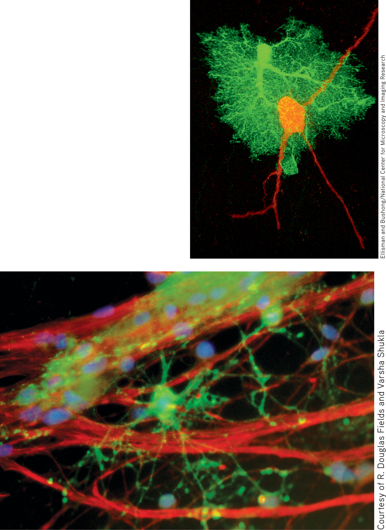
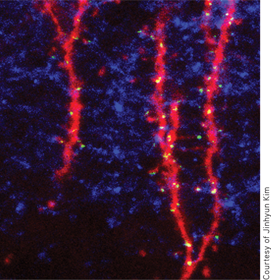
Bottom: This colored micrograph shows the first stages of myelin formation by an oligodendrocyte (green). Like a spider spinning a web, the oligodendrocyte sends tendrils out to neighboring axons (red) and wraps layers of myelin around them in a spiral-shaped pattern.
Courtesy of R. Douglas Fields and Varsha Shukla
Along with neurons, the human nervous system is made up of other specialized cells, called glial cells or simply glia (see photo below). Glial cells are the most abundant cells in the human brain, outnumbering neurons by about 10 to 1. Glia is Greek for “glue,” and although they don’t actually glue neurons together, glia do provide structural support for neurons throughout the nervous system.
glial cells or glia
(GLEE-ull) The support cells that assist neurons by providing structural support, nutrition, and removal of cell wastes; glial cells manufacture myelin.
There are several different kinds of glial cells, each with its own specialized function (Fields, 2013). Microglia remove waste products from the nervous system, including dead and damaged neurons. Astrocytes, shown in the photo below, provide connections between neurons and blood vessels. Astrocytes are also involved in brain development and the communication of information among neurons (Clarke & Barres, 2013).
Two other types of glial cells, oligodendrocytes in the brain and Schwann cells in the rest of the nervous system, form the myelin sheath, a white fatty covering that is wrapped around the axons of some, but not all, neurons. In much the same way that insulating plastic on electrical wires prevents interference when wires contact each other, myelin helps insulate one axon from the axons of other neurons. Rather than forming a continuous coating of the axon, however, the myelin sheath occurs in segments that are separated by small gaps. The small gaps are called the nodes of Ranvier, or simply nodes (see FIGURE 2.2). Neurons whose axons are wrapped in myelin communicate their messages up to 50 times faster than do unmyelinated neurons (Fields, 2013).
myelin sheath
(MY-eh-lin) A white, fatty covering wrapped around the axons of some neurons that increases their communication speed.
The importance of myelin becomes readily apparent when it is damaged. For example, multiple sclerosis is a disease that involves the degeneration of patches of the myelin sheath. This degeneration causes the transmission of neural messages to be slowed or interrupted, resulting in disturbances in sensation and movement. Muscle weakness, loss of coordination, and speech and visual disturbances are some of the symptoms that characterize multiple sclerosis.
Communication Within the Neuron
THE ACTION POTENTIAL
Essentially, the function of neurons is to transmit information throughout the nervous system. But exactly how do neurons transmit information? We’ll first describe communication within a neuron, and then, in the following section, we’ll describe communication between neurons.
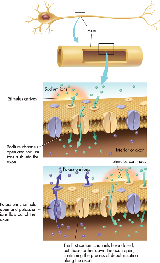
In general, messages are gathered by the dendrites and cell body and then transmitted along the axon in the form of a brief electrical impulse called an action potential. The action potential is produced by the movement of electrically charged particles, called ions, across the membrane of the axon. Some ions are negatively charged, others positively charged.
action potential
A brief electrical impulse by which information is transmitted along the axon of a neuron.
Think of the axon membrane as a gatekeeper that carefully controls the balance of positive and negative ions on the interior and exterior of the axon. As the gatekeeper, the axon membrane opens and closes ion channels that allow ions to flow into and out of the axon.
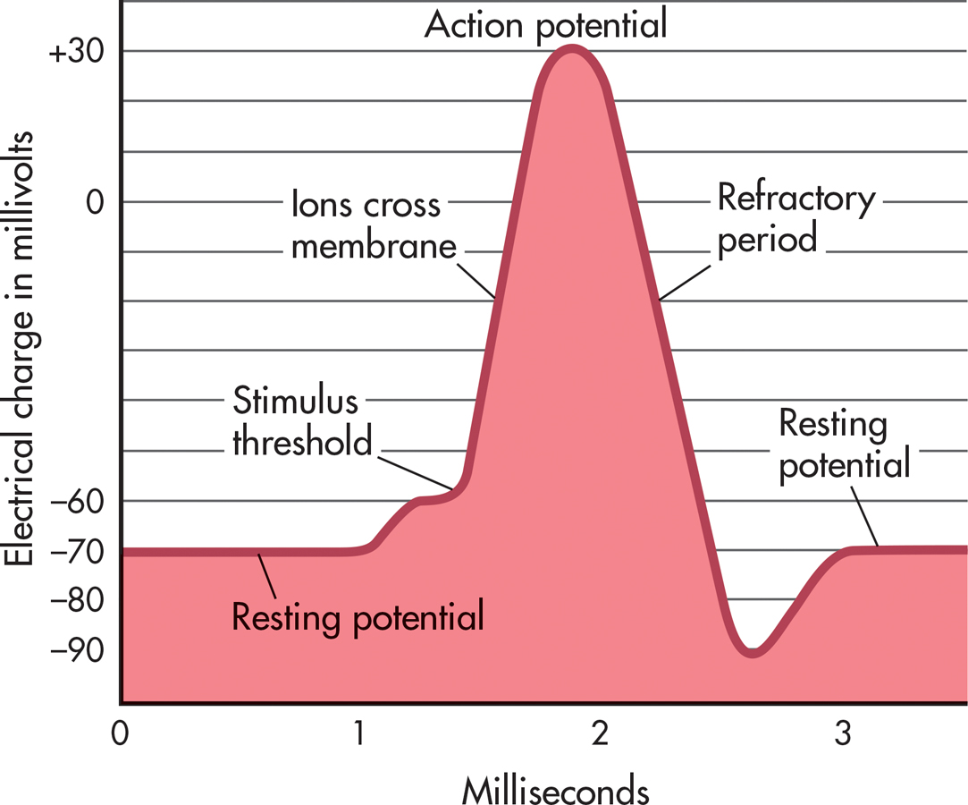
Each neuron requires a minimum level of stimulation from other neurons or sensory receptors to activate it. This minimum level of stimulation is called the neuron’s stimulus threshold. While waiting for sufficient stimulation to activate it, the neuron is said to be polarized. This means that there is a difference in the electrical charge between the inside and the outside of the axon.
stimulus threshold
The minimum level of stimulation required to activate a particular neuron.
More specifically, there is a greater concentration of negative ions inside the neuron. Thus, the axon’s interior is more negatively charged than is the exterior fluid surrounding the axon. The negative electrical charge is about –70 millivolts (thousandths of a volt) (see FIGURE 2.4). The –70 millivolts is referred to as the neuron’s resting potential.
resting potential
The state in which a neuron is prepared to activate and communicate its message if it receives sufficient stimulation.
In this polarized, negative-inside/positive-outside condition, there are different concentrations of two particular ions: sodium and potassium. While the neuron is in resting potential, the fluid surrounding the axon contains a larger concentration of sodium ions than does the fluid within the axon. The fluid within the axon contains a larger concentration of potassium ions than is found in the fluid outside the axon.
An action potential is triggered when the neuron is sufficiently stimulated by other neurons or sensory receptors. First, the neuron depolarizes: At each successive axon segment, sodium ion channels open for a mere thousandth of a second. The sodium ions rush to the axon interior from the surrounding fluid, and then the sodium ion channels close. Less than a thousandth of a second later, the potassium ion channels open, allowing potassium to flow out of the axon and into the fluid surrounding it. Then the potassium ion channels close (see FIGURE 2.3). This sequence of depolarization and ion movement continues down the entire length of the axon.
As this ion exchange occurs, the relative balance of positive and negative ions separated by the axon membrane changes. The electrical charge on the inside of the axon momentarily changes to a positive electrical charge of about +30 millivolts. The result is a brief positive electrical impulse that progressively occurs at each segment down the axon—the action potential.
Although it’s tempting to think of the action potential as traveling in much the same way as electricity travels through a wire, that’s not what takes place in the neuron. The axon is actually a poor conductor of electricity. At each successive segment of the axon, the action potential is regenerated in the same way in which it was generated in the previous segment—by depolarization and the movement of ions.
Once the action potential is started, it is self-sustaining and continues to the end of the axon. In other words, there is no such thing as a partial action potential. Either the neuron is sufficiently stimulated and an action potential occurs, or the neuron is not sufficiently stimulated and an action potential does not occur. This principle is referred to as the all-or-none law.
After the action potential, the neuron enters a refractory period, lasting a thousandth of a second or less, during which the neuron cannot fire. Instead, the neuron repolarizes and reestablishes the negative-inside/positive-outside condition. Like depolarization, repolarization occurs progressively at each segment down the axon. This process reestablishes the resting potential conditions so that the neuron is capable of firing again. The graph in FIGURE 2.4 depicts the complete sequence from resting potential to action potential and back to resting potential.
Action potentials are generated in mere thousandths of a second. Thus, a single neuron can potentially generate hundreds of neural impulses per second. Just how fast do neural impulses zip around your body? The fastest neurons in your body communicate at speeds of up to 270 miles per hour. In the slowest neurons, messages creep along at about 2 miles per hour. This variation in communication speed is due to two factors: the axon diameter and the myelin sheath. The larger the axon’s diameter, the faster it conducts action potentials. And, myelinated neurons communicate much faster than unmyelinated neurons, because the action potential “jumps” from node to node rather than progressing down the entire length of the axon.
Communication Between Neurons
BRIDGING THE GAP
KEY THEME
Communication between neurons takes place at the synapse, the junction between two adjoining neurons.
KEY QUESTIONS
How is information communicated at the synapse?
What is a neurotransmitter, and what is its role in synaptic transmission?
What are seven important neurotransmitters, and how do psychoactive drugs affect synaptic transmission?
The primary function of a neuron is to communicate information to other cells, most notably other neurons. The point of communication between two neurons is called the synapse. At this communication junction, the message-sending neuron is referred to as the presynaptic neuron. The message-receiving neuron is called the postsynaptic neuron. For cells that are specialized to communicate information, neurons have a surprising characteristic: They don’t touch each other. The presynaptic and postsynaptic neurons are separated by a tiny, fluid-filled space, called the synaptic gap, which is only 20 to 40 nanometers wide. How small is that? For comparison, the thickness of a single sheet of paper is about 100,000 nanometers.
synapse
(SIN-aps) The point of communication between two neurons.
synaptic gap
(sin-AP-tick) The tiny space between the axon terminal of one neuron and the dendrite of an adjoining neuron.
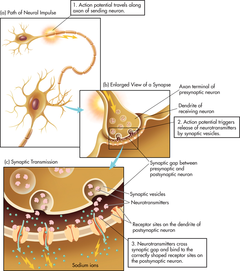
How do neurons communicate? In most cases, when the presynaptic neuron is activated, it generates an action potential that travels to the end of the axon. At the end of the axon are several small branches called axon terminals. Floating in the interior fluid of the axon terminals are tiny sacs called synaptic vesicles (see FIGURE 2.5). The synaptic vesicles hold special chemical messengers manufactured by the neuron, called neurotransmitters.
axon terminals
The branches at the end of the axon that contain tiny pouches, or sacs, called synaptic vesicles.
synaptic vesicles
(sin-AP-tick VESS-ick-ullz) The tiny pouches or sacs in axon terminals that contain chemicals called neurotransmitters.
neurotransmitters
Chemical messengers manufactured by a neuron.
When the action potential reaches the axon terminals, some of the synaptic vesicles “dock” on the axon terminal membrane, then release their neurotransmitters into the synaptic gap. These chemical messengers cross the synaptic gap and attach to receptor sites on the dendrites of the receiving or post-synaptic neuron. This journey across the synaptic gap takes just a few millionths of a second. The entire process of transmitting information at the synapse is called synaptic transmission.
synaptic transmission
(sin-AP-tick) The process through which neurotransmitters are released by one neuron, cross the synaptic gap, and affect adjoining neurons.
What happens to the neurotransmitter molecules after they’ve attached to the receptor sites of the postsynaptic neuron? Most often, they detach from the receptor and are reabsorbed by the presynaptic neuron so they can be recycled and used again. This process is called reuptake. Reuptake also occurs with many of the neurotransmitters that failed to attach to a receptor and were left floating in the synaptic gap. Neurotransmitter molecules that are not reabsorbed or that remain attached to the receptor site are broken down or destroyed by enzymes.
reuptake
The process by which neurotransmitter molecules detach from a postsynaptic neuron and are reabsorbed by a presynaptic neuron so they can be recycled and used again.
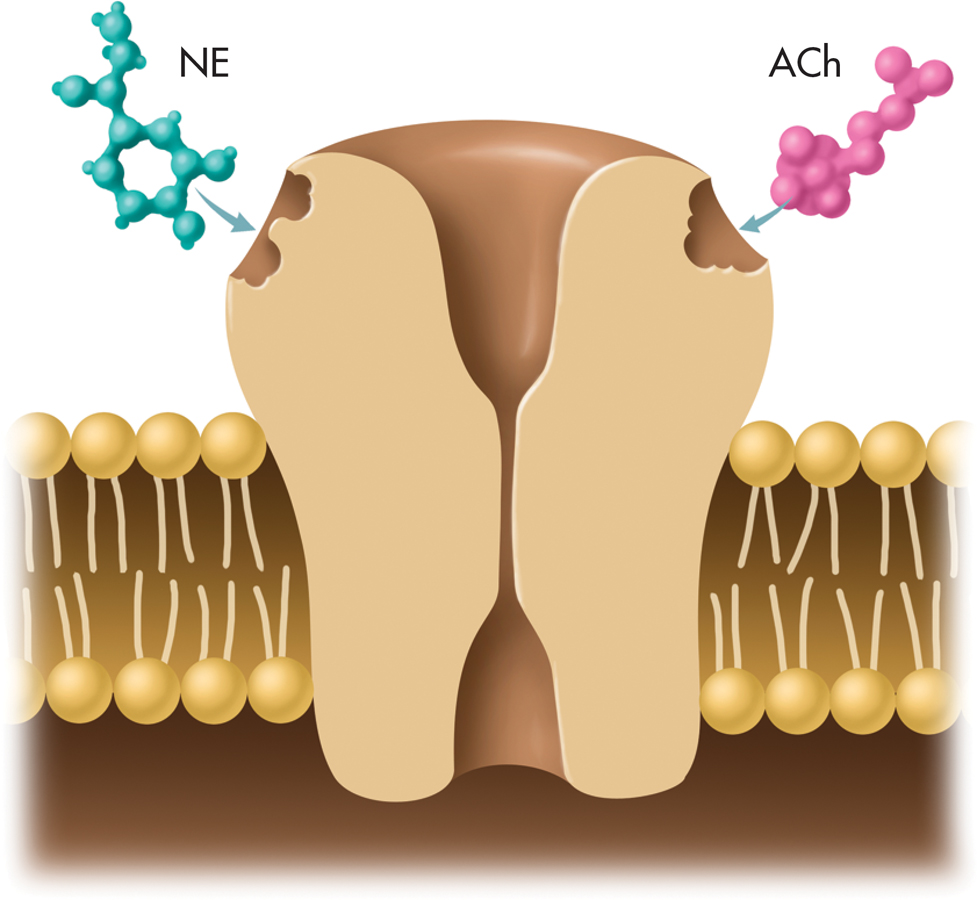
Some neurons produce only one type of neurotransmitter, but others manufacture three or more. Each neurotransmitter has a chemically distinct shape. Like a key in a lock, a neurotransmitter’s shape must precisely match that of a receptor site on the postsynaptic neuron for the neurotransmitter to affect that neuron (see FIGURE 2.6). And, the postsynaptic neuron can have many differently shaped receptor sites on its dendrites and other surfaces. Thus, a given neuron may be able to receive several different neurotransmitters.
Depending upon the receptor to which it binds, a neurotransmitter communicates either an excitatory or an inhibitory message to a postsynaptic neuron. An excitatory message increases the likelihood that the postsynaptic neuron will activate and generate an action potential. An inhibitory message decreases the likelihood that the postsynaptic neuron will activate.
When released by a presynaptic neuron, neurotransmitters cross hundreds, even thousands, of synaptic gaps and affect the intertwined dendrites of adjacent neurons. Because the receiving neuron can have thousands of dendrites that intertwine with the axon terminals of many presynaptic neurons, the number of potential synaptic interconnections between neurons is truly mind-boggling. Each neuron in the brain communicates directly with an average of 1,000 other neurons (Hyman, 2005). However, some specialized neurons have as many as 100,000 connections with other neurons. Thus, there are up to 100 trillion synaptic interconnections in your brain (Eroglu & Barres, 2010). That’s the number 10 followed by 14 zeros!
Neurotransmitters and Their Effects
Your ability to perceive, feel, think, move, act, and react depends on the delicate balance of neurotransmitters in your nervous system. Yet neurotransmitters are present in only infinitesimal amounts in brain tissue—roughly equivalent to a pinch of salt dissolved in an Olympic-sized swimming pool.
Researchers have linked abnormal levels of specific neurotransmitters to various physical and behavioral problems (see TABLE 2.1). However, the connection between a particular neurotransmitter and a particular effect is not a simple one-to-one relationship. Most behaviors are the result of the complex interaction of different neurotransmitters. Further, neurotransmitters sometimes have different effects in different areas of the brain.

Summary of Important Neurotransmitters
| Neurotransmitter | Primary Roles | Associated Disorder |
|---|---|---|
| Acetylcholine | Learning, memory Muscle contractions |
Alzheimer’s disease |
| Dopamine | Movement Thought processes Rewarding sensations |
Parkinson’s disease Schizophrenia Drug addiction |
| Serotonin | Emotional states Sleep Sensory perception |
Depression |
| Norepinephrine | Physical arousal Learning, memory Regulation of sleep |
Depression, stress |
| Glutamate | Excitatory messages | Seizures Alzheimer’s disease |
| GABA | Inhibitory messages | Anxiety disorders |
| Endorphins | Pain perception Positive emotions |
Opioid addiction |
IMPORTANT NEUROTRANSMITTERS
Acetylcholine, the first neurotransmitter discovered, is found in all motor neurons. It stimulates muscles to contract, including the heart and stomach muscles. Whether it is as simple as the flick of an eyelash or as complex as a back flip, all movement involves acetylcholine.
acetylcholine
(uh-seet-ull-KO-leen) Neurotransmitter that causes muscle contractions and is involved in learning and memory.
Acetylcholine is also found in many neurons in the brain, and it is important in memory, learning, and general intellectual functioning. People with Alzheimer’s disease, which is characterized by progressive loss of memory and deterioration of intellectual functioning, have a severe depletion of several neurotransmitters in the brain, most notably acetylcholine.
The neurotransmitter dopamine is involved in movement, attention, learning, and pleasurable or rewarding sensations. Evidence suggests that the addictiveness of many drugs, including cocaine and nicotine, is related to their ability to increase dopamine activity in the brain (Volkow & others, 2011a, b).
dopamine
(DOPE-uh-meen) Neurotransmitter involved in the regulation of bodily movement, thought processes, and rewarding sensations.

The degeneration of the neurons that produce dopamine in one brain area causes Parkinson’s disease, which is characterized by rigidity, muscle tremors, poor balance, and difficulty in initiating movements. Symptoms can be alleviated by a drug called L-dopa, which converts to dopamine in the brain.

Excessive brain levels of dopamine are sometimes involved in the hallucinations and perceptual distortions that characterize the severe mental disorder called schizophrenia. Some antipsychotic drugs that relieve schizophrenic symptoms work by blocking dopamine receptors and reducing dopamine activity in the brain. Unfortunately, because the drugs reduce dopamine in several different areas of the brain, long-term use sometimes produces symptoms that are very similar to those of Parkinson’s disease. In the chapters on psychological disorders (Chapter 14) and therapies (Chapter 15), we’ll discuss schizophrenia, dopamine, and antipsychotic drugs in more detail.
The neurotransmitters serotonin and norepinephrine are found in many different brain areas. Serotonin is involved in sleep, sensory perceptions, moods, and emotional states, including depression (Deneris & Wyler, 2012). Some antidepressant drugs, like Prozac, increase the availability of serotonin in certain brain regions. Norepinephrine is implicated in the activation of neurons throughout the brain and helps the body gear up in the face of danger or threat. Norepinephrine also plays a key role in the regulation of sleep, learning, and memory retrieval (McCarley, 2007). Like serotonin and dopamine, norepinephrine dysfunction is implicated in some psychological disorders, especially depression (Goddard & others, 2010).
serotonin
(ser-uh-TONE-in) Neurotransmitter involved in sensory perceptions, sleep, and emotions.
norepinephrine
(nor-ep-in-EF-rin) Neurotransmitter involved in learning, memory, and regulation of sleep; also a hormone manufactured by adrenal glands.
The most abundant neurotransmitters in the brain are two closely related neurotransmitters, glutamate and gamma-aminobutyric acid, abbreviated GABA. In a delicate balancing act, glutamate conveys excitatory messages and GABA communicates inhibitory messages. Like a dimmer switch, GABA regulates the level of neural activity in the brain. Too much GABA impairs learning, motivation, and movement, but too little GABA can lead to seizures (McCarthy, 2007). Alcohol makes people feel relaxed and less inhibited partly by increasing GABA activity and decreasing glutamate, reducing overall brain activity.
glutamate
Neurotransmitter that usually communicates an excitatory message.
GABA (gamma-aminobutyric acid)
Neurotransmitter that usually communicates an inhibitory message.
Glutamate is involved in learning, memory, and sensory processes (Morris, 2013). Too much glutamate can over-stimulate the brain, causing seizures and cell death. Glutamate is also implicated in Alzheimer’s disease, neurological diseases, and schizophrenia.
Endorphins are another important class of neurotransmitter. Chemically similar to morphine, heroin, and other opioid drugs, endorphins are hundreds of times more potent and are released in response to stress, trauma, and pain. Endorphins are implicated in the pain-reducing effects of acupuncture, an ancient Chinese medical technique that involves inserting needles at various locations in the body (Kemmer, 2007; Zhao, 2008). Also associated with positive mood, endorphins may cause “runner’s high” (see photo).
endorphins
(en-DORF-inz) Neurotransmitters that regulate pain perceptions.
How Drugs Affect Synaptic Transmission
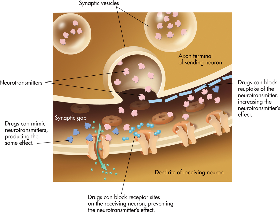
Much of what is known about different neurotransmitters has been learned from observing the effects of drugs and other substances. Many drugs, especially those that affect moods or behavior, work by interfering with the normal functioning of neurotransmitters in the synapse (Volkow & others, 2011a, b).
!launch!
Some drugs increase or decrease the amounts of neurotransmitters released by neurons. For example, the venom of a black widow spider bite causes acetylcholine to be released continuously by motor neurons, causing severe muscle spasms. Drugs may also affect the length of time the neurotransmitters remain in the synaptic gap, either increasing or decreasing the amount available to the postsynaptic receptor.
One way in which drugs can prolong the effects of the neurotransmitters is by blocking the reuptake of the neurotransmitters by the sending neuron. For example, there is a category of antidepressant medications that are referred to as SSRIs, which stands for selective serotonin reuptake inhibitors. SSRIs include such trade-name medications as Prozac, Zoloft, and Paxil. Each of these medications inhibits the reuptake of serotonin, increasing the availability of serotonin in the brain. Similarly, the illegal drug cocaine produces its exhilarating rush by interfering with the reuptake of dopamine (Volkow & others, 2011a, b).
Drugs can also mimic specific neurotransmitters. An agonist is a drug or other chemical that binds to a receptor. Agonist drugs are chemically similar to a specific neurotransmitter and produce the same effect. For example, nicotine is a stimulant because it is chemically similar to acetylcholine. It occupies acetylcholine receptor sites, stimulating skeletal muscles and causing the heart to beat more rapidly.
agonist
Drug or other chemical substance that binds to a receptor site and triggers a response in the cell.
Alternatively, a drug can act as an antagonist by blocking the effect of neurotransmitters. A drug may fit into receptor sites and prevent neurotransmitters from acting. For example, the drug curare blocks acetylcholine receptor sites, causing virtually instantaneous paralysis. The brain sends signals to the motor neurons, but the muscles can’t respond because the motor neuron receptor sites are blocked by the curare. Similarly, the drug naloxone is an opioid antagonist. By blocking endorphin receptors, it can quickly reverse the effects of heroin, oxycodone, or other opioid drugs (Rich & others, 2011). FIGURE 2.7 summarizes the effects of drugs on synaptic transmission.
antagonist
A drug or other chemical substance that blocks a receptor site and inhibits or prevents a response in the receiving cell.
CONCEPT REVIEW 2.1
The Neuron
Match the part of the neuron in the list with its function in neural communication.
Question
axon dendrite action potential synaptic gap neurotransmitters myelin sheath | Chemical messengers manufactured by a neuron Receives information from other neurons Fluid-filled space between sending and receiving neurons Brief electrical charge that communicates information within the neuron A fluid-filled tube that carries information to other neurons, glands, and muscles White, fatty covering surrounding an axon that increases the speed of neural communication |
Test your understanding of Introduction to Neuroscience and the Neuron with
 .
.