4.3 Sleep
KEY THEME
Modern sleep research began with the invention of the EEG and the discovery that sleep is marked by distinct physiological processes and stages.
KEY QUESTIONS
What characterizes sleep onset, the NREM sleep stages, and REM sleep?
What is the typical progression of sleep cycles? How do sleep patterns change over the lifespan?
What evidence suggests that we have a biological need for sleep?
From Aristotle to Shakespeare to Freud, history is filled with examples of scholars, writers, and scientists who have been fascinated by sleep and dreams. But prior to the twentieth century, there was no objective way to study the internal processes that might be occurring during sleep. Instead, sleep was largely viewed as a period of restful inactivity in which dreams sometimes occurred.
The Dawn of Modern Sleep Research

The invention of the electroencephalograph by German psychiatrist Hans Berger in the 1920s gave sleep researchers an important tool for measuring the rhythmic electrical activity of the brain (Stern, 2001). These rhythmical patterns of electrical activity are referred to as brain waves. The electroencephalograph produces a graphic record called an EEG, or electroencephalogram. By studying EEGs, sleep researchers firmly established that brain-wave activity systematically changes throughout sleep.
electroencephalograph
(e-lec-tro-en-SEFF-uh-low-graph) An instrument that uses electrodes placed on the scalp to measure and record the brain’s electrical activity.
EEG (electroencephalogram)
The graphic record of brain activity produced by an electroencephalograph.
Along with brain activity, today’s sleep researchers monitor a variety of other physical functions during sleep. Eye movements, muscle movements, breathing rate, airflow, pulse, blood pressure, amount of exhaled carbon dioxide, body temperature, and breathing sounds are just some of the body’s functions that are measured in contemporary sleep research (Carskadon & Dement, 2005).
What happens during sleep? Most people tend to think of sleep as an “either/or” condition—the brain is either awake and active or asleep and idle. In fact, sleep researchers know that the sleeping brain doesn’t just shut down during sleep. The sleeping brain remains active, although its patterns of activity are distinctly different from the patterns displayed by the waking brain.
Research also disputes the notion that sleep is a global, brain-wide state (Colwell, 2011). It turns out that during sleep, some brain areas, and even some brain neurons, remain active, while others do not (Nir & others, 2011; Vyazovskiy & others, 2011).
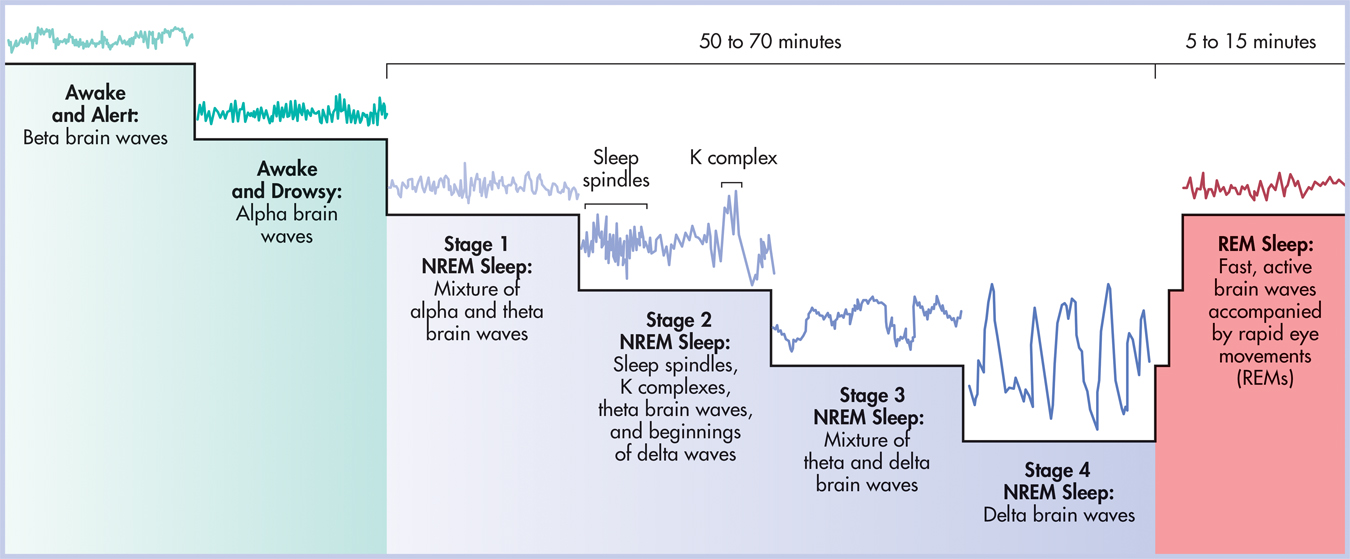
Sleep researchers distinguish between two basic types of sleep. REM sleep, or rapid-eye-movement sleep, is often called active sleep or paradoxical sleep because it is associated with heightened body and brain activity during which dreaming consistently occurs. NREM sleep, or non-rapid-eye-movement sleep, is often referred to as quiet sleep because the body’s physiological functions and brain activity slow down during this period of slumber. Usually pronounced as “non-REM sleep,” it is further divided into four stages, as we’ll describe shortly.
REM sleep
Type of sleep during which rapid eye movements (REM) and dreaming usually occur and voluntary muscle activity is suppressed; also called active sleep or paradoxical sleep.
NREM sleep
Quiet, typically dreamless sleep in which rapid eye movements are absent; divided into four stages; also called quiet sleep.
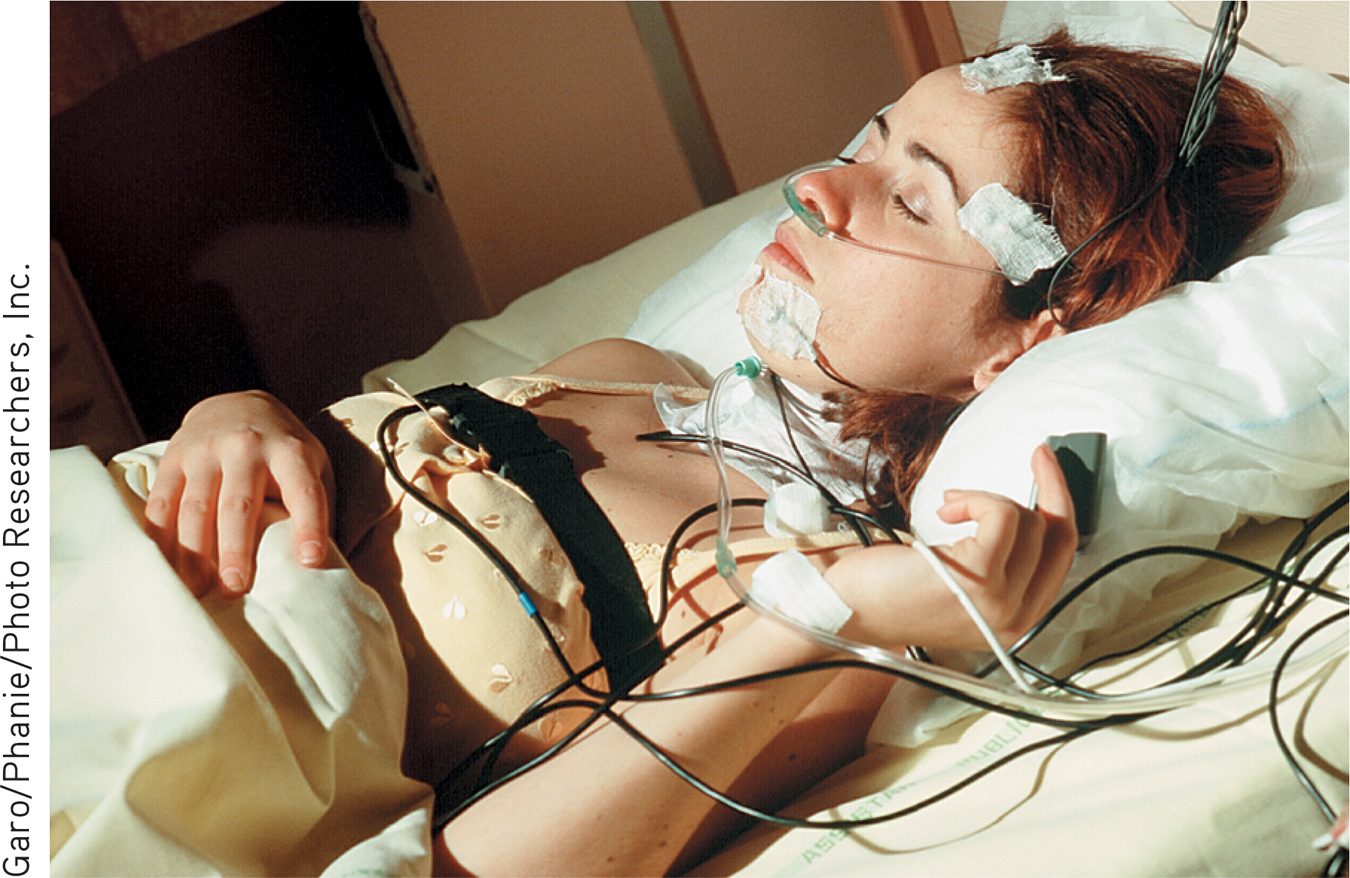
The Onset of Sleep and Hypnagogic Hallucinations
Awake and reasonably alert as you prepare for bed, your brain generates small, fast brain waves, called beta brain waves. After your head hits the pillow and you close your eyes, your muscles relax. Your brain’s electrical activity gradually gears down, generating slightly larger and slower alpha brain waves.
beta brain waves
Brain-wave pattern associated with alert wakefulness.
alpha brain waves
Brain-wave pattern associated with relaxed wakefulness and drowsiness.
During this drowsy, presleep phase, you may experience odd but vividly realistic sensations. You may hear your name called or a loud crash, feel as if you’re falling, floating, or flying, or see kaleidoscopic patterns or an unfolding landscape. These brief, vivid sensory phenomena that occasionally occur during the transition to light sleep are called hypnagogic hallucinations. Some hypnagogic hallucinations can be so vivid or startling that they cause a sudden awakening (Vaughn & D’Cruz, 2005). Hypnagogic imagery may also reflect daily activities and preoccupations. For example, about a third of participants who learned a new video game, Alpine Racer II, reported hypnagogic images related to the skiing game (Schwartz, 2010; Wamsley & others, 2010).
hypnagogic hallucinations
(hip-na-GAH-jick) Vivid sensory phenomena that occur during the onset of sleep.

IN FOCUS
What You Really Want to Know About Sleep
Why do I yawn?
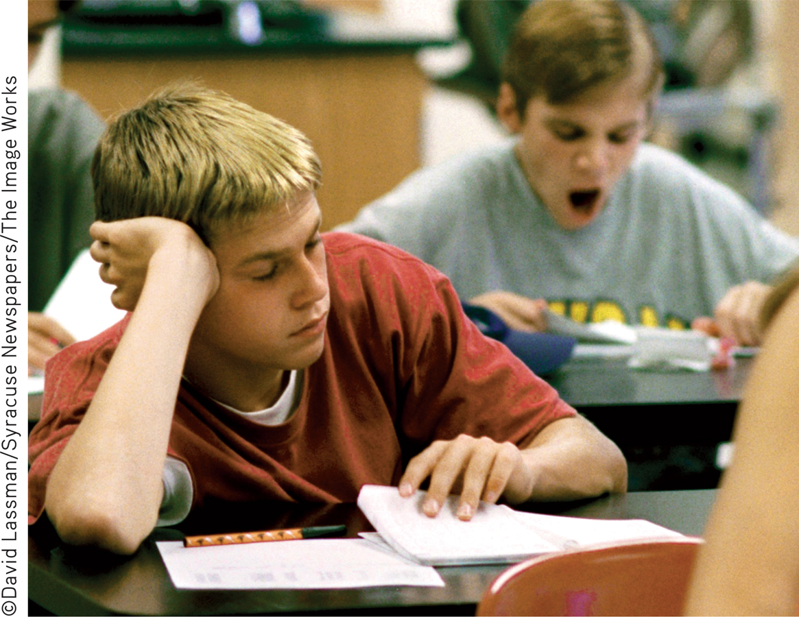
Researchers aren’t certain. But the notion that too little oxygen or too much carbon dioxide causes yawning is not supported by research. Some evidence suggests that yawning regulates and increases your level of arousal. Yawning is typically followed by an increase in activity level. Hence, you frequently yawn after waking up in the morning, while attempting to stay awake in the late evening, or when you’re bored.
Is yawning contagious?

Seeing, hearing, or thinking about yawning can trigger a yawn. More than half of adults will yawn when they’re shown videos of other people yawning. Blind people will yawn more frequently in response to audio recordings of yawning. Some psychologists believe that contagious yawning is related to our ability to feel empathy for others. (Have you yawned yet?) Interestingly, chimpanzees and macaques, both highly social animals, display contagious yawning. And so do domestic dogs, which in a recent study were shown to “catch” yawns from human strangers. From an evolutionary perspective, such observations lend support to the idea that contagious yawning may have evolved as an adaptive social cue, allowing groups to signal and coordinate times of activity and rest. In support of that view, a new study found that chimpanzees yawn more after viewing iTouch videos of other chimpanzees yawning—but only if the chimps were members of their own social group.
Why do I get sleepy?
A naturally occurring compound in the body called adenosine may be the culprit. In studies with cats, prolonged wakefulness sharply increases adenosine levels, which reflect energy used for brain and body activity. As adenosine levels shoot up, so does the need for sleep. Slow-wave NREM sleep reduces adenosine levels. In humans, the common stimulant drug caffeine blocks adenosine receptors, promoting wakefulness.
Sometimes in the morning when I first wake up, I can’t move. I’m literally paralyzed! Is this normal?
REM sleep is characterized by paralysis of the voluntary muscles, which keeps you from acting out your dreams. In a relatively common phenomenon called sleep paralysis, the paralysis of REM sleep carries over to the waking state for up to 10 minutes. If preceded by an unpleasant dream or hypnagogic experience, this sensation can be frightening. Sleep paralysis can also occur as you’re falling asleep. In either case, the sleep paralysis lasts for only a few minutes. So, if this happens to you, relax—voluntary muscle control will soon return.
sleep paralysis
A temporary condition in which a person is unable to move upon awakening in the morning or during the night.
Do deaf people who use sign language sometimes “sleep-sign” during sleep?
Yes.
Do the things people say when they talk in their sleep make any sense?
Sleeptalking typically occurs during NREM stages 3 and 4. There are many anecdotes of spouses who have supposedly engaged their sleeptalking mates in extended conversations, but sleep researchers have been unsuccessful in having extended dialogues with people who chronically talk in their sleep. As for the truthfulness of the sleeptalker’s utterances, they’re reasonably accurate insofar as they reflect whatever the person is responding to while asleep. By the way, not only do people talk in their sleep, but they can also sing or laugh in their sleep. In one case we know of, a little boy sleep-sang “Frosty the Snowman.”
MYTH  SCIENCE
Is it true that you can have coherent conversations with people who are talking in their sleep?
Sources: Campbell & de Waal (2011); Campbell & others (2009); Cartwright (2004); Empson (2002); Joly-Mascheroni & others (2008); Landolt (2008); Platek & others (2005); Pressman (2007); Rétey & others (2005); Romero & others, 2013; Takeuchi & others (2002).
Probably the most common hypnagogic hallucination is the vivid sensation of falling, which is often accompanied by a myoclonic jerk—an involuntary muscle spasm of the whole body that jolts the person completely awake. Also known as sleep starts, these experiences can seem really weird (or embarrassing) when they occur, but they are normal events that sometimes occur during sleep onset (Mahowald, 2005).
The First 90 Minutes of Sleep and Beyond
The course of a normal night’s sleep follows a relatively consistent cyclical pattern. As you drift off to sleep, you enter NREM sleep and begin a progression through the four NREM sleep stages (see FIGURE 4.3). Each progressive NREM sleep stage is characterized by corresponding decreases in brain and body activity. On average, the progression through the first four stages of NREM sleep occupies the first 50 to 70 minutes of sleep.
STAGE 1 NREM
As the alpha brain waves of drowsiness are replaced by even slower theta brain waves, you enter the first stage of sleep. Lasting only a few minutes, stage 1 is a transitional stage during which you gradually disengage from the sensations of the surrounding world. During stage 1 NREM, you can quickly regain conscious alertness if needed. Although hypnagogic experiences can occur in stage 1, less vivid mental imagery is common, such as imagining yourself engaged in some everyday activity. These imaginings lack the unfolding, storylike details of a true dream.
STAGE 2 NREM
Stage 2 represents the onset of true sleep. Stage 2 sleep is defined by the appearance of sleep spindles, brief bursts of brain activity that last a second or two, and K complexes, single high-voltage spikes of brain activity (see FIGURE 4.3). Other than these occasional sleep spindles and K complexes, brain activity continues to slow down considerably. Breathing becomes rhythmical. Slight muscle twitches may occur. Theta waves are predominant in stage 2, but larger, slower brain waves, called delta brain waves, also begin to emerge. During the 15 to 20 minutes initially spent in stage 2, delta brain-wave activity gradually increases.
sleep spindles
Short bursts of brain activity that characterize stage 2 NREM sleep.
K complex
Single but large high-voltage spike of brain activity that characterizes stage 2 NREM sleep.
STAGE 3 AND STAGE 4 NREM
In combination, stages 3 and 4 are often referred to as slow-wave sleep (SWS). Both stages are defined by the amount of delta brain-wave activity. When delta brain waves represent more than 20 percent of total brain activity, the sleeper is said to be in stage 3 NREM. When delta brain waves exceed 50 percent of total brain activity, the sleeper is said to be in stage 4 NREM.
During the 20 to 40 minutes spent in the night’s first episode of stage 4 NREM, delta waves eventually come to represent 100 percent of brain activity. At that point, heart rate, blood pressure, and breathing rate drop to their lowest levels. Not surprisingly, the sleeper is almost completely oblivious to the world. Noises as loud as 90 decibels may fail to wake him. However, his muscles are still capable of movement. For example, sleepwalking typically occurs during stage 4 NREM sleep.
It can easily take 15 minutes or longer to regain full waking consciousness from stage 4. It’s even possible to answer your cell phone or respond to a text message, carry on a conversation for several minutes, and hang up without ever leaving stage 4 sleep—and without remembering having done so the next day. When people are briefly awakened by sleep researchers during stage 4 NREM and asked to perform some simple task, they often don’t remember it the next morning.

Thus far, the sleeper is approximately 70 minutes into a typical night’s sleep and immersed in deeply relaxed stage 4 NREM sleep. At this point, the sequence reverses. In a matter of minutes, the sleeper cycles back from stage 4 to stage 3 to stage 2 and enters a dramatic new phase: the night’s first episode of REM sleep.
REM SLEEP
During REM sleep, the brain becomes more active, generating smaller and faster brain waves. Visual and motor neurons in the brain activate repeatedly, just as they do during wakefulness. Dreams usually occur during REM sleep. Although the brain is very active, voluntary muscle activity is suppressed, which prevents the dreaming sleeper from acting out her dreams.
REM sleep is accompanied by considerable physiological arousal. The sleeper’s eyes dart back and forth behind closed eyelids—the rapid eye movements. Heart rate, blood pressure, and respirations can fluctuate up and down, sometimes extremely. Muscle twitches occur. In both sexes, sexual arousal may occur, which is not necessarily related to dream content.
This first REM episode tends to be brief, about 5 to 15 minutes. From the beginning of stage 1 NREM sleep through the completion of the first episode of REM sleep, about 90 minutes have elapsed.
BEYOND THE FIRST 90 MINUTES
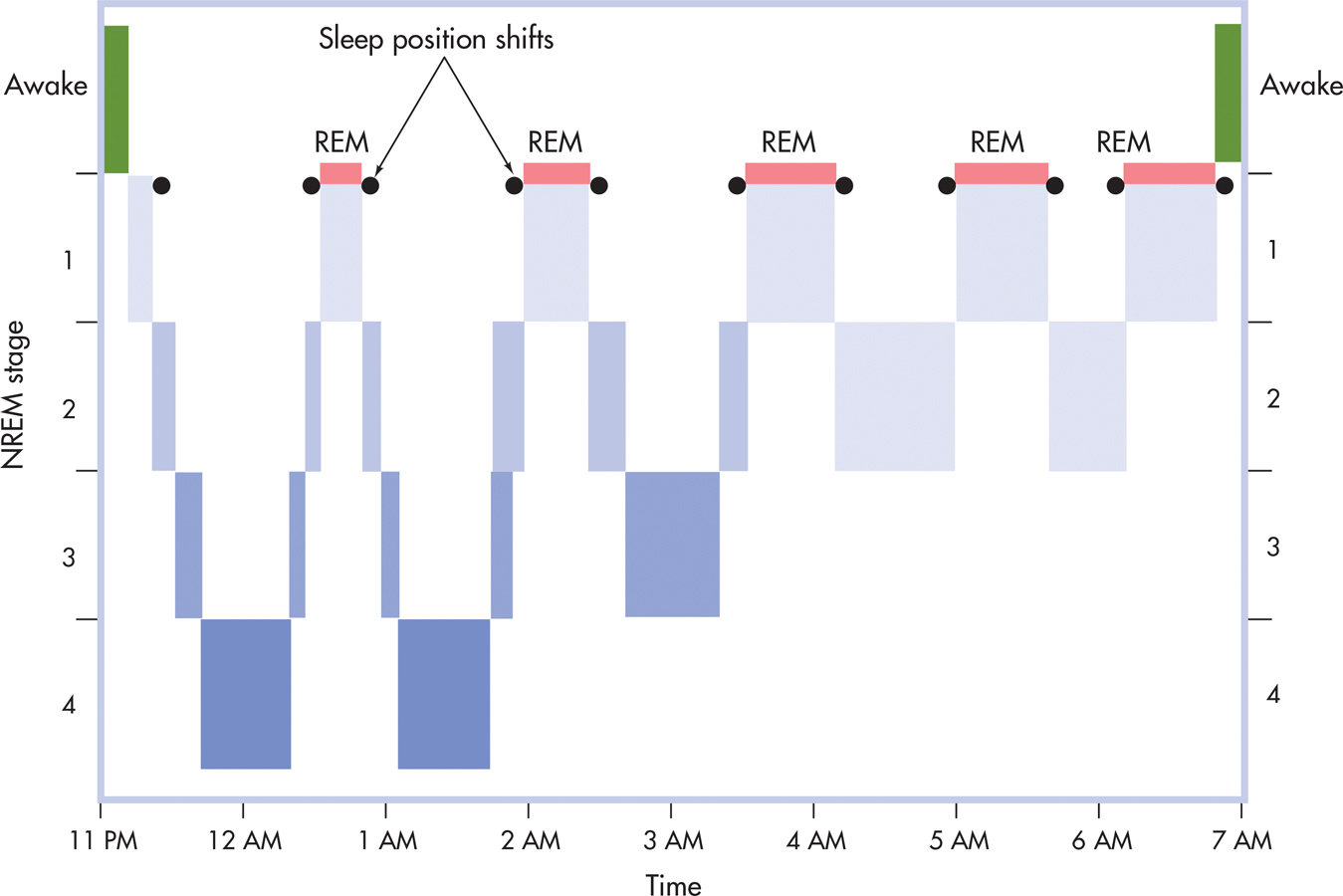
Throughout the rest of the night, the sleeper cycles between NREM and REM sleep. Each sleep cycle lasts about 90 minutes on average, but the duration of cycles may vary from 70 to 120 minutes. Usually, four more 90-minute cycles of NREM and REM sleep occur during the night. Just before and after REM periods, the sleeper typically shifts position.
The progression of a typical night’s sleep cycles is depicted in FIGURE 4.4. Stages 3 and 4 NREM, slow-wave sleep, usually occur only during the first two 90-minute cycles. As the night progresses, REM sleep episodes become increasingly longer, and less time is spent in NREM. During the last two 90-minute sleep cycles before awakening, NREM sleep is composed primarily of stage 2 sleep and periods of REM sleep that can last as long as 40 minutes. In a later section, we’ll look at dreaming and REM sleep in more detail.
CHANGING SLEEP PATTERNS OVER THE LIFESPAN
As any parent knows, sleep patterns change over the lifespan. Circadian rhythms seem to develop before birth. Consistent daily variations in movement, heart rate, and other variables are evident by the fifth month of prenatal development. During the third trimester of prenatal development, active (REM) and quiet (NREM) sleep cycles emerge. In the final weeks, REM and NREM sleep are clearly distinguishable in the fetus (Mirmiran & others, 2003).
The newborn sleeps about 16 hours a day, though not all at once. Up to 8 hours—or 50 percent—of the newborn’s sleep time is spent in REM sleep. The rest is spent in quiet sleep that is very similar to NREM stages 1 and 2. It’s not until about the third month of life that the deep, slow-wave sleep of NREM stages 3 and 4 appears.
The typical 90-minute sleep cycles gradually emerge over the first few years of life. The infant’s sleep during the first months of life is characterized by shorter 60-minute sleep cycles, producing up to 13 sleep cycles per day. By age 2, the toddler is experiencing 75-minute sleep cycles. By age 5, the typical 90-minute sleep cycles of alternating REM and NREM sleep are established (Grigg-Damberger & others, 2007).
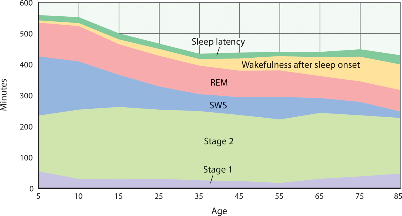
From childhood through late adulthood, the pattern of a typical night’s sleep evolves and changes (see FIGURE 4.5). Total sleep time decreases, as does the percentage of a night’s sleep spent in deeper slow-wave sleep. This is offset by gradual increases in the percentage of time spent each night in lighter NREM stages 1 and 2. The percentage of a night’s sleep devoted to REM sleep increases during childhood and adolescence, remains stable throughout adulthood, and then decreases during late adulthood (Ohayon & others, 2004).
Why Do We Sleep?
It may seem obvious: We sleep because we’re tired and need rest. Thus, it may surprise you to learn that sleep researchers aren’t really sure why we sleep. New research by Lulu Xie and her colleagues (2013) revealed that sleep initiates a process that clears metabolic waste products from the brain. But sleep serves other functions as well (see Underwood, 2013). Along with providing needed rest to muscles and body, sleep is thought to maintain immune system function, improve brain function, enhance learning and consolidate memory, and help regulate moods and emotion (Roth & others, 2010; Walker & Van der Helm, 2009).

Nature Production/Nature Picture Library
However, these explanations don’t account for the enormous variability in sleep patterns across different species. Some researchers take the view that the sleep patterns exhibited by different animals, including humans, are the result of evolutionary adaptation (Roth & others, 2010; Siegel, 2009). According to this view, different sleep patterns evolved as a way of conserving energy and preventing a particular species from interacting with the environment when doing so is most hazardous.
For example, animals with few natural predators, such as gorillas and lions, sleep as much as 15 hours a day. In contrast, grazing animals, such as cattle and horses, tend to sleep in short bursts that total only about 4 hours per day (Siegel, 2005). Lions, much like their evolutionary cousins, the domestic cat, sleep long and deeply, but giraffes, their prey, sleep very little. Similarly, hibernation patterns also reflect evolutionary adaptation, coinciding with periods during which food is relatively scarce and environmental conditions pose the greatest threat to survival.
Rather than an all-encompassing theory explaining why we sleep, today’s sleep researchers recognize that sleep serves a host of vital functions. Studying sleep from multiple perspectives—psychological, physiological, and neurological—yields a dynamic understanding of how sleep occurs and is regulated, and how it contributes to optimal functioning (Datta & MacLean, 2007).
SLEEP AND MEMORY FORMATIONLET ME SLEEP ON IT!
One important function of sleep deserves special mention. Sleep plays a critical role in strengthening new memories and in integrating new memories with existing memories (Walker, 2009, 2010). Sleep seems to be especially important in preserving emotional memories and memories of details that are relevant to personal goals and preoccupations (Stickgold & Walker, 2013).
Sleep researchers are finding that the strengthening and enhancement of new memories during sleep is a very active process. That process seems to work like this: New memories formed during the day are reactivated during the 90-minute cycles of sleep that occur throughout the night. This process of repeatedly reactivating these newly encoded memories during sleep strengthens the neuronal connections that contribute to forming long-term memories (Wamsley & others, 2010; Yang & others, 2014). And, along with helping solidify new memories, sleep is also critical to integrating the new memories into existing networks of memories (Stickgold & Walker, 2013).
THE EFFECTS OF SLEEP DEPRIVATION
The importance of sleep is demonstrated by sleep deprivation studies. After being deprived of sleep for just one night, research subjects develop microsleeps, which are episodes of sleep lasting only a few seconds that occur during wakefulness. People who go without sleep for a day or more also experience disruptions in mood, mental abilities, reaction time, perceptual skills, and complex motor skills (Bonnet, 2005).
But what about getting less than your usual amount of sleep? Sleep restriction studies reduce the amount of time that people are allowed to sleep to as little as four hours per night.
Sleep restriction produces numerous impairments and changes, not the least of which is an increased urge to sleep. Concentration, vigilance, reaction time, memory skills, and the ability to gauge risks are diminished. Motor skills—including driving skills—decrease, producing a greater risk of accidents. As discussed in the Focus on Neuroscience box “The Sleep-Deprived Emotional Brain,” moods, especially negative moods, become much more volatile (Harvey, 2011). Harmful changes occur in hormone levels, including stress hormone levels (Balbo & others, 2010). The immune system’s effectiveness is diminished, increasing susceptibility to colds and infections. Finally, metabolic changes occur, including changes linked to obesity and diabetes (Van Cauter & others, 2008).
When sleep-deprived, people tend to consume more calories and gain weight (Markwald & others, 2013). One explanation is that sleep deprivation tends to increase the appeal of high-calorie foods. As compared to food choices made when they were well rested, people who were sleep-deprived tended to prefer high-caloric treats like potato chips and desserts over nourishing but low-calorie foods like apple slices (Greer & others, 2013).
If sleep restriction continues for multiple nights, all of these changes become more pronounced. Unfortunately most people are not good at judging the extent to which their performance is impaired by inadequate sleep. People tend to think they are performing adequately, but in fact, their abilities and reaction time are greatly diminished (M. Walker, 2008).
Sleep researchers have also selectively deprived people of different components of normal sleep. To study the effects of REM deprivation, researchers wake sleepers whenever the monitoring instruments indicate that they are entering REM sleep. After several nights of being selectively deprived of REM sleep, the subjects are allowed to sleep uninterrupted. What happens? They experience REM rebound—the amount of time spent in REM sleep increases by as much as 50 percent. Similarly, when people are selectively deprived of NREM stages 3 and 4, they experience NREM rebound, spending more time in NREM sleep (Borbély & Achermann, 2005; Tobler, 2005). Thus, it seems that the brain needs to experience the full range of sleep states, making up for missing sleep components when given the chance.
REM rebound
A phenomenon in which a person who is deprived of REM sleep greatly increases the amount of time spent in REM sleep at the first opportunity to sleep without interruption.
FOCUS ON NEUROSCIENCE
The Sleep-Deprived Emotional Brain
Whether they are children or adults, people often react with greater emotionality when they’re not getting adequate sleep (Zohar & others, 2005). Is this because they’re simply tired, or do the brain’s emotional centers become more reactive in response to sleep deprivation?
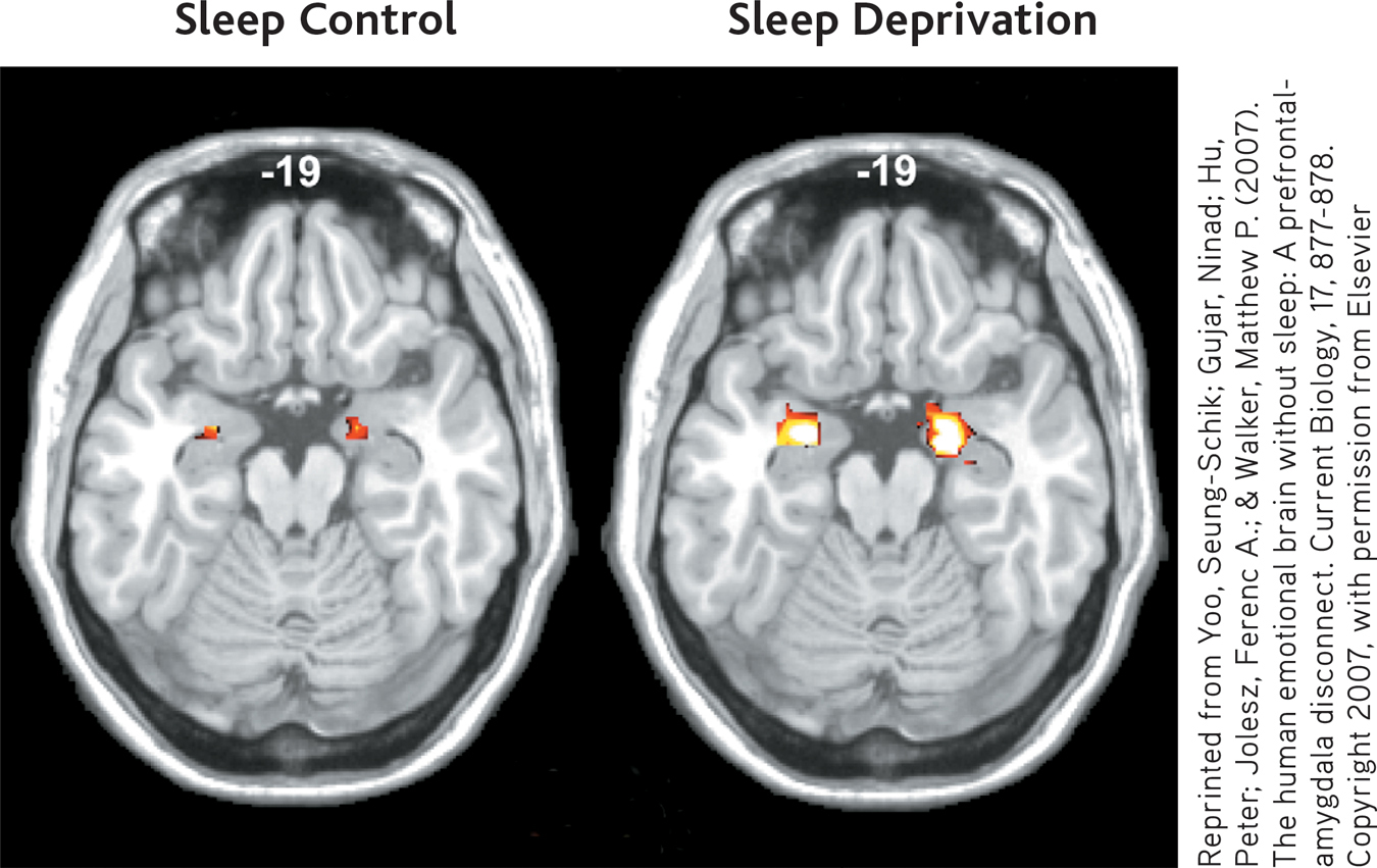
To study this question, researcher Seung-Schik Yoo and his colleagues (2007) deprived some participants of sleep for 35 hours while other participants slept normally. Then, all of the participants observed a series of images ranging from emotionally neutral to very unpleasant and disturbing images while undergoing an fMRI brain scan.
Compare the two fMRI scans shown here. The orange and yellow areas indicate the degree of activation in the amygdala, a key component of the brain’s emotional centers. Compared to the adequately rested participants, the amygdala activated 60 percent more strongly when the sleep-deprived participants looked at the aversive images.
Yoo’s research clearly shows that the sleep-deprived brain is much more prone to strong emotional reactions to negative stimuli. And, new research shows that sleep deprivation also affects how we respond to positive stimuli. The same team of researchers found that sleep-deprived participants responded with heightened activity in the brain’s reward circuits to images of pleasant scenes, such as cute bunnies or ice cream sundaes (Gujar & others, 2011). The implication? Unrealistically positive responses may lead sleep-deprived people to engage in risky or addictive behavior.
As sleep scientist Matthew Walker (2011) observes, “When functioning correctly, the brain finds the sweet spot on the mood spectrum. But the sleep-deprived brain will swing to both extremes, neither of which is optimal for making wise decisions.”
So, when you’re consistently operating on too little sleep, monitor your emotional reactions so that you don’t overreact and say or do something that you will regret later—such as after you’ve caught up on your sleep.
CONCEPT REVIEW 4.1
Circadian Rhythms and Stages of Sleep
Fill in the blanks with the missing terms.
Question 4.1
| 1. | Anthony participated in a sleep research study in which he spent two months in a laboratory isolated from sunlight and other natural light/dark cues. Eventually, his body clock drifted to a -hour daily cycle. |
Question 4.2
| 2. | When Anthony left the sleep research study, his body clock was reset by exposure to , and his returned to a normal 24-hour cycle. |
Question 4.3
| 3. | You’ve been asleep for about 10 minutes and are experiencing brief bursts of brain activity, called sleep spindles. This means that you are in sleep. |
Question 4.4
| 4. | Compared to his 80-year-old grandfather, 7-year-old Vance spends more of each night in sleep. |
Question 4.5
| 5. | During a sleep study, the lab assistant wakes you and asks you to recite the alphabet backwards. You do so, but remember nothing about this event when asked the next morning. You were probably awakened during . |