9.3 Prenatal Development
KEY THEME
During the prenatal stage, the single-celled zygote develops into a full-term fetus.
KEY QUESTIONS
What are the three stages of prenatal development?
How does the brain develop?
What are teratogens?
At conception, chromosomes from the biological mother and father combine to form a single cell—the fertilized egg, or zygote. Over the relatively brief span of nine months, that single cell develops into the estimated trillion cells that make up a newborn baby. This prenatal stage has three distinct phases: the germinal period, the embryonic period, and the fetal period (see photos).
prenatal stage
The stage of development before birth; divided into the germinal, embryonic, and fetal periods.
The Germinal and Embryonic Periods
The germinal period, also called the zygotic period, represents the first two weeks of prenatal development. During this time, the zygote undergoes rapid cell division before becoming implanted on the wall of the mother’s uterus. Some of the zygote’s cells will eventually form the structures that house and protect the developing fetus and provide nourishment from the mother. By the end of the two-week germinal period, the single-celled zygote has developed into a cluster of cells called the embryo.
germinal period
The first two weeks of prenatal development.
The embryonic period begins with week 3 and extends through week 8. During this time of rapid growth and intensive cell differentiation, the organs and major systems of the body form. Genes on the sex chromosomes and hormonal influences also trigger the initial development of the sex organs.
embryonic period
The second period of prenatal development, extending from the third week through the eighth week.
Protectively housed in the fluid-filled amniotic sac, the embryo’s lifeline is the umbilical cord. Extending from the placenta on the mother’s uterine wall to the embryo’s abdominal area, the umbilical cord delivers nourishment, oxygen, and water, and carries away carbon dioxide and other wastes. The placenta is actually a disk-shaped, vascular organ that prevents the mother’s blood from directly mingling with that of the developing embryo. Acting as a filter, the placenta prevents many harmful substances that might be present in the mother’s blood from reaching the embryo.
The placenta cannot, however, filter out all harmful agents from the mother’s blood. Harmful agents or substances that can cause abnormal development or birth defects are called teratogens. Generally, the greatest vulnerability to teratogens occurs during the embryonic stage, when major body systems are forming. But many substances, including cocaine, prescription and over-the-counter drugs, cigarette smoke, and alcohol, can damage the developing fetus at any stage before birth. Known teratogens include:
Exposure to radiation
Toxic chemicals and metals, such as mercury, PCBs, and lead
Viruses and bacteria, such as German measles (rubella), syphilis, genital herpes, and human immunodeficiency virus (HIV)
Prescription painkillers and other prescription and nonprescription drugs
Addictive drugs, including heroin, sedatives, cocaine, amphetamines, and methamphetamine
Maternal smoking and exposure to second-hand smoke
Alcohol
teratogens
Harmful agents or substances that can cause malformations or defects in an embryo or fetus.
Alcohol deserves special mention. Although drinking alcohol at any point during pregnancy can harm the fetus, the greatest risk is during the first trimester of prenatal development, when the brain and major organs are developing. Excessive drinking can cause fetal alcohol syndrome, which is characterized by physical and mental problems (Sokol & others, 2003). Symptoms include abnormal facial features, poor coordination, learning disabilities, behavior problems, and intellectual disability. Binge drinking is especially harmful to the developing fetus, but most researchers believe that there is no safe level of alcohol use during pregnancy.
Finally, the mother’s psychological state can affect the unborn child. Chronic stress, depression, and anxiety are associated with low birth weight and premature birth (see Dunkel Schetter, 2011). Poor nutrition, lack of sleep, and other unhealthy behaviors can also affect the unborn child’s growth and development.
By the end of the embryonic period, the embryo has grown from a cluster of a few hundred cells no bigger than the head of a pin to over an inch in length. Now weighing about an ounce, the embryo looks distinctly human, even though its head accounts for about half its body size.
Question 9.4
f5MYI4H9rWajO2bSvg1Kka0UxwJlhuTbBs4ukLcxkAgiJaBjj/QJZtnlum3ebt/vPNEtzfxCRb/8Uck/id0LKNxsIBMPRfOyjeQV513ZGi85Oz22AsqxQkol9UsU7O+NTkhdxlHkSeHMkXLbhr18Xf8AHsC6zEJITI6IRCqERiCDAeVyV4K0jSAxXOcG0hu/0dVmp2go4d93HVIt8dTywdVFNW3tEgrMMj+b2xJvYFeNoX4nGQF20a8cK79LRSf3zM6ND+q7CNvPznvf3pwa5klwSxELvtapOlkrP83s7anJYL5yDqlL6c/qylWfGPkZdmirQfROuTnQWnyZZgxRXhWxlDtGJ5Pk9R6hL+2TivhFUVGeKA5bHzu/aWTH1GDhqPH+nH3gxKk9WxD4KHRCNSVzTzcxUUf42dNT8Cl1EYIEkM5vMnYJgT5smn4DLk9RI6IQMcYwRDEWtcEYKgG2ZZaC474Bt3JFQeXGoo3qTDscMY4CsYMXoA==Prenatal Brain Development
By three weeks after conception, a sheet of primitive neural cells has formed. Just as you might roll a piece of paper to make a tube, this sheet of neural cells curls to form the hollow neural tube. The neural tube is lined with stem cells, which are cells that can divide indefinitely, renew themselves, and give rise to a variety of other types of cells. At four weeks, this structure is about the size of a grain of salt. At seven weeks it is about a quarter-inch long.
stem cells
Undifferentiated cells that can divide and give rise to cells that can develop into any one of the body’s different cell types.
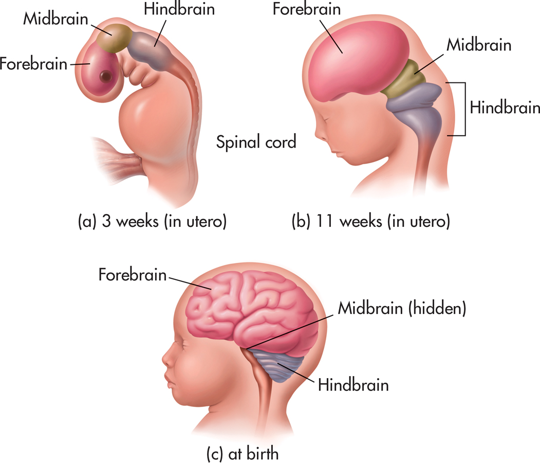
The neural stem cells divide and multiply, producing other specialized cells that eventually give rise to neurons and glial cells. Gradually, the top of the neural tube thickens into three bulges that will eventually form the three main regions of the brain: the hindbrain, midbrain, and forebrain (see FIGURE 9.2). As the neural tube expands, it develops the cavities, called ventricles, that are found at the core of the fully developed brain. The ventricles are filled with cerebrospinal fluid, which cushions and provides nutrients for the brain and spinal cord.
During peak periods of brain development, new neurons are being generated at the rate of 250,000 per minute (McDonald, 2009). The developing brain cells multiply, differentiate, and begin their migration to their final destination. Guided by the fibers of a special type of glial cell, the newly generated neurons travel to specific locations (Bystron & others, 2008). They join with other developing neurons and begin forming the structures of the developing nervous system.
Question 9.5
3pImpmKRAmdoqeD0LVqkuzpZdDDUpRnVgUxiNLcSSWrJ+UqRBuoVwRpd71mmax3onsZjinXnaI8lhWcTO+ONpsklOf+fu1lTbMAvO3gnd3oSFmyrT+qsnfN8/TDGqIJvQG8IeKsLWQtlLcjQA9QTRAi6+kKIpD7+elVcqDpZ1C5xm+gx9WOCCJAAT6oAohqM3IqANEleJ17qMDw9N9k3V7aOj8Kl6MmChGJE8XW/GzetjxTFpLCPalZsDzRoAKMWk8lAUHsv7h1lhLVnzzBh4mzSqogykS+KfdWxFCQzRj0pCI99vKrgOitpqzHYm8DTkhz5tA==The Fetal Period
The third month heralds the beginning of the fetal period—the final and longest stage of prenatal development. The main task during the next seven months is for body systems to grow and reach maturity in preparation for life outside the mother’s body. By the end of the third month, the fetus can move its arms, legs, mouth, and head. The fetus becomes capable of reflexive responses, such as fanning its toes if the sole of the foot is stroked and squinting if its eyelids are touched. During the fourth month, the mother experiences quickening—she can feel the fetus moving.
fetal period
The third and longest period of prenatal development, extending from the ninth week until birth.
The fetal brain is constantly changing, forming as many as 2 million synaptic connections per second. Connections that are used are strengthened, while connections that remain unused are eventually pruned or eliminated. The fetus now has distinct sleep–
During the final two months of the fetal period, the fetus will double in weight, gaining an additional three to four pounds of body fat. This additional body fat will help the newborn adjust to changing temperatures outside the womb. It also contributes to the newborn’s chubby appearance. As birth approaches, growth slows and the fetus’s body systems become more active.
At birth, the newborn’s brain is only about one-fourth the size of an adult brain, weighing less than a pound. After birth, the neurons grow in size and continue to develop new dendrites and interconnections with other neurons. Myelin forms on axons in key areas of the brain, such as those involved in motor control (Jakovcevski & others, 2009). Axons also grow longer, and the branching at the ends of the axons becomes more dense. But the process of neural development has only begun. The development of dendrites and synapses, as well as the extension of axons, continues throughout the lifespan.
Test your understanding of Genetics and Prenatal Development with
 .
.