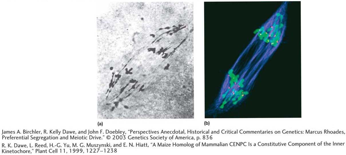
Analysis of maize chromosomes, then and now. Maize chromosomes are large and easily visualized by light microscopy. (a) An image from Marcus Rhoades (1952). (b) This image is comparable to that in part a except that the spindle is shown in blue (stained with antibodies to tubulin), the centromeres are shown in red (stained with antibodies to a centromere- associated protein), and the chromosomes are shown in green.
[(a) James A. Birchler, R. Kelly Dawe, and John F. Doebley, “Perspectives Anecdotal, Historical and Critical Commentaries on Genetics: Marcus Rhoades, Preferential Segregation and Meiotic Drive.” © 2003 Genetics Society of America, p. 836. (b) R. K. Dawe, L. Reed, H.-G. Yu, M. G. Muszynski, and E. N. Hiatt, “A Maize Homolog of Mammalian CENPC Is a Constitutive Component of the Inner Kinetochore,” Plant Cell 11, 1999, 1227- 1238 .]
[Leave] [Close]