Chapter 4. From GloFish to human disease models: Forward and reverse genetics in zebrafish

Introduction

From GloFish to human disease models: Forward and reverse genetics in zebrafish

by Karl J. Clark, Ph.D. & Stephen C. Ekker, Ph.D.

Introduction

The zebrafish (Danio rerio) is not only a popular tropical fish pet but also an important model organism for vertebrate development, genetics, and disease. Zebrafish have certain features—including a large family size, external development, and a relatively low cost of maintenance and production—that make them useful in the laboratory. Scientists have discovered some naturally occurring mutations in the zebrafish that affect its visual appearance. For example, “leopard” zebrafish make spots instead of stripes. Along with studying natural mutations, how can scientists introduce genetic mutations that interest them in zebrafish? To use the zebrafish as a genetic model for studying vertebrate development and model human disease, researchers had to develop efficient methods to disrupt gene function in the zebrafish. What are some of the methods used by researchers to study these fascinating processes in zebrafish? Here we discuss advancements in forward and reverse genetic mutagenesis screening, including chemical and insertional mutagenesis, and targeted gene knockdown and knockout approaches. With these newly developed genetic manipulation tools, the zebrafish is rapidly increasing in popularity as an animal model for human disease.

The Model Organism Zebrafish

Figure 1a: Two influential scientists that who helped establish zebrafish and other teleost species as biological model organisms.
Oppenheimer photo courtesy of Dr. Marnie Halpern. Streisinger photo from Nature Reviews Genetics 3, 717–724 (2002). doi: 10.1038/nrg892 http://www.nature.com/nrg/journal/v3/n9/full/nrg892.html, File name: 3-4_F1a_Clark_Ekker.TIF and 3-4_F1b_Clark_Ekker.TIF

First described by Hamilton in 18221, the zebrafish is native to the flood plains of India and lives in diverse water habitats2,3. Zebrafish were imported to Europe in the early 1900s as an aquarium fish4. Within twenty to thirty years, zebrafish became highly popular pets among hobbyists because they are easy to care for and because they have beautiful, diverse natural pigment patterns. Along with the features that made zebrafish popular pets, these animals also have many attributes of excellent model systems for scientific inquiry. Zebrafish are small (2- to 3-cm-long adults), hardy fish that produce eggs in high numbers and with great frequency. Like most fish, zebrafish are egg layers, and their embryos develop outside of the mother, allowing researchers to study early developmental processes using high resolution, noninvasive approaches.

Figure 1b: Two influential scientists that who helped establish zebrafish and other teleost species as biological model organisms.
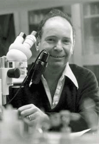
Figure 1 Source: Figure 3 (Panel A only) from Garcia-Cao, I. et al. Super p53 mice exhibit enhanced DNA damage response, are tumor resistant and age normally. Embo J 21, 6225-35 (2002), doi: 10.1093/emboj/cdf595, http://www.nature.com/emboj/journal/v21/n22/fig_tab/7594814a_F3.html File name: 3-4_F1a_Clark_Ekker.TIF and 3-4_F1b_Clark_Ekker.TIF

Since the 1930s, zebrafish, and other teleost species, have been used as model systems for understanding the developmental basis of vertebrate embryology. Notably, Dr. Jane Marion Oppenheimer (Figure 1a) published a series of seven critically important papers using fish. In one study, she demonstrated that a developmental structure called the dorsal organizer (or Spemann organizer) is responsible for initiating the body plan in vertebrates5. This developmental structure is functionally conserved between fish and amphibians5.

Figure 2: Wild-type and homozygous golden mutant adult zebrafish.
Images courtesy of Dr. Keith Cheng. File name: 3-4_F2_Clark_Ekker in jpg and eps format

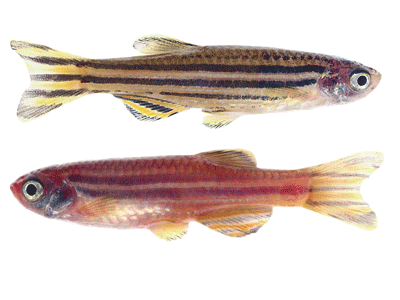
Dr. George Streisinger (Figure 1b) and colleagues at the University of Oregon launched the modern era of zebrafish genetics. They recognized the strengths of zebrafish in developmental biology and established zebrafish as a model system for vertebrate genetics6. In the 1970s, Streisinger purchased a “golden” zebrafish, one of the first mutant zebrafish strains, from a local pet shop. This “golden” zebrafish produces pigment similar to normal striped zebrafish, but the pigment intensity is much lighter (Figure 2). The recessive nature of the “golden” allele provided a simple, visible phenotype that served as a test allele for the development of new genetic tools7.

1.Hamilton (formerly Buchanan), F. An account of the fishes found in the River Ganges and its branches. Edinburgh, UK: Constable and Co., 1822.

2.Engeszer, R. E. et al. Zebrafish in the wild: A review of natural history and new notes from the field. Zebrafish 4, 21–40 (2007). doi: 10.1089/zeb.2006.9997

3.Spence, R. et al. The behaviour and ecology of the zebrafish, Danio rerio. Biological Reviews of the Cambridge Philosophical Society 83, 13–34 (2008). doi: 10.1111/j.1469-185X.2007.00030.x

4.Stansch, K. Die exotischen zierfische in wort und bild. Braunschweig, Germany: Gustav, Wenzel & Sohn, 1914, 349.

5.Oppenheimer, J. M. Structures developed in amphibians by implantation of living fish organizer. Proceedings of the Society for Experimental Biology and Medicine 34, 461–463 (1936). doi: 10.3181/00379727-34-8659C

6.Grunwald, D. J. & Eisen, J. S. Headwaters of the zebrafish—emergence of a new model vertebrate. Nature Reviews Genetics 3, 717–724 (2002). doi: 10.1038/nrg892

7.Streisinger, G. et al. Production of clones of homozygous diploid zebra fish (Brachydanio rerio). Nature 291, 293–296 (1981). doi: 10.1038/291293a0

Naturally Occurring Mutations in Zebrafish

Zebrafish display a number of naturally occurring mutations. Many fish carrying these mutations were isolated from the wild and are now sold in pet stores. These naturally occurring mutations include pigmentation mutations such as leopard and brass and developmental mutations such as the long fin allele.

Forward Genetics Approaches in Zebrafish

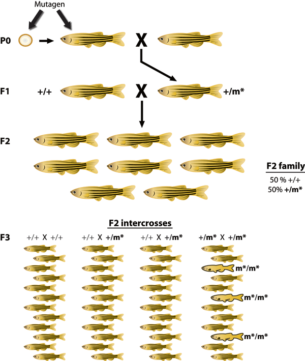
How can a scientist identify a gene responsible for a phenotype observed in zebrafish? As discussed in Chapter 2 of your textbook, forward genetics is an approach that starts with a phenotype of interest and identifies the gene or genes responsible for the phenotype. Over the years, researchers have developed a wide range of techniques to produce new mutations in zebrafish. The first method involved gamma ray irradiation of zebrafish sperm or early zebrafish embryos. Gamma rays are an effective mutagen (an agent that produces mutations), and they disrupt genes in numerous ways. Gamma ray irradiation produces both small (single-gene) and large (multigene) deletions as well as chromosomal inversions or translocations.

Because gamma ray irradiation-induced alterations often affect multiple genes or chromosomal regions, researchers realized that the identification of a gene responsible for an observed mutant phenotype would be difficult or in some cases impossible using this strategy. To overcome this hurdle, researchers introduced mutations in the fish genome using a chemical called N-ethyl-N-nitrosourea (ENU), which is a more precise mutagen at the molecular level. ENU typically produces small, single nucleotide mutations. Research groups in Boston, Massachusetts, and Tübingen, Germany, used ENU and followed the general scheme of an F3 mutant screen (Figure 3) to produce the first extensive collection of vertebrate ENU mutants8,9. This collection of zebrafish ENU mutants included approximately six hundred independent genetic loci. This landmark study was published in a special issue of Development in 1996. This collection of mutants was associated with diverse phenotypes and identified new genes critical for embryonic patterning, organogenesis, cellular survival, axon pathfinding, craniofacial development, and more.

Figure 3: Forward mutagenesis in zebrafish.
Generated by Illustrator. File name: 3-4_F3_Clark_Ekker in jpg and eps formats.

To understand the genetic basis of a specific phenotype, researchers typically carry out genetic mapping and positional cloning experiments. These approaches allow researchers to pinpoint the location of the associated gene and identify the responsible mutation. These experiments are much easier to conduct if the genome sequence of the model organism is known. Once scientists recognized the genetic potential of the zebrafish, they began sequencing the zebrafish genome in 2000 at the Sanger Center, UK. The final draft sequence of the zebrafish genome was released in 2011. As the zebrafish genome was sequenced, hundreds of zebrafish mutations were isolated. For example, in 2005, scientists learned that the “golden” phenotype was caused by a mutation in the SLC24A5 gene10. Could this gene tell us anything about humans? Interestingly, a mutation in the human SLC24A5 gene is associated with light skin color and is most often found in people of northern and western European descent. This observation highlights the strong degree of conservation of gene function between zebrafish and humans and provides support for using zebrafish as a model system for medical science.

Genetic engineering of the zebrafish initially started with a low efficiency but simple method of injecting DNA solutions into fertilized zebrafish eggs. In rare cases, this process results in integration of the injected DNA into zebrafish chromosomes. This method was used to generate the first transgenic pets—GloFish—whose new coloration comes from introduced transgenes that permit the expression of blue, green, yellow, and red fluorescent proteins from coral reef animals11. Although these fluorescent proteins are most apparent using a UV light, in GloFish the proteins are expressed at sufficient levels to be visible to the naked eye in normal lighting conditions.

Scientists developed two additional technologies—retroviral integration and DNA transposons—to modify the zebrafish genome. Scientists engineered pseudotyped-retroviruses to infect zebrafish embryos and a broad range of host cell types. Once inside the cell, the retrovirus can efficiently integrate into the host cell genome. Scientists performed a large-scale retroviral integration screen and identified numerous new mutants with the DNA sequence of the virus serving as a molecular tag of the mutation. This approach greatly facilitated the molecular characterization of the disrupted gene12,13.

The development of mobile DNA elements called transposons (discussed in Chapter 15 of your textbook) made microinjection and integration of DNA into the zebrafish genome highly efficient and accessible to a broad range of researchers14. With DNA transposon-based approaches, scientists now had an effective way to insert mutations into the zebrafish genome without using viruses. Transposon-based mutagenesis offers several new molecular options beyond that of retroviral mutagenesis. For example, transposon-based mutagenesis can be used to generate new zebrafish strains that express a fluorescent tag fused to the disrupted protein, which allows researchers to track the normal expression pattern of the protein encoded by the disrupted gene. In one example, scientists inserted a highly conserved gene called myom3 into zebrafish and observed its expression in the skeletal muscle (Figure 4a)15. This dominant fluorescent tag also allows for the immediate, noninvasive genotyping of animals to identify mutant carriers versus wild-type siblings at the single gene level. Scientists can use this approach to detect nonvisible differences due to genetic mutations, such as changes in behavior. Indeed, this approach facilitated a forward genetic screen for vertebrate genes involved in the response to the addictive component of tobacco, nicotine16. The mutant called Humphrey Bogart (hbog/gabbr1.2; Figure 4b) results in a viable genetic background with homozygous mutant animals displaying only one third of the normal locomotive response to nicotine in zebrafish larvae (Figure 4c, bottom panel).

Figure 4: RFP expression and mutations from protein-trap transposons.
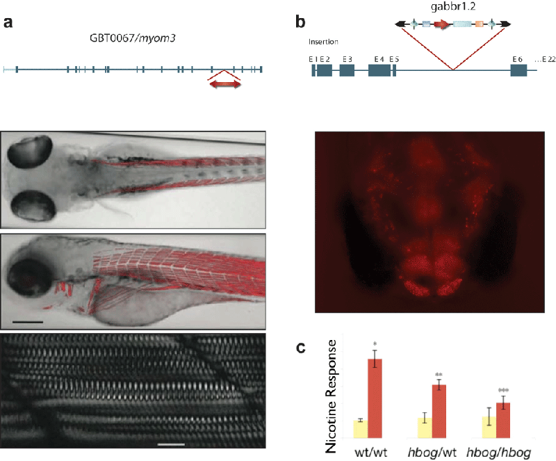
(a) From Figure 3C of Clark, K. J. et al. In vivo protein trapping produces a functional expression codex of the vertebrate proteome. Nature Methods 8, 506–512 (2011). doi: 10.1038/nmeth.1606 http://www.nature.com/nmeth/journal/v8/n6/full/nmeth.1606.html (b) New figures corresponding to data from Petzold, A. M. et al. Nicotine response genetics in the zebrafish. Proceedings of the National Academy of Sciences of the United States of America 106, 18662–18667 (2009). doi: 10.1073/pnas.0908247106 http://www.pnas.org/content/106/44/18662.short, file name: 3-4_F4_Clark_Ekker in jpg and eps format

8.Haffter, P. et al. The identification of genes with unique and essential functions in the development of the zebrafish, Danio rerio. Development 123, 1–36 (1996).

9.Driever, W. et al. A genetic screen for mutations affecting embryogenesis in zebrafish. Development 123, 37–46 (1996).

10.Lamason, R. et al. SLC24A5, a putative cation exchanger, affects pigmentation in zebrafish and humans. Science 310, 1782–1786 (2005). doi: 10.1126/science.1116238

11.glofish.com. <http://www.glofish.com>

12.Gaiano, N. et al. Insertional mutagenesis and rapid cloning of essential genes in zebrafish. Nature 383, 829–832 (1996). doi: 10.1038/383829a0

13.Amsterdam, A. et al. Identification of 315 genes essential for early zebrafish development. Proceedings of the National Academy of Sciences of the United States of America 101, 12792–12797 (2004). doi: 10.1073/pnas.0403929101

14.Ni, J. et al. Transposon tools hopping in vertebrates. Briefings in Functional Genomics and Proteomics 7, 444–453 (2008). doi: 10.1093/bfgp/eln049

15.Clark, K. J. et al. In vivo protein trapping produces a functional expression codex of the vertebrate proteome. Nature Methods 8, 506–512 (2011). doi: 10.1038/nmeth.1606

16.Petzold, A. M. et al. Nicotine response genetics in the zebrafish. Proceedings of the National Academy of Sciences of the United States of America 106, 18662–18667 (2009). doi: 10.1073/pnas.0908247106

Reverse Genetics Approaches in Zebrafish

Figure 5: Custom zinc finger nuclease mutagenesis.
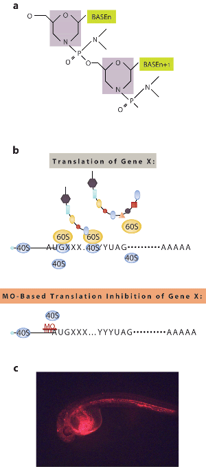
Figure 5 Source: (a) and (b) are from Figure 1a and Figure 2a of Ekker, S. C. & Larson, J. D. Morphant technology in model developmental systems. Genesis 30, 89–93 (2001). doi: 10.1002/gene.1038 http://onlinelibrary.wiley.com/doi/10.1002/gene.1038/abstract. Figure 6c is an image from Figure 3J of Nasevicius, A. & Ekker, S. C. Effective targeted gene “knockdown” in zebrafish. Nature Genetics 26, 216–220 (2000). doi: 10.1038/79951

As discussed in Chapter 2 and Section 14.7 of your textbook, reverse genetics is an approach that starts with a gene of unknown function and then identifies its function or associated phenotypes. The most widely used reverse genetic tool in zebrafish is morpholino antisense technology, which is used to knock down gene expression (Figure 5a)17. Morpholinos are synthetic nucleotide sequences that have a unique backbone chemistry making them undetectable and unmodifiable by cells. Unlike siRNA-based approaches for gene knockdown (discussed in Sections 8.1 and 8.5 of your textbook), these molecules work by binding to RNA and interfering with its translation or splicing (Figure 5b). By injecting morpholinos into fertilized zebrafish eggs, translation of a specific mRNA can be inhibited efficiently for several days during a developmental time period that includes much of zebrafish organogenesis. Morpholinos have been used to determine the functions of unknown genes and model diverse human genetic diseases in zebrafish (Figure 5c represents one such example, the blood defect porphyria)18.

17.Bill, B. R. et al. A primer for morpholino use in zebrafish. Zebrafish 6, 69–77 (2009). doi: 10.1089/zeb.2008.0555

18.Nasevicius, A. & Ekker, S. C. Effective targeted gene “knockdown” in zebrafish. Nature Genetics 26, 216–220 (2000). doi: 10.1038/79951

Generating Zebrafish Mutants Using High-Throughput Genome-wide Approaches

One goal of the zebrafish research community is using a combination of forward and reverse genetic approaches to produce a mutation in every gene of the organism. ENU mutants are currently being screened using high-throughput sequencing studies to look for base changes in individual genes19. Additional mutant zebrafish strains are also being made using both retroviral and transposon insertional strategies15.

Figure 6: Morpholino-based gene knockdown technology in zebrafish.
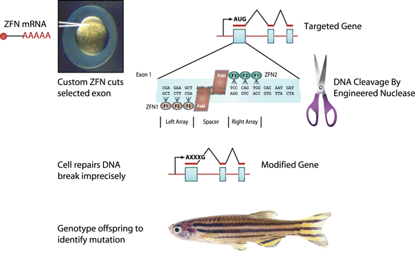
Figure 6 Source: Adapted from Ekker, S. C. Zinc finger-based knockout punches for zebrafish genes. Zebrafish 5, 121-123 (2008). doi: 10.1089/zeb.2008.9988 http://www.liebertonline.com/doi/abs/10.1089/zeb.2008.9988, file name: 3-4_F5_Clark_Ekker in jpg and eps format

Until recently, the most commonly used mutagenesis approaches were largely random or semirandom in nature. Today, however, scientists are using new approaches that target specific gene sequences to create double-strand breaks in selected DNA sequences in zebrafish20. Using this approach, sequence-specific DNA-binding protein domains such as custom-designed zinc fingers are fused to a DNA cutting domain from the DNA restriction enzyme FokI (Figure 6). Following the creation of a double-strand DNA break, the DNA tries to repair itself through the error-prone process of nonhomologous end-joining (discussed in Section 16.4 of your textbook). The resulting insert or deletion can produce a frame-shift mutation in the protein coding region, which results in a mutated protein product.

19.Moens, C. B. et al. Reverse genetics in zebrafish by TILLING. Briefings in Functional Genomics and Proteomics 7, 454–459 (2008). doi: 10.1093/bfgp/eln046

15.Clark, K. J. et al. In vivo protein trapping produces a functional expression codex of the vertebrate proteome. Nature Methods 8, 506–512 (2011). doi: 10.1038/nmeth.1606

20.Ekker, S. C. Zinc finger-based knockout punches for zebrafish genes. Zebrafish 5, 121–123 (2008). doi: 10.1089/zeb.2008.9988.

Zebrafish as a Model System: An Ever-Expanding Field

The high conservation of gene function between zebrafish and humans has led to the exponential use of zebrafish in the scientific literature, beginning with the publication of the first large-scale collection of genetic mutants in 19968, 9 (Figure 7). The extensive new reverse and forward genetic tools in addition to the completed zebrafish genome assures the continued use for this outstanding model system for years to come.

Figure 7: Zebrafish publication rate.
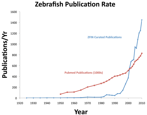
Figure 7 Source: Curated zebrafish publication data were retrieved from http://zfin.org in July 2011, and Pubmed publication data were retrieved from http://www.ncbi.nlm.nih.gov/pubmed in July 2011 and analyzed by the authors.

8.Haffter, P. et al. The identification of genes with unique and essential functions in the development of the zebrafish, Danio rerio. Development 123, 1–36 (1996).

9.Driever, W. et al. A genetic screen for mutations affecting embryogenesis in zebrafish. Development 123, 37–46 (1996).

Summary

The zebrafish has a diverse history from aquarium pets to embryology. Recent advances in molecular genetics include the popular use of the zebrafish as a model system for understanding human health. Forward genetic screening using both chemical and insertional mutagens has identified an array of new genetic loci for vertebrate development, disease, and behavior. Reverse genetic approaches, including both targeted gene knockdown and knockouts, now open the door for hypothesis-based testing based on comparative gene sequences. The ever-expanding collection of new tools available to researchers has resulted in an exponential growth of scientific publications using this model system.

References

1.Hamilton (formerly Buchanan), F. An account of the fishes found in the River Ganges and its branches. Edinburgh, UK: Constable and Co., 1822.

2.Engeszer, R. E. et al. Zebrafish in the wild: A review of natural history and new notes from the field. Zebrafish 4, 21–40 (2007). doi: 10.1089/zeb.2006.9997

3.Spence, R. et al. The behaviour and ecology of the zebrafish, Danio rerio. Biological Reviews of the Cambridge Philosophical Society 83, 13–34 (2008). doi: 10.1111/j.1469-185X.2007.00030.x

4.Stansch, K. Die exotischen zierfische in wort und bild. Braunschweig, Germany: Gustav, Wenzel & Sohn, 1914, 349.

5.Oppenheimer, J. M. Structures developed in amphibians by implantation of living fish organizer. Proceedings of the Society for Experimental Biology and Medicine 34, 461–463 (1936). doi: 10.3181/00379727-34-8659C

6.Grunwald, D. J. & Eisen, J. S. Headwaters of the zebrafish—emergence of a new model vertebrate. Nature Reviews Genetics 3, 717–724 (2002). doi: 10.1038/nrg892

7.Streisinger, G. et al. Production of clones of homozygous diploid zebra fish (Brachydanio rerio). Nature 291, 293–296 (1981). doi: 10.1038/291293a0

8.Haffter, P. et al. The identification of genes with unique and essential functions in the development of the zebrafish, Danio rerio. Development 123, 1–36 (1996).

9.Driever, W. et al. A genetic screen for mutations affecting embryogenesis in zebrafish. Development 123, 37–46 (1996).

10.Lamason, R. et al. SLC24A5, a putative cation exchanger, affects pigmentation in zebrafish and humans. Science 310, 1782–1786 (2005). doi: 10.1126/science.1116238

11.glofish.com. <http://www.glofish.com>

12.Gaiano, N. et al. Insertional mutagenesis and rapid cloning of essential genes in zebrafish. Nature 383, 829–832 (1996). doi: 10.1038/383829a0

13.Amsterdam, A. et al. Identification of 315 genes essential for early zebrafish development. Proceedings of the National Academy of Sciences of the United States of America 101, 12792–12797 (2004). doi: 10.1073/pnas.0403929101

14.Ni, J. et al. Transposon tools hopping in vertebrates. Briefings in Functional Genomics and Proteomics 7, 444–453 (2008). doi: 10.1093/bfgp/eln049

15.Clark, K. J. et al. In vivo protein trapping produces a functional expression codex of the vertebrate proteome. Nature Methods 8, 506–512 (2011). doi: 10.1038/nmeth.1606

16.Petzold, A. M. et al. Nicotine response genetics in the zebrafish. Proceedings of the National Academy of Sciences of the United States of America 106, 18662–18667 (2009). doi: 10.1073/pnas.0908247106

17.Bill, B. R. et al. A primer for morpholino use in zebrafish. Zebrafish 6, 69–77 (2009). doi: 10.1089/zeb.2008.0555

18.Nasevicius, A. & Ekker, S. C. Effective targeted gene “knockdown” in zebrafish. Nature Genetics 26, 216–220 (2000). doi: 10.1038/79951

19.Moens, C. B. et al. Reverse genetics in zebrafish by TILLING. Briefings in Functional Genomics and Proteomics 7, 454–459 (2008). doi: 10.1093/bfgp/eln046

20.Ekker, S. C. Zinc finger-based knockout punches for zebrafish genes. Zebrafish 5, 121–123 (2008). doi: 10.1089/zeb.2008.9988.

Quiz

Question 4.1

ctBYg4lptTmYIYDbRHuyEe3OsnQHCpl98sVRExNkiOTTqwUw5l4mQ6SfiPHxenERh9ugSwFyx+snRs71vMVefiokoeF6eSy3FkcVN7fkaH3KaGineIY9ehylPVn94V9l4TQIeq5oD16U5FeQlJJC6ZCm7pyAK3vKnk/dZlZa9VQLlanJaQsG23R5G/Tv+BmBkm39FYIuG9IVzbdU4x0U8LqrtYvzgqM7BC9D0ol0ZqqzCb+y91SbEn4KAiDeaHVsOgT8kirvXvR43yGdw8ozi2fzE0qmdPBnPvzRENHC5KArCSAhcIz2HAhY+Td/lyJOpjLpDpKcq/9o3xIzaIQipvy+4fobdvIi2xDVBd5yQ3w0S2aN7NR9O6BlrwgKI0crKzCDOg/cl/WXmdR5uP646S+Q6h3lvp0d02WUbOMjrA0jbZng1G8XsuhcW2RDmMLvnn1mS1kB/hKVVM8z9wY8bf+qQEo2TMJaVvb1CeU21Hibp17eh4zEXMxH1xoMlZcNxYJBUVDbNe2VI8RZ5AkRva4hDGEo8RNxb+17NHWDWGVRaT07+z/g4qmRrEEu9hFh0IJTE82pHNWj8S1LNSq2iB3wYCNfsVbe37c+3CR0J1106azl2ca41izC0R/U1mJUsQcFy3HXCFNlfDaV1+avk+43FshuqeZoH+u9ZaX1Zj5pnhttLTse51JWbWW5mvwjO78gxACMJ+qRoijTnpKIp6r01yvDTg3wu4YkvcA8UWyUj5PXrBsw3fVLXav7u8Q9ow3H9NI7yrExf0T+GbrjLizBRbykDB/qvbsscgjQ8LW0Bcrvc2/3oEKZHNxbA+tApcvCOeck266ngA66D5T1ox8hjaEb97tHaT7ZgRAsjJSxKlYoSq5Lm9a6pF31SbRsIbqihiWsq4kau4CuqdXoUQpap9BvPShXdSQzR+o9U7kbOB+MeRg4KkIBRaXtcEll5d+f6pIP5qbevTjuhOAZ4mrAAZfgzdyPZNpivQ8AsRUVgDXu/fwTae0qcdEqPlApnql36VoKmwYAIlE9ZDppmD5la1gbN0vfMzoO1H50jijaXVnedgltyaoG+03iaW21LWd8QT5CXaQbgi1129pr5hZprVY4Gln8KeAjAA==
1. As discussed in this article and throughout your textbook, genetic model organisms have been very important in our understanding of many aspects of biological function. Which of the following statements about genetic model organisms is TRUE?

Question 4.2

ViztDpyVyIcAhyyH33bVES+nYML1ZJEplrMSD79a/9NO7nLj+maeZuykoAZ/jn+jDU+B4HMVI7gf/uIbbJPlFpaqGasakLs4EwPo4rfAmwjiBuxgQrwnyxG2UmO5fRgsy/W9h4nBUBK68qtpqlVq8wtFrnpToI1vG5GIAw0ybAqydWmSxJltE+wtnKWfy+doJWEx7urJ2jfQiYgN/5TrgglUsknKbaatug+RdfonouPUx6yUbZCgf+rrj5YvdlOO0l5VN/SV8ezQUNM/rEPBEdKgUjvTCrZq8sC58aX2/L7LgYpVbXdGnAHfosAVO470EFGg2OqIE8dvfp40I7X8qVXSeje33aYLwx9Ho86KpMehDQWItn/nRtMdu0ceolyzcCsK3oSQoxnQg15aCA/OORA97S+rOSmR4jQlFIi10d7wRvfvwSog2auxODURRbYRCNk/4Kj3xi6A3g2yQSu/AZBAjadaLuFHKZWcoxtzm50bsTC7VfNUl02d5CFCeHa3UFrnkZ9C7Gl7aItwUaj4Wr0tFfc5zHArbRFfWttjoFYkXgFYnNujfwuwHKrFxeN8ByYFuBeeVyd7qWQKfHMYFr9+sYlI9AuyqZRdfc7kyEbd2SPI1VycZORt0pd4Q7WkFM8VvA3bLuTJP+5/jVGNWYSp4xJL3zUc00R2E8GNNUg6Rhb1t/ia3jPvEdBFJ8v4iFtV2upzXxkKsKKz7dc59KHbZHSFmWJHOViUelPwQdTz5KdEJ2MHPU0xOD5mKktonOXux838kpk6uXj6mCgcw0kXUVs3lWf252aBiRD8y13JZIHYkFGu39W3N8pJuAzNSCTFUBvn/GnQNVQMxO8J9icYmCTPwzL6FxKKuJul/1BTJAMbTXqBpNwnn78NNvaGMdPs1iJG0Mw8jlpU3Xu5JtugZtCWwYPDBlt0UGJ+Yip3yNJSpiFTmTKsr9yZv/waEHhV3XdrCG8iIIAKhslgi0cZs+oaJcra3OeX5nNw75a5DJQA9EVmjIF/YrpZs8b4LjDDpDa4Hm7o+cHKWh5jlCfkVv9WnzIPJB4vbHKQDMs/xm0olq3VETZdBqvBR0HGfN1QKZPX2ayDhnl9f+lZqhZn0Ki2gBn/p3vL7QdbrwVMtVdmByoPp1Yb5Wehw2bvQGLfmn+qbOx7arFk
2. Transposable element (TE)-mediated mutagenesis offers a number of benefits beyond classical chemical or radiation-based mutagenesis. Which of the following is a (are) benefit(s) of transposon-mediated mutagenesis?

Question 4.3

msRVf93cgJwpV+/llBIpfu1NvLv/O/CIKE/TGrBql3Ee/XnD5I/U18ash/Xb18o3jI2aG8mDJuArRlM7/hcQNpvrvlrbrxjkFyH8cZBJmcw5iC0+h6meIB7vbyzp4VZUCVDAgS2NrRCQZofJKG0WNAhGBrYoRPfEb2nVRuDR+kD3DdkulsEtfvdrv/cni2Vq2tcHq+dBp9PmF0g2LQOFH/62EZvReHvPqA5DkvbKMsARFIO6bsk4tmvvVdOSzZH3y4PaxZRooy9txDjbOpUvdzu5sM1KO9zHXKLQ7uiBn2/DPew6irYhb81mco9+z0kUftjqKQuPeC/ICKgUbkvsTJqpIubQBgmqnqIZWmWV2GQDSBut27JbfXkLkU64+vD5846490O765kYhgf5BdO1m7ic2pXerWmbZVzS6H1u3RxpKqywAP3vDhrFAPRs1Dks3PqcYztaaKhy1yI7fBDxGLZnRlEkWqtd2iWpLcf9g968YzfaqnCW7fwiAfxmEOXKaY+ZxOO/U27AoyIQcroTnEGGIQukZLUoTshYXYmK2/q62ceKN8iYgiqj4yJNQNrkA93w5eEOa9lpU9HV0V2gtA4mkpulqAmuRh2EhYYKIkyW3L2YgUeTzD6cuxmjSHyFTlA4nYDkCEfYDFguw+ar6Vpzsl49vFH/IPFVkhMTlVwuD4XKDLxDjskqmsbxtVnZTbPLrwbAqoGkvmIydnfTScIoT+O9Bty8AeLswN9CbzxXE634WAPBzVYGbAUOXbpoOchQ4w==
3. As described in this paper and in your textbook, targeted gene disruption techniques have been established for most genetic model organisms. These techniques generally harness endogenous mechanisms, including those of gene regulation through mRNA targeting or DNA repair. Which of the following techniques rely on endogenous pathways of DNA repair?

Question 4.4

p7xR427wDK8oJf5RP4uriubKWQGHv/mijpMYN0lQdhPXSTAr0Yp9HliNDWndWLga+243pY0d2inU+AKQLW1AvstbPeS2yqjCLuHuz/Y47wGx0CmWgwYiJd5h7FdQSHIzU5jRD5I1oD+1amuQQju9aaUC28U7kqALC8NuKsUpFfxNkwIa/o88tLNdRTcDeJO+FKozSTw+kR0KGmhnxc02m6Kg5x9w0NwavxoJGL2H0U0wmXNEwnGuof1Pyov2EVoKWib1CZOEUlIjn5KL9AMfYiNZ/1eJEKgfMkCC0dbN0HUzug98IflUVt2eMFaa7PTMkVngAIvgN/aIuylvp6nRF/FT1Jcd6OAh3CUTZ2iYpVhlxXZ0hG3F4EF8IsX1fIQwRvA0Ryxt0vszb6b49UtK4Qe717uNmcwlA32RjE7ueznITykax4oEJ+KviuB4OMyOnYaRN+hxpPDykOWSIN/AkjlUBd9JoQBgRoONJdaRzJ0zKgk/eMIBMmkXD2C7RPquPq6i8N47OmmELOf4T1ueYm007kY/57TdNbgPrRbkyRPqQcm1UMMYIwHjIp52ejSMtvQ5kFzgfjuGWqPJU1DuVZ85CBbTfLT+kC5AEg8TvIqvzRWBe0FRdAJPlRgpcwKTzY5W+LyYJvjJnvBC1xv4GyOiAtjlfWfGbyPulVHh6/bBcmc2bsuRu16X48fiZpdiMnPrut6ZewA+hJFjPiGfCyb8UAkSxthYVZPQ5A==
4. The paper text and Figure 3 discuss the isolation of recessive mutants through forward mutagenesis. How would this scheme change if one were attempting to isolate dominant mutations?

Question 4.5

sJaY886V/uwaNJ7fgkwcTvtj8SjqWTqmO5elJe1z8YRBK0oi2lNNu3MhO8FSgwCwGqAlgw8JYnuMkWKSIIbhCSOv134A963RzxqS6IJaad+oPXFhrj8USgivWM3AZyfEm2F9UUpxoX2UxEz2TxDNc3P1sTHdaoKzrhJQ6L3LK+WIEBcC/56jRO4GUmU5Y3Jb5LN+G4qBMMdHl9NfBzWj4vPPuTECU3ENy/vhiN4kf1ouMYOwNjBon48U1TEwSGL3ryUHslBLH3yd8rBACKpfIGmBE+9gDb5ivC4F33CfjEYf2GCsJuOSZkSJXyqp7qMtMoiOC1VOQEGNunjTdAMJoOnYD3oByQNU9AyYoL7B+G10ZKXcINNgi8UXaztKXruA44fIUUChhQpIJryQPWsfnpGl6VsYhnDwhPGtV6i6sDymsgSl4vpPHPwStFzHQK3xYmYUtB5cK3y33gtmRZ/e2zzVVxbA+3TbTexKm2cgI67TQfO/i5JSgdYUp51JWBMr1+b+na5Lq6UAVxmXieqAo0Dg+R6wsXRYfibI9MRMa0xRL1VHlkfpJb3FysSLsUf4Ns/g0N2Vaqv0PPo5PdiyJk5uLqFmifG6ZERTSXzQ/42VwYWGJE3kxizdYEjdPrN0UMUtayHBivosWHoQ3X+0I/xSqW2PGNxJ+xvUNjvsGc2+fr6y82X0FshgPqLOoiLzBORbuQyGpkfnwK9eb5ZWFsueNqadb0o/ztkWWs0ppYFBEyVV+i3UUmqdROfboJs1zcKNiZ2LCDfsJQSZ0xqUqaO5rI+EmvGZlYWpvnKFyFP3f2PGyKd3AVlXnNltYQA5HFi3BhaIGl6q7r16m4448juEa8DaL2vJ+FjZMGKXb8Rc+z2pgvMqwOW2f2BziAx4XFE05Lcd+g7GG5svTMmDPWtn9mT/fCPNa8IAh5zw1UaBGXxALbNF63bx53fqZRNFRZevssaCXBWBsLTCwsQfrDZi2cnrLCivK6NvkuTVwG4I+ltuOvES4e2TH3fSOgmrrQMY8W9MWMvJdXFyV1Onzmdlq3KG4NbAlj8N5AogR739lKEv0fz4Q+MpqUq97rChcSsrnDfhVChiMn2Fhppkgxu8TvCqdjDVDJbvnTrP0ZOKgOLTxAYNAOnalnZoQfZULFS31XcuWSGTdPxQ37XOjBevQSNj2FxO0QSoe1UCk6U159qmpwiyOz8T01EEqr2ZnI6GffIpYR+7K2e6P7grnjUQLaRXkeTfYqWiRUhXxMaV2x8kU+gb4bFse9gfG5aMWFX00cYyGlmBtfqpT6HynQ5fz09xfIHlWLrkECRjCBL+E2KM8nNvELUMtnrg/vaSLU/WTLtx72eyOhLKRAdJ25B16H9RhJjE5emunY6mB5P7ZqEZe1GRxbNEpKiBzx87dTDWIQqSbdPNOtAkvK6iAeVTvUDfFl94+SMjWKot8L+dfSMjlRQvz4ETcoFjpWGtLstMJp2etqSv/xyT4MfU1b/8wcPs2K11sIbG7h2NonP1CuyDdoY+02UoUMOxKUrR
5. Hydroxymethylbilane synthase (HMBS) deficiency is one of the causes of acute intermittent porphyria, a disease that has been suggested to be one of the orgins of legends of vampirism as affected individuals both have skin sensitivity to light and increased desire for iron-rich foods. To identify a stable mutation in the zebrafish version of HMBS, you decide to employ directed mutagenesis using a sequence-specific zinc finger DNA binding sequence. How could you identify and/or confirm mutants resulting from your zinc finger-mediated targeted mutagenesis of HMBS?

Question 4.6

ZrY3WFNVFyYnH52KPf9urHc7BwFHpbvlWZREoZKSnkSeZ64QiqtghEzCmKU/ySUdhrXdlj43I9KX4XF/cQ2Zg+/KJlpubgJrnQDpXWIHlyszQ9Oaazh00SdfrvCyPci6JD7oLroijvyrLnyGfGNOUNV66nnyBBBM0iSdEgG5p1RymKdoqIIGsYgB6X//r1ER8Cz9+57aykZjtwL/LRFr5wZUdqcnXrP983CwM+tgqkrgSOeeZ+/bJ/4YcxpuJcmzxVXZbbAOIFDUcgaVH7/t+jS8ac7zeNxJqtfmbk5AYOhJNFrMok02fcvideictlKRqv2W2zRFlFqTZ1/nqHSSVNXn29bbgOAXadWAoAAZ9y47MJVdNfpR5NOEejpesefzveeEKSIDet21bEPJFIK8Obdig+9f+xxXvHgCMaiGbz/RmVFxKFqelipnRH0jirPTBVr8X9TvPMf6mre3L+00GCG0cHkdLncRmiEtr3zugFRSp4/xBHvBvnVrFjcs6MCO1IAiEMbA5rBKkkbmFuRStye9v8iUdyrWwWN/sw+OU2dTPoANoFkym8+TscpRjAL/MTjK9y3nGDQlWvOyiH76SbmDo8bFugTVmCaIwIkCOl2S4ICPw6jb/vX/eRm8ztYJrzNAVsO5Sca2Nqcx2HGu/0h1C7pxgP+D1keH3Od9wh7FsDSQPFL8wrhYeYpYA7kEhDP1e0S/dQRD8b4iW7v/DcLkB7B48UYLt8Ies5v1zP4BYtGn0Pb0oPEpWHVqXiI4EAo9a/Q4K/Swe4wyqEvScf652N4Wcl1PcBWT9etjOgkB2NHVqwm9nUOuXajJQkYMMYTfTtPIcJS07eiahv4xm7YIPqh2Kc25S5dX7ER1+NUP/GvxU3WVwtiBNM738+IYAKn5dlZiTfm8T8qrqdVjrfvTZ2iuXg5H97QALQruo9UVo/gIVrPUbfHDhRGDSIYLSmDO3gxjK+tZ7LedFgcrzZVr/PnK/YLdovMcDESuBBDmxLeZZarEVETMpSYSYJkpl7snaA16oZPhUUfCQBa+TaTeCMuu0Anais1/SfVLO8ZGWkHy/DMbhG35Q/EBMS3T8UZQSXkY9b3aK07RKOoG8yd5PZDV40OgA7WYYhqUHePhGH3i16qILvaLYrFVGlop+lTRDTZK6vqAz/vBJCaOe3+khPRkFgTUZG7HekUI1fPTet33kBaM8yOJ6q96fYJTTtGdmvmlE2ki8uXAcfPLkh9w768HB2651CBIHg8Aur/9XBYWir/KXLBsVkaNXdIn3N06Uyo1be9nKOaxG+aDRgkM/AIcwYm223h6iWIdTUwlB3ezSxJ3Z7NGMevwZoiX5dD9PUH75/RNb9cbTRXjuuWRbFJqczVWBNXuTi6Ie5ZiFFjxR+nY+pYgTCm5baNZAdSj6SdSNVTMKf8z54pivIAV9Uvw+NXCFz442LA4wZ8b4mdVkLXn+igwK/dEweA5FvyW9Dw+R8oJcPKsIOW1AhyWs3oWw5Ig3FbM0m7XDCe6iL7wU+Nmzn8kGHhJHUMFbEg7konwgtqq1PPh1EUzIpRM9qsGuZ6g54lFeF6EzXu3U0huQcrgIwHrkYK/ipTP49bOVJJGJzUgh4xW7PIhRPlfJ/m71nBHIKXYpYUbCgJu+cAp5QoUso3ukdU2i+paRTbo8kDJcDpHYfEP0zMxHsxcrog31v22H3jykCyM6I2GBS+J5UFaPt7LajbZW38WL2zsiR+vYkuOBawav7/0ct70Q5NQrb06IsJQ3fVNSwPBwpAeuU20k5vKJ041PSLv5w6aIC+QSLV71Os+0k9/qYCQlVYY66eUHkEBtTQQqESpLk5+Z9AJ9ucWL7ywu+pO46GNZ1yrpnMblTWg78VwiYh04A47N/3igLVPegRwWslK7MIzYWsra00bk2QFFrVdjA3AdEnlqcOK84DZKU/YwQcJLs+WQMfhUMJ8j84QVQLdfqkWXXu1KaE+ZeGtkgjyednWMdqDIFwKRWWTDCPnTwX10oHF+GCTv6LFv61KTnwRk3tj88PgwVIYhnJoVRydCwyX9/GZXSsmZ6aY99LdX/ga13dkT4F9gUsp62sHufIbTVxVhh/7L2V1G7OGi58AoK5AtWe1XCYXN1olc2Hsk9Wlme4mTOZlMc/afmtHXUJmH/Zt2EiCru+dCGFdvBswepVeqWGPFL/CBMMmH9Bdzr1IhaaNeAOy+SsEocYRLqbROIW9FJLQYoqGETR3pl/DK1bLlvVIQFL4YRnVpZsMw+P0bBCRszSGghFEG+fJOoMFl94ZSTfDunAsw6H6DXmUg85AUoYv3v1BNm1rTameKDA+tQagyWEExbrcKuH81QCCwFxV4RDfBaIDRx8lvePd0xtPcUKWlzm8DnIs0w2cwxQ+a2rbRy+toIacafiqnw+bfe0Wh8d9e7MRTABBmh28h3KTHeuKF+rU1UhT0LHgNXW2mmR+7/nkhy9BzN/hLuIUefsC8la/yLSsxzam+oSq2dy66yFhxm/6EcHFY9tO4tFod4+0siYCtdzRd1c8Jnka5Cb3Nh/8wOlTnI5mEwLdYilml+X+6OIlIkgTkgd9bSWULhuhlPFF7BM5oo2GaIw=
6. In a novel screen to identify zebrafish mutants affected in left versus right motor neurons, F3 fish from ENU mutagenesis (see Figure 3)