Chapter 5. Polyglutamine diseases: from expansion mutations to targets for therapy

Introduction

Polyglutamine diseases: from expansion mutations to targets for therapy

Carlo Rinaldi, M.D. & Kenneth H. Fischbeck, M.D.

Introduction

Certain genes contain sequences of the three nucleotides cytosine-adenine-guanine (CAG) repeated over and over, resulting in long stretches of glutamine residues (called polyglutamine tracts) in the encoded proteins (discussed in Section 16.2 of your textbook). In 1991, La Spada and colleagues provided the first evidence that these CAG trinucleotide repeats may be linked to human disease. They discovered that the neurodegenerative disease spinal and bulbar muscular atrophy (SBMA) is associated with an abnormally long CAG repeat in the androgen receptor (AR) gene1. In the years that followed, the same kind of CAG trinucleotide repeat expansion was found to occur in eight other genes, leading to nine different neurodegenerative diseases. The list of CAG repeat expansion-associated diseases now includes SBMA; Huntington disease (HD); spinocerebellar ataxia (SCA) types 1, 2, 3, 6, 7, and 17; and dentatorubral-pallidoluysian atrophy. Because the trinucleotide CAG encodes the amino acid glutamine, these nine inherited neurodegenerative disorders are associated with expanded polyglutamine tracts in the disease proteins, and they are collectively known as polyglutamine diseases (Table 1). How do the polyglutamine repeats contribute to disease? And how does polyglutamine repeat expansion occur?

TABLE 1

Polyglutamine toxicity might also contribute to other trinucleotide expansion diseases. For example, in the neurodegenerative disease spinocerebellar ataxia 8 (SCA8), which was found to be caused by a CTG repeat expansion, animal studies have shown evidence for transcription of the opposite strand, where CTG is read as CAG2. This bidirectional expression may also apply to other trinucleotide repeat disorders, such as myotonic dystrophy (DM1)3 and Huntington disease–like 2 (HDL2)4. Studies in human samples are needed to confirm these findings.

1.La Spada, A. R. et al. Androgen receptor gene mutations in X-linked spinal and bulbar muscular atrophy. Nature 352, 77–79 (1991). doi: 10.1038/352077a0

2. Moseley, M. L. et al. Bidirectional expression of CUG and CAG expansion transcripts and intranuclear polyglutamine inclusions in spinocerebellar ataxia type 8. Nature Genetics 38, 758–769 (2006). doi :10.1038/ng1827

3.Cho, D. H. et al. Antisense transcription and heterochromatin at the DM1 CTG repeats are constrained by CTCF. Molecular Cell 20, 483–489 (2005). doi: 10.1016/j.molcel.2005.09.002

4.Wilburn, B. et al. An antisense CAG repeat transcript at JPH3 locus mediates expanded polyglutamine protein toxicity in Huntington's disease-like 2 mice. Neuron 70, 427–440 (2011). doi: 10.1016/j.neuron.2011.03.021

The Polyglutamine Diseases

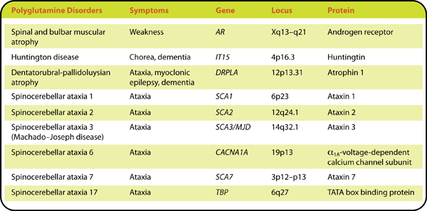
Although the genes associated with SBMA, HD, DRPLA, and SCA types 1, 2, 3, 6, 7, and 17 are structurally and functionally distinct, these polyglutamine diseases share certain key characteristics. They are all progressive, often fatal disorders that typically begin in adulthood and worsen over a period of 10 to 30 years. The diseases occur only when the length of the repeat exceeds a threshold number, ranging from about 20 to 50 CAGs. With the exception of SBMA, which is X-linked, the polyglutamine diseases are all transmitted in an autosomal dominant manner, suggesting that the mutant proteins may be toxic, and their toxicity has been confirmed in cell culture experiments and animal models.

Individuals with longer CAG repeats have more severe symptoms and disease onset earlier in life. For example, in HD, the median age of onset decreases from 67 years for patients with 39 CAGs to 27 years for patients with 50 CAGs5. Furthermore, the severity of the disease may increase from one generation to the next because the length of the CAG repeat is unstable and tends to increase with transmission. This phenomenon is known as “anticipation.”

How can this tendency toward expansion of the CAG repeats be explained? The characteristic instability of repeats can be caused by mistakes that occur during DNA replication, including strand slippage, misalignment, and stalling (Figure 1)6. Single-strand loops can form in repeat-containing DNA, allowing the two strands to be displaced (i.e., slipped). Such slippage can result in the addition of trinucleotides to the newly synthesized DNA strand.

Figure 1: CAG repeat expansion during DNA replication.
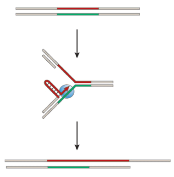
Figure 1 Source: Modified version of Figure 3 from Nature 447, 932–940 (2007). doi: 10.1038/nature05977, file name: File name: 3-5_ F1_Rinaldi_Fischbeck.jpg

5.Brinkman, R. R. et al. The likelihood of being affected with Huntington disease by a particular age, for a specific CAG size. American Journal of Human Genetics 60, 1202–1210 (1997).

6.Mirkin, S. M. Expandable DNA repeats and human disease. Nature 447, 932–940 (2007). doi: 10.1038/nature05977

Protein Aggregation

Expanded polyglutamine tracts are sticky and make the disease-associated proteins prone to aggregation. Mutant polyglutamine proteins accumulate as nuclear inclusions in the brains of HD and SCA patients and in cell culture and mouse models of these and other polyglutamine diseases (Figure 2)7, 8, 9. The inclusions are not necessary for the disease mechanism, and they may be protective10, 11, 12. Regardless, they are a pathological hallmark of the polyglutamine diseases. Also, in vitro experiments have shown that polyglutamine proteins with longer repeats aggregate more easily and are associated with increased cellular toxicity13, which led to the hypothesis that the tendency of the disease proteins to aggregate is related to the neurodegeneration. This tendency is consistent with other, more common neurodegenerative disorders that are associated with the accumulation of aggregation-prone proteins, including Alzheimer′s and Parkinson′s diseases.

Figure 2: Nuclear inclusions in polyglutamine disease.
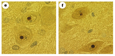
Figure 2 Source: Figure 1 (panels e and f only) from Skinner, P. J. et al. Ataxin-1 with an expanded glutamine tract alters nuclear matrix-associated structures. Nature 389, 971–974 (1997). doi: 10.1038/40153 http://www.nature.com/nature/journal/v389/n6654/fig_tab/389971a0_F1.html, File name: 3-5_ F2_Rinaldi_Fischbeck in jpg and eps format

7.DiFiglia, M. et al. Aggregation of huntingtin in neuronal intranuclear inclusions and dystrophic neurites in brain. Science 277, 1990–1993 (1997). doi: 10.1126/science.277.5334.1990

8.Davies, S. W. et al. Formation of neuronal intranuclear inclusions underlies the neurological dysfunction in mice transgenic for the HD mutation. Cell 90, 537–548 (1997). doi: 10.1016/S0092-8674(00)80513-9

9.Paulson, H. L. et al. Intranuclear inclusions of expanded polyglutamine protein in spinocerebellar ataxia type 3. Neuron 19, 333–344 (1997). doi: 10.1016/S0896-6273(00)80943-5

10. Klement, I. A. et al. Ataxin-1 nuclear localization and aggregation: Role in polyglutamine-induced disease in SCA1 transgenic mice. Cell 95, 41–53 (1998). doi: 10.1016/S0092-8674(00)81781-X

11. Saudou, F. et al. Huntingtin acts in the nucleus to induce apoptosis but death does not correlate with the formation of intranuclear inclusions. Cell 95, 55–66 (1998). doi: 10.1016/S0092-8674(00)81782-1

12. Arrasate, M. et al. Inclusion body formation reduces levels of mutant huntingtin and the risk of neuronal death. Nature 431, 805–810 (2004). doi: 10.1038/nature02998

13. Hackam, A. S. et al. The influence of huntingtin protein size on nuclear localization and cellular toxicity. Journal of Cell Biology 141, 1097–1105 (1998). doi: 10.1083/jcb.141.5.1097

How do Polyglutamine Disease Proteins Cause Cell Damage?

Many hypotheses have been put forward to explain the mechanism of polyglutamine disease (Figure 3). Here are some of the more likely possibilities.

Figure 3: Figure 3: Polyglutamine disease mechanisms.
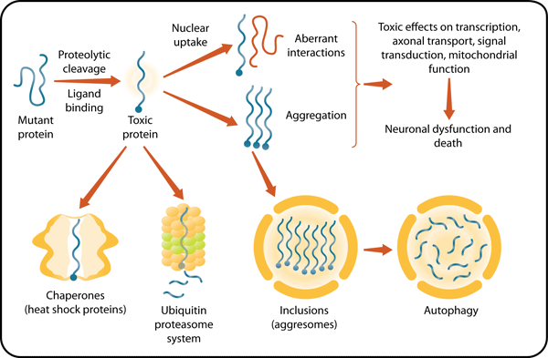
Figure 3 Source: Original designed by author and illustrator. Published previously in Pennuto, M. & Fischbeck, K. H. Therapeutic prospects for polyglutamine disease. In Ramirez-Alvarado, M., Kelly, J. W. & Dobson, C. M. (eds.), Protein Misfolding Diseases: Current and Emerging Principles and Therapies. Hoboken, NJ: John Wiley & Sons, 2010. File name: 3-5_ F3_Rinaldi_Fischbeck in jpg and eps format

Transcriptional Dysregulation

One way to explain the toxic effects of polyglutamine proteins is that they accumulate in the nucleus and have abnormal interactions with nuclear proteins, which in turn disrupt transcription14. Many proteins that normally regulate transcription are found in nuclear inclusions and have been shown to interact with polyglutamine expanded proteins15. The mutant proteins may prevent these transcriptional regulators from carrying out their normal functions.

14.Zoghbi, H. Y . & Orr, H. T. Glutamine repeats and neurodegeneration. Annual Review of Neuroscience 23, 217–247 (2000). doi: 10.1146/annurev.neuro.23.1.217

15.Yamada, M., Tsuji, S. & Takahashi, H. Pathology of CAG repeat diseases. Neuropathology 20, 319–325 (2000). doi: 10.1111/j.1440-1789.2000.00354.x

Mitochondrial Dysfunction

Several findings show that polyglutamine diseases are associated with mitochondrial dysfunction. For example, mitochondria isolated from HD patients and a mouse model of HD have defects in their membrane potential and depolarization16. Mutant forms of huntingtin may disrupt mitochondrial function by decreasing the expression of the transcriptional coactivator PGC-1alpha, which regulates mitochondrial biogenesis and respiration17.

16.Panov, A. V. et al. Early mitochondrial calcium defects in Huntington's disease are a direct effect of polyglutamines. Nature Neuroscience 5, 731–736 (2002). doi:10.1038/nn884

17.Cui, L. et al. Transcriptional repression of PGC-1alpha by mutant huntingtin leads to mitochondrial dysfunction and neurodegeneration. Cell 127, 59–69 (2006). doi:10.1016/j.cell.2006.09.015

Impairment of the Ubiquitin-Proteasome System

The proteasome is a protein complex that degrades unneeded or damaged proteins. Proteins are targeted for proteasome-mediated degradation when a small protein called ubiquitin is attached to them by a ubiquitin ligase enzyme. The cellular inclusions associated with polyglutamine diseases are ubiquitinated and contain proteasome components, suggesting that the ubiquitin-proteasome system (UPS) may be sequestered by polyglutamine expanded proteins9, 18. In addition, ataxin-3, the protein associated with the polyglutamine disease SCA3 has been shown to bind to and remove attached ubiquitins from proteins that are targeted for UPS-mediated degradation19, 20, 21. Studies of the polyglutamine diseases SCA7 and HD, however, have not shown a role for UPS defects22, 23. Further studies are needed to confirm whether impairment of the UPS contributes to polyglutamine disease pathogenesis.

9.Paulson, H. L. et al. Intranuclear inclusions of expanded polyglutamine protein in spinocerebellar ataxia type 3. Neuron 19, 333–344 (1997). doi: 10.1016/S0896-6273(00)80943-5

18.Chai, Y. et al. Evidence for proteasome involvement in polyglutamine disease: Localization to nuclear inclusions in SCA3/MJD and suppression of polyglutamine aggregation in vitro. Human Molecular Genetics 8, 673–682 (1999). doi: 10.1093/hmg/8.4.673

19.Burnett, B., Li, F. & Pittman, R. N. The polyglutamine neurodegenerative protein ataxin-3 binds polyubiquitylated proteins and has ubiquitin protease activity. Human Molecular Genetics 12, 3195–3205 (2003). doi: 10.1093/hmg/ddg344

20.Chai, Y. et al. Poly-ubiquitin binding by the polyglutamine disease protein ataxin-3 links its normal function to protein surveillance pathways. Journal of Biological Chemistry 279, 3605–3611 (2004). doi: 10.1074/jbc.M310939200

21.Donaldson, K. M. et al. Ubiquitin-mediated sequestration of normal cellular proteins into polyglutamine aggregates. PNAS 100, 8892–8897 (2003). doi: 10.1073/pnas.1530212100

22.Bowman, A. B. et al. Neuronal dysfunction in a polyglutamine disease model occurs in the absence of ubiquitin-proteasome system impairment and inversely correlates with the degree of nuclear inclusion formation. Human Molecular Genetics = 14, 679–691 (2005). doi: 10.1093/hmg/ddi064

23.Bett, J. S. et al. The ubiquitin-proteasome reporter GFPu does not accumulate in neurons of the R6/2 transgenic mouse model of Huntington's disease. PLoS One 4, e5128 (2009). doi: 10.1371/journal.pone.0005128

The Role of Autophagy

Autophagy is a process by which cellular components, including damaged or surplus organelles and protein aggregates, are delivered to the lysosome for degradation. In humans, defects in autophagy have been directly associated with neurodegeneration, cancer, and inflammatory diseases24. Signs of decreased efficiency of autophagy have been reported in several polyglutamine diseases, which may be contributing to the disease mechanism25.

24.Kundu, M. & Thompson, C. B. Autophagy: Basic principles and relevance to disease. Annual Review of Pathology 3, 427–455 (2008). doi: 10.1146/annurev.pathmechdis.2.010506.091842

25.Martinez-Vicente, M. et al. Cargo recognition failure is responsible for inefficient autophagy in Huntington′s disease. Nature Neuroscience 13, 567–576 (2010). doi: 10.1038/nn.2528

Other Mechanisms

Other cellular processes have been implicated in the toxicity of mutant polyglutamine proteins. For example, altered cellular signaling, intracellular calcium homeostasis26, 27, and axonal transport28, 29 have been found in several polyglutamine disorders.

26.Zeron, M. M. et al. Increased sensitivity to N-methyl-D-aspartate receptor-mediated excitotoxicity in a mouse model of Huntington's disease. Neuron 33, 849–860 (2002). doi: 10.1016/S0896-6273(02)00615-3

27.Tang, T. S. et al. Disturbed Ca2+ signaling and apoptosis of medium spiny neurons in Huntington's disease. Proceedings of the National Academy of Sciences of the United States of America 102, 2602–2607 (2005). doi: 10.1073/pnas.0409402102

28.Gunawardena, S. et al. Disruption of axonal transport by loss of huntingtin or expression of pathogenic polyQ proteins in Drosophila. Neuron 40, 25–40 (2003). doi: 10.1016/S0896-6273(03)00594-4.

29.Szebenyi, G. et al. Neuropathogenic forms of huntingtin and androgen receptor inhibit fast axonal transport. Neuron 40, 41–52 (2003). doi: 10.1016/S0896-6273(03)00569-5

The Protein Context

Although long polyglutamine tracts alone may be toxic and cause pathology in cell culture and animal models30, 31, the protein context is also important. For example, the toxicity of the mutant AR is dependent on the binding of androgens in animal models of SBMA32, and transgenic mice with high expression levels of the wild-type AR gene have a degenerative phenotype33. These findings suggest that polyglutamine expansion in SBMA may lead to toxicity by increasing normal androgen-dependent AR activity, perhaps by stabilizing the mutant protein.

Protein context is also important in the polyglutamine disease SCA1, which is associated with the ataxin-1 protein. For example, phosphorylation of the serine residue at position 776 in ataxin-1 is necessary for the toxicity of the mutant protein33. Transgenic mice expressing high levels of ataxin-1 with a normal polyglutamine repeat length but with an amino acid substitution at position 776 that mimics phosphorylation have pathological changes that are indistinguishable from those seen in mice that express high levels of mutant ataxin-1 with a polyglutamine expansion34. Similarly, phosphorylation at serines 13 and 16 in the huntingtin protein and at serines 215 and 792 in the AR protein plays an important role in HD and SBMA models, respectively35, 36.

Polyglutamine expansions may enhance normal or toxic activities associated with certain conformations of the protein (causing gain of function) and weaken the activities associated with others (causing loss of function). For example, a combination of loss and gain of function explains the clinical picture of SBMA, where patients often have signs of androgen insensitivity (breast enlargement and reduced fertility) combined with signs of motor neuron degeneration (weakness and muscle atrophy) due to acquired functions of the AR protein that are toxic to motor neurons.

30.Ordway, J. M. et al. Ectopically expressed CAG repeats cause intranuclear inclusions and a progressive late onset neurological phenotype in the mouse. Cell 91, 753–763 (1997). doi: 10.1016/S0092-8674(00)80464-X

31.Marsh, J. L. et al. Expanded polyglutamine peptides alone are intrinsically cytotoxic and cause neurodegeneration in Drosophila. Human Molecular Genetics 9, 13–25 (2000). doi: 10.1093/hmg/9.1.13

32.Katsuno, M. et al. Testosterone reduction prevents phenotypic expression in a transgenic mouse model of spinal and bulbar muscular atrophy. Neuron 35, 843–854 (2002). doi: 10.1016/S0896-6273(02)00834-6

33.Monks, D. A. et al. Overexpression of wild-type androgen receptor in muscle recapitulates polyglutamine disease. Proceedings of the National Academy of Sciences of the United States of America 104, 18259–1864 (2007). doi: 10.1073/pnas.0705501104

34.Emamian, E. S. et al. Serine 776 of ataxin-1 is critical for polyglutamine-induced disease in SCA1 transgenic mice. Neuron 38, 375–387 (2003). doi: 10.1016/S0896-6273(03)00258-7

35.Duvick, L. et al. SCA1-like disease in mice expressing wild-type ataxin-1 with a serine to aspartic acid replacement at residue 776. Neuron 67, 929–935 (2010). doi: 10.1016/j.neuron.2010.08.022

36.Gu, X. et al. Serines 13 and 16 are critical determinants of full-length human mutant huntingtin induced disease pathogenesis in HD mice. Neuron 64, 828–840 (2009). doi: 10.1016/j.neuron.2009.11.020

SUMMARY: Unanswered Questions and Routes to Therapy

Our understanding of the mechanisms underlying polyglutamine diseases has broadened over the last 20 years. This increased understanding has allowed the identification of potential therapeutic targets—including the UPS, transcription factors, molecular chaperones (such as heat shock proteins) that protect cells from toxic proteins, and enzymes that cleave the disease proteins—but many questions remain. Why do these widely expressed polyglutamine-expanded proteins selectively damage the nervous system? Which cellular pathways are most vulnerable to the toxic insults of polyglutamine-expanded proteins? How do these responses account for the clinical features of the diseases? And, ultimately, how can this knowledge be harnessed to develop safe and effective treatments for polyglutamine diseases?

Our understanding of polyglutamine disease mechanisms helps us design assays to identify compounds that can be tested in disease-specific cell culture systems, animal models, and finally in clinical trials in patients with these progressive neurodegenerative diseases.

References

1.La Spada, A. R. et al. Androgen receptor gene mutations in X-linked spinal and bulbar muscular atrophy. Nature 352, 77–79 (1991). doi: 10.1038/352077a0

2. Moseley, M. L. et al. Bidirectional expression of CUG and CAG expansion transcripts and intranuclear polyglutamine inclusions in spinocerebellar ataxia type 8. Nature Genetics 38, 758–769 (2006). doi :10.1038/ng1827

3.Cho, D. H. et al. Antisense transcription and heterochromatin at the DM1 CTG repeats are constrained by CTCF. Molecular Cell 20, 483–489 (2005). doi: 10.1016/j.molcel.2005.09.002

4.Wilburn, B. et al. An antisense CAG repeat transcript at JPH3 locus mediates expanded polyglutamine protein toxicity in Huntington's disease-like 2 mice. Neuron 70, 427–440 (2011). doi: 10.1016/j.neuron.2011.03.021

5.Brinkman, R. R. et al. The likelihood of being affected with Huntington disease by a particular age, for a specific CAG size. American Journal of Human Genetics 60, 1202–1210 (1997).

6.Mirkin, S. M. Expandable DNA repeats and human disease. Nature 447, 932–940 (2007). doi: 10.1038/nature05977

7.DiFiglia, M. et al. Aggregation of huntingtin in neuronal intranuclear inclusions and dystrophic neurites in brain. Science 277, 1990–1993 (1997). doi: 10.1126/science.277.5334.1990

8.Davies, S. W. et al. Formation of neuronal intranuclear inclusions underlies the neurological dysfunction in mice transgenic for the HD mutation. Cell 90, 537–548 (1997). doi: 10.1016/S0092-8674(00)80513-9

9.Paulson, H. L. et al. Intranuclear inclusions of expanded polyglutamine protein in spinocerebellar ataxia type 3. Neuron 19, 333–344 (1997). doi: 10.1016/S0896-6273(00)80943-5

10. Klement, I. A. et al. Ataxin-1 nuclear localization and aggregation: Role in polyglutamine-induced disease in SCA1 transgenic mice. Cell 95, 41–53 (1998). doi: 10.1016/S0092-8674(00)81781-X

11. Saudou, F. et al. Huntingtin acts in the nucleus to induce apoptosis but death does not correlate with the formation of intranuclear inclusions. Cell 95, 55–66 (1998). doi: 10.1016/S0092-8674(00)81782-1

12. Arrasate, M. et al. Inclusion body formation reduces levels of mutant huntingtin and the risk of neuronal death. Nature 431, 805–810 (2004). doi: 10.1038/nature02998

13. Hackam, A. S. et al. The influence of huntingtin protein size on nuclear localization and cellular toxicity. Journal of Cell Biology 141, 1097–1105 (1998). doi: 10.1083/jcb.141.5.1097

14.Zoghbi, H. Y . & Orr, H. T. Glutamine repeats and neurodegeneration. Annual Review of Neuroscience 23, 217–247 (2000). doi: 10.1146/annurev.neuro.23.1.217

15.Yamada, M., Tsuji, S. & Takahashi, H. Pathology of CAG repeat diseases. Neuropathology 20, 319–325 (2000). doi: 10.1111/j.1440-1789.2000.00354.x

16.Panov, A. V. et al. Early mitochondrial calcium defects in Huntington's disease are a direct effect of polyglutamines. Nature Neuroscience 5, 731–736 (2002). doi:10.1038/nn884

17.Cui, L. et al. Transcriptional repression of PGC-1alpha by mutant huntingtin leads to mitochondrial dysfunction and neurodegeneration. Cell 127, 59–69 (2006). doi:10.1016/j.cell.2006.09.015

18.Chai, Y. et al. Evidence for proteasome involvement in polyglutamine disease: Localization to nuclear inclusions in SCA3/MJD and suppression of polyglutamine aggregation in vitro. Human Molecular Genetics 8, 673–682 (1999). doi: 10.1093/hmg/8.4.673

19.Burnett, B., Li, F. & Pittman, R. N. The polyglutamine neurodegenerative protein ataxin-3 binds polyubiquitylated proteins and has ubiquitin protease activity. Human Molecular Genetics 12, 3195–3205 (2003). doi: 10.1093/hmg/ddg344

20.Chai, Y. et al. Poly-ubiquitin binding by the polyglutamine disease protein ataxin-3 links its normal function to protein surveillance pathways. Journal of Biological Chemistry 279, 3605–3611 (2004). doi: 10.1074/jbc.M310939200

21.Donaldson, K. M. et al. Ubiquitin-mediated sequestration of normal cellular proteins into polyglutamine aggregates. PNAS 100, 8892–8897 (2003). doi: 10.1073/pnas.1530212100

22.Bowman, A. B. et al. Neuronal dysfunction in a polyglutamine disease model occurs in the absence of ubiquitin-proteasome system impairment and inversely correlates with the degree of nuclear inclusion formation. Human Molecular Genetics = 14, 679–691 (2005). doi: 10.1093/hmg/ddi064

23.Bett, J. S. et al. The ubiquitin-proteasome reporter GFPu does not accumulate in neurons of the R6/2 transgenic mouse model of Huntington's disease. PLoS One 4, e5128 (2009). doi: 10.1371/journal.pone.0005128

24.Kundu, M. & Thompson, C. B. Autophagy: Basic principles and relevance to disease. Annual Review of Pathology 3, 427–455 (2008). doi: 10.1146/annurev.pathmechdis.2.010506.091842

25.Martinez-Vicente, M. et al. Cargo recognition failure is responsible for inefficient autophagy in Huntington′s disease. Nature Neuroscience 13, 567–576 (2010). doi: 10.1038/nn.2528

26.Zeron, M. M. et al. Increased sensitivity to N-methyl-D-aspartate receptor-mediated excitotoxicity in a mouse model of Huntington's disease. Neuron 33, 849–860 (2002). doi: 10.1016/S0896-6273(02)00615-3

27.Tang, T. S. et al. Disturbed Ca2+ signaling and apoptosis of medium spiny neurons in Huntington's disease. Proceedings of the National Academy of Sciences of the United States of America 102, 2602–2607 (2005). doi: 10.1073/pnas.0409402102

28.Gunawardena, S. et al. Disruption of axonal transport by loss of huntingtin or expression of pathogenic polyQ proteins in Drosophila. Neuron 40, 25–40 (2003). doi: 10.1016/S0896-6273(03)00594-4.

29.Szebenyi, G. et al. Neuropathogenic forms of huntingtin and androgen receptor inhibit fast axonal transport. Neuron 40, 41–52 (2003). doi: 10.1016/S0896-6273(03)00569-5

30.Ordway, J. M. et al. Ectopically expressed CAG repeats cause intranuclear inclusions and a progressive late onset neurological phenotype in the mouse. Cell 91, 753–763 (1997). doi: 10.1016/S0092-8674(00)80464-X

31.Marsh, J. L. et al. Expanded polyglutamine peptides alone are intrinsically cytotoxic and cause neurodegeneration in Drosophila. Human Molecular Genetics 9, 13–25 (2000). doi: 10.1093/hmg/9.1.13

32.Katsuno, M. et al. Testosterone reduction prevents phenotypic expression in a transgenic mouse model of spinal and bulbar muscular atrophy. Neuron 35, 843–854 (2002). doi: 10.1016/S0896-6273(02)00834-6

33.Monks, D. A. et al. Overexpression of wild-type androgen receptor in muscle recapitulates polyglutamine disease. Proceedings of the National Academy of Sciences of the United States of America 104, 18259–1864 (2007). doi: 10.1073/pnas.0705501104

34.Emamian, E. S. et al. Serine 776 of ataxin-1 is critical for polyglutamine-induced disease in SCA1 transgenic mice. Neuron 38, 375–387 (2003). doi: 10.1016/S0896-6273(03)00258-7

35.Duvick, L. et al. SCA1-like disease in mice expressing wild-type ataxin-1 with a serine to aspartic acid replacement at residue 776. Neuron 67, 929–935 (2010). doi: 10.1016/j.neuron.2010.08.022

36.Gu, X. et al. Serines 13 and 16 are critical determinants of full-length human mutant huntingtin induced disease pathogenesis in HD mice. Neuron 64, 828–840 (2009). doi: 10.1016/j.neuron.2009.11.020

37.Palazzolo, I. et al. Akt blocks ligand binding and protects against expanded polyglutamine androgen receptor toxicity. Human Molecular Genetics 16, 1593–1603 (2007). doi: 10.1093/hmg/ddm109

Quiz

Question 5.1

vTQlihtHEE7iOYNZrJmhjrKH9RRQ54PA4fQcqPTxlHFm1Jr8zYvZu0W6oam6DA9IhL+V0AHjtEVBaPrSiqwBx2hsreZnFTQuM5jWCNa45rU70vyGJmrPVZsdwN5uc0warmotD4pIcXaohbzzB+seQ0UGAAlKgp5a6drR1x8p0t/hlbq7qzZx/ZQYCUjCKxP/3lmxG7YkOVJreMcZwqRHYpebzFI8o/bTcKaV/eNOD+MzTy/ETXQL3bfJwG8eVR7OJg6Bk71kmoTm5aUbQKVsXDOl1kaXdwfGRjl1ZgHXT5mhROtsmQAc4K8f/I6DoCJQgCnya1pZ/KycoOjKsRvz47v5ky0BJI7QNI4txN3m5wIBCawNu3yhMaya51RNmrJLHPmigNcFs0mNVcRL7eMKKzZjgoZK4qFhoixyV4zbfssnWA+k+BSfe70dOaO/FzZOWdjKO0aXAADM4AHoLbQwRABPolmplAMUKHXpiUXzjvmlRY+igKGUZEsxM+otnC9gLhMFYhoYXs42K8bjDOav0Xs4pz0U5tF5QMOaz+Nxp1tUxdYWwp8xGA==
1. Which of the following statements could explain why there is often a threshold number of polyglutamine repeats necessary before a disease state begins?

Question 5.2

g+hZLQsD3UGCUE7pKt1IVhM+6Q9ag/Vty12QEBsbgN5XUQ9ZihTELkMvtGghJAiFSRaJEWR7Bg5pKH4FFJhDEtvPgagy9re4u0Ofl4lGFmHgjB9AuAa+4rHSOi+hfpsMqut/IOLroMx12k8y1bN4bou/+OIk/06Rh3s9Pdmso1+mX7QooxxhgkAZPTpA/7BrlsI1TrTEgGxuRKl2PBLVyH3tWuzgBZPel/Cmir16vwYQL0H3ww0UzCpJS5JY456nHuQrEVp5cA3Iwpe2AFAboXHlT5jRmUR0sEW18CATwH0nJKbpTTzVEobpm4hQm33P3eWCQnT7ldZBuzV8P7Jic+u33r2MshUfO0q1LO5BLVRisi4qSER1lKUVoTF89pgGiCEzuKCIO5q/3ThwXIOev0NSTqwETzWRRptq17G5mvRlzOfzL+7hi9a3PWVW4f5QXIuw7Wel/ihbrOQd+JLKxU9wxwZO0umfjGVgq5hEcGLs+NhZLmZSpZnUzKIhRtYBCs3rwwXzSbFt52OHrcj/ULrG3fO7RxciE4E8Xzs0YbNnjgu3PF6xzvQShmSi4nvNOYI0+XLcAq1uebspfs53n+kdmNVBuM9hGGECN8M7P0E06Udnza2fGqdSwFiaV1+KewwFUIOCV2jR07wGZ8ZX/bs779+Rh2CW
2. Which of the following results would NOT suggest that aggregation causes disease?

Question 5.3

bX0b93GzRqoAZ0SkPR8T7lb270jsxppWeKbDh52sfdjQiJkbJUDzW80i7ZAE9+3e0Jov+cjAalyat1/zJwr8nCZXd13HcdCzcK63p4LiZDw75osveHbFR2FIUzI11FBESHDnQ8Z4HYw9vptykWRLBJVe/BDSg3nr45+Kp7e9407KfwJ4s75kudujqyJ8lK196COVYpnE0GOqHhtC48VtNEwiTnBigu9vGUpPShaoBGNKPLvcAjAOEZmzQ0NIOZXcBiwIEw7DCpr8Z8ibbLIURFlGOy+eJbDocF1AxRWgsLRwXG+QFhb3z5OhDitraCLiTVdJQGGBhCc3ATG9DIG2ZyEy1uiiytvX/E9cehRi84CmreBigzDmKrtrFdoxxvVbMw44SvnSrbSLXcheY7YF9DO281Shx/DbhiL4gKq33pkMYb+bx92l+Z4/rwEhIYynb1D4vgq4v0YnO8td26HGpnSjIrIodup2rQWxGvmmZrLtTiZVLkxoe5jIONMvJCL0GFc/SzqqPZp1bczduwqrkUD9fEWHyx31xJuxUOxEtOXzxRSwMQlBjX6nkK98m5VE
3. Which result is consistent with the model that the ubiquitin-proteasome system is involved in spinocerebellar ataxia type 7 (SCA7) neurodegenerative disease?

Question 5.4

oOM0hZxtyBxOz3tOIt7q9xXjY3NnUqddERmNzmAjL90dHv17DtSMtXIeQvTFCjh+Kya0cy24I6QtCH90Nr2S05RnYjmucH9f7slEZzdWj354OCs8X76ReD6MA6558YDxIL23pGoOAuAidN2r3OIIF9qk+P4e08D2lxYeVGM8IWUU99ogBJG/XRW6TFRDkWokfr+e7RbVlqaODgAAAR2thC3Xdp/CEILkb29+eGbrFYNuhxHl4nfvZ8QA3pw04OGGc+bKdtJ+PWhWH4WCR8PKDT13EZho8puOW2wWNbh1/Whrdg+UK/B1uGOUg1fL2XQO0Y/udKupDpRm9ZLYgQNXrcWheb1tu4bGtsRe3Cy2zyb7VlhVF9xSUJh2onfqR90A86/YTAGOHehSJ/y68ItY+6O7JCz9qBXHf4nzZ+nHjrfitqvf5fHf9gKgFem0yl+s8YNqlBwtSHl/geC3nLgDG5SntA3ibYs2SPoP6fIgfibU5sXQGiJSHrlgn0SA373a
4. How might polyglutamine expansions cause loss of function to the resulting protein?

Question 5.5

KVF95JUsD7v7leI7c/JNi1weS2Aujm6cPh2N6YNGV18IAR3yLiNq5rIbrKw/QpgO4FnRpsH5HWJKcvhgoZyljpcEeiA0+zSI1njJ8N4D54zpq8+0mc5CjL0S98zBklQN0fror61Gu/HnyevMuZnDxMO3fbP9u55hI9WVHBR8Q5M2CboZaPbAg40e1EstXJ8Ck4qjeOr8S61gEm6SBza/Oyl4Sw5CZ8CKlSem6F/VwKWw85pBt/8vTP7TXDC/t33FcmuhWj3mrPSbLoOsBznMjaWi4ojQmItabIj78ei/4cdRzbnfh+15GpuxtvEUnFC7IiECZLLwjBqlISmeZWDnW9sGQuT9o+Ji/7751SwMm5UtH+P4xeAAU8mnSAXoXywhZcQjd8Ccz8x6PGhnsvaVoJi6weFJXGr1gu53u1yAD3lyVu+kXbeSmGc51HevmB8IZe57viWhyMfRMVXRB1X3rPtPjSmTJBNT5xXeLy8K4q6Zpsp3iaLoBRX/BJf0v3J6TIC2GflsMS9kyZHMVliBDNhGFPUnP5Qe/NTAUGNu95MBuEHnCzi413P1u/B5+b49fsnc6rSOZaZ4HNBPjK9AMKGNslVXGl1UouMs9jKA3A8rJE6kqEpP7A==
5. Which of the following statements is NOT a reason that it is feasible to screen for drugs and develop treatments for polyglutamine diseases:

Question 5.6

hBUpkI6p8Mw5arjNsajS3vhQm2+e+Ra5CmfU+ybU7Hkponw8bB16LsNMPGkmMPxKPZNHOColknC+GSjaF51eCxuWNwRP5FMWdwDeA7fVBcCSNCDAcmIROzhP5PcmDWtNqY47954yrZMxnJQhAq+KMejwsUylw+MJjK2l+4nKB1RaM3ZZCZw8/Rgokmy0lGhmE/e/tUKl+m5Kss8QjZE43oUFUpWaqhOAlc1JA+lRoTD1163aw81TPwmbr8AXOhjEVxsvkT02UmVhuzN3sFqxS+0Yt9uY7j2e0PckE3kUOmadVdd3yIEVBjxHa8dUbOoRgGdSuzYyFtYaAaQxeVrWSjsBjloQYAUlkq3y7xDTUDe6Sq7/A/BtQXDoPWOUWQNlXO0uCFoG9u7AYhQiYqJwADaaq0v02OBrybaRhYw4rvGAFMwlFiBx+feT5O1i0N9R5Xv2i3sy0Uko5E4W3MU5U0U0mNYHZLSl+aRMviMfyV9dRP2/53V0CSHJ2mrCKTK+/Ja0ysYJ+ANDjIoSI9Ud6rBhRCLcBLCz24VcUgheEwUG77eqNRk0O1Zv3/lqoiY4ZrcB0nD/3qodx68mh94jiA4MxKDiOwvqWySzayefbdCb/yLCZoF/B9uEXYJxZwT4Kzq+hoDT3y+jOm2q8y5yYF454+oNCiR75HPBXwZWdtNPC/rTD03oAm9+emgo9AbxggV8Is20gjW/+cWe
6. How might polyglutamine proteins affect transcription?

Question 5.7

rTJHozfcJ/kcYISwsG92SLuV4K3dA1ly35SCCu3oU+hrlgJfixZzwFjgpHmJKboBCa9fXG86Awbi5FM/h10gpRGiHV8W49mVm01wUiQquqqkUXoiLJJ7Ut074fnV8lafJobBxDaH2lfNQrZVaJn/zndb5Zm1N6Sckh+b5aUbDjjcX5RMDQflgRqUQVRDdmukTEAtxBhGMWK/K4xuaX5SWGw6bBfJO5j0QOZeNQC4ykk7EjPgnWrnO8pJz5CUH0YvKY0StoPPly5yEePX4Yl7mRNZJyimstE2lHmjlvT0Ppt89lnrjiY4yrP5OfYYP9gowH091q5B8Ysu1v7qwUil1CqCVg2BsRVUYPGwpdQl2hDQRWIDwKIrt5sPzGxCtrgeIg/MLvXENcu6XkcmGBP1ChoCLF2gjm3wAWkpxlNMe9N0zEqIgvDCgVJ24PYMC6aI5qyS2bUYN0Km3VQK8ketM3C8eKTllg+1Ylt6/52dVYoOrniYpZW5ZwT8eIdW+FX0uOmlEnnNjy0mSPdzK1lo3c7sxLFcOKgnV3YWc8UXP08pA++ma4p9NsFNsgBQ0cCibzbPRCUkF3n6IH+ngTXEO7B7TMFwFU2F7JRO7zvBzBBfSxL/BtesrKzZZwvLqF3cHBv/x5h3vOg0UUwd3XFo7rGkDohV108m9F7DNg==
7. How does transcription contribute to the formation of polyglutamine expansions?