Chapter 1. Anatomy of the Brain

1.0.1 Anatomy of the Brain

true
Begin
Anatomy of the Brain
brainstem
Central structure of the brain (including the hindbrain, midbrain, thalamus, and hypothalamus) that is responsible for most life-sustaining, unconscious behavior.
hindbrain
Evolutionarily the oldest part of the brain; contains the pons, medulla, reticular formation, and cerebellum, the structures that coordinate and control most voluntary and involuntary movements.
midbrain
Central part of the brain; contains neural circuits for hearing and seeing as well as for orienting movements.
afferent
Conducting toward a CNS structure.
efferent
Conducting away from a CNS structure.
pons
This structure, immediately rostral to the medulla, forms a physical bridge between the cerebellum and the rest of the brain. The pons regulates various autonomic functions, receives sensory information from the head and neck, controls various involuntary movements, and plays an important role in the regulation of sleep.
diencephalon
The between brain, which integrates sensory and motor information on its way to the cerebral cortex.
tectum
Roof (area above the ventricle) of the midbrain; its functions are sensory processing, particularly visual and auditory, and the production of orienting movements.
tegmentum
Floor (area below the ventricle) of the midbrain; a collection of nuclei with movement-related, species-specific, and pain perception functions.
orienting movement
Movement related to sensory inputs, such as turning the head to see the source of a sound.
cerebral cortex
Heavily folded and layered tissue that is the outer structure of the forebrain; composed of neocortex and allocortex.
thalamus
Diencephalon structure through which most sensory information from the sensory systems is organized, integrated, and projected into the appropriate region of the neocortex.
hypothalamus
Diencephalon structure that contains many nuclei associated with temperature regulation, eating, drinking, and sexual behavior.
dorsal
toward the back
superior
above
inferior
below
nuclei
A group of neurons forming a cluster that can be identified using special stains
forebrain
Evolutionarily the most recent addition to the brain; coordinates advanced cognitive functions such as thinking, planning, and language; contains the allocortex, neocortex, and basal ganglia.
allocortex
Part of the cerebral cortex (“other bark”), composed of three or four layers; plays a role in controlling motivational and emotional states as well as in certain forms of memory.
neocortex
Most recently expanded outer layer (“new bark”) of the forebrain, typically composed of six layers of gray matter. Its name is a misnomer, as it actually isn’t newer because it arose at the same time during evolution as other forms of the cortex. It is also called isocortex because it is almost always six-layered, with few exceptions.
basal ganglia
Subcortical forebrain nuclei that coordinate voluntary movements of the limbs and body; connected to the thalamus and to the midbrain.
Parkinson disease
Disorder of the motor system correlated with a loss of dopamine from the substantia nigra and characterized by tremors, muscular rigidity, and a reduction in voluntary movement.
Dopamine
Amine neurotransmitter involved in coordinating movement, attention, learning, and reinforcing behaviors
Periaqueductal gray matter
Nuclei in the midbrain that surround the cerebral aqueduct joining the third and fourth ventricles; PAG neurons contain circuits for species-typical behaviors (e.g., female sexual behavior) and play an important role in the modulation of pain.
ventral
toward the belly
red nucleus
The red nucleus plays a role in coordinating movement of the arms and hands.
substantia nigra
The substantia nigra connects to the basal ganglia and is critical for the initiation of voluntary motor movement. Parkinson disease is a movement disorder caused by the deterioration of dopamine cells in this area of the tegmentum.
periaqueductal gray matter
This distinct collection of cells that surrounds the cerebral aqueduct plays a role in both species-typical behaviors, such as female sexual behavior, and the modulation of pain.
reticular formation
This diffuse collection of cells that extend from the medulla (caudally) to the midbrain (rostrally) is involved in arousal, attention, and wakefulness.

Anatomy of the Brain

By: Dr. Daniel Hummer, Morehouse College

1.1 Anatomy of the Brain

Anatomy of the Brain

The human brain is composed of several major components, each containing numerous smaller structures. What role do these various structures play in producing behavior? This activity will help familiarize you with the names and functions of prominent areas of the hindbrain and midbrain, which are both part of the brainstem, and the forebrain, which sits on top.

After completing this activity, you should be able to:

  • Identify and describe structures of the hindbrain.
  • Identify and describe structures of the midbrain.
  • Identify and describe structures of the forebrain.

This activity relates to the following principles of nervous system function:

  • Principle 1: The Nervous System Produces Movement in a Perceptual World the Brain Constructs
  • Principle 4: The CNS Functions on Multiple Levels
  • Principle 5: The Brain Is Symmetrical and Asymmetrical
  • Principle 6: Brain Systems Are Organized Hierarchically and in Parallel
  • Principle 7: Sensory and Motor Divisions Permeate the Nervous System
  • Principle 9: Brain Functions Are Localized and Distributed

1.2 The Hindbrain

The Hindbrain

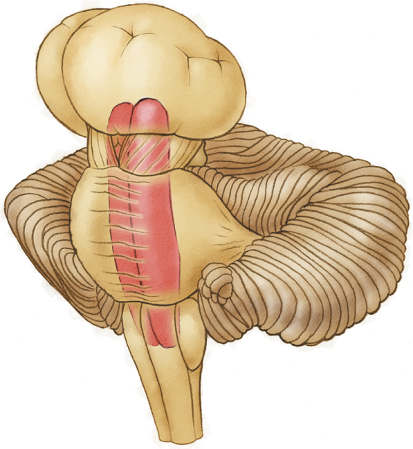
The brainstem, which includes the hindbrain and midbrain, begins where the spinal cord enters the skull. It receives afferent signals from the body so that a sensory world can be constructed and sends efferent signals out to direct movement.

Rotate the figure (by either moving the image with your mouse/touchpad or using the arrow buttons) to view the hindbrain from various perspectives and to learn more about the hindbrain and its components.

1.3 Test Your Knowledge: The Hindbrain

Test Your Knowledge: The Hindbrain
true

Before proceeding, check your knowledge of the hindbrain structures by matching the colored circle on the drawing with the appropriate term.

Note: you must choose all correct answers before proceeding.
Image

1.4 The Midbrain and Diencephalon

The Midbrain and Diencephalon

The midbrain is another major brainstem component that is located superior to the pons and inferior to the diencephalon. The midbrain is composed of two main structures, the tectum and the tegmentum, which together process sensory information and facilitate movement related to it, called orienting movement.

The diencephalon integrates and relays sensory and motor information to the cerebral cortex. Two of its principle structures are the thalamus and hypothalamus.

Explore the diencephalon and midbrain, as well as their components. Rotate the figure (by either moving the image with your mouse/touchpad or using the arrow buttons) to view the midbrain from various perspectives.

1.5 The Midbrain: Tegmentum

The Midbrain: Tegmentum

Distinct nuclei within the tegmentum serve a variety of important functions, from the initiation and control of movement to the processing of pain information.

Let’s learn about each component of the tegmentum.

1.6 Test Your Knowledge: The Midbrain

Test Your Knowledge: The Midbrain
true

Test your knowledge of the midbrain by matching each structure to the corresponding feature or function. You will have three attempts to correctly match all items.

1.7 The Forebrain

The Forebrain
true

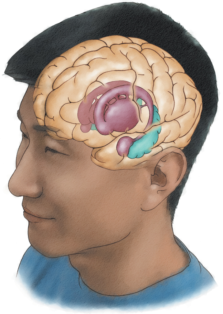
The forebrain sits on top of and surrounds the brainstem and is the largest region of the mammalian brain. It includes two principle structures: the cerebral cortex, made up of the allocortex and neocortex, and the basal ganglia.

Now, we’ll explore each component of the forebrain. First, explore the neocortex, allocortex, and basal ganglia, and then use the Next button to view some of the structures within each of these regions. You must explore all of the regions before you move on.

Rotate the figure (by either moving the image with your mouse/touchpad or using the arrow buttons) to view the forebrain from various perspectives and learn more about the forebrain components.

1.8 Test Your Knowledge: The Forebrain

Test Your Knowledge: The Forebrain
true
Finish

Before proceeding, let's check your knowledge of the forebrain structures by matching the colored circle in the drawing with the correct term.
Image

1.9 Summary

Summary

Congratulations! You have successfully completed this activity on brain anatomy. In this activity, you reviewed the names and functions of prominent areas of the hindbrain, midbrain, and forebrain.

Your instructor may now have you take a short quiz about this activity. Good luck!

Question

q6XWQJflWtVkaFwYK6NEneWW7Y2E1zzqMCTXSg==
true
Viewed final slide of activity